You have no items in your shopping cart.
Myelin Basic Protein antibody
SKU: orb2276388
Description
Images & Validation
−Item 1 of 3
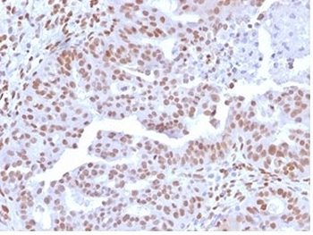
| Tested Applications | IHC |
|---|---|
| Dilution Range | 1:100-1:200 |
| Reactivity | Human |
| Application Notes |
Key Properties
−| Host | Rabbit |
|---|---|
| Clonality | Monoclonal |
| Isotype | IgG |
| Clone No. | MSVA-390R |
| Immunogen | Recombinant fragment (around aa 150-250) of human MBP (exact sequence is proprietary) |
| Conjugation | Unconjugated |
Storage & Handling
−| Storage | Maintain refrigerated at 2-8°C for up to 2 weeks. For long term storage store at -20°C in small aliquots to prevent freeze-thaw cycles. |
|---|---|
| Expiration Date | 12 months from date of receipt. |
| Disclaimer | For research use only |
Alternative Names
−GDB; Golli MBP; myelin basic protein; Hemopoietic MBP; HMBPR; HUGO; MLD; Myelin A1 Protein, basic; Myelin Deficient; Myelin membrane encephalitogenic protein; SHI; Shiverer; SP
Similar Products
−- Item 1 of 13
MBP Rabbit Polyclonal Antibody [orb783422]
IF, IHC-Fr, IHC-P, WB
Bovine, Canine, Equine, Porcine, Rabbit, Sheep
Human, Mouse, Rat
Rabbit
Polyclonal
Unconjugated
50 μl, 100 μl, 200 μl - Item 1 of 6
PRMT7 Antibody [orb1410050]
FC, IF, WB
Human
Mouse
Monoclonal
Unconjugated
20 μg, 100 μg, 100 μg (without BSA and Azide) - Item 1 of 4
PRMT7 Antibody [orb1410049]
FC, IF, WB
Human
Mouse
Monoclonal
Unconjugated
20 μg, 100 μg, 100 μg (without BSA and Azide) - Item 1 of 6
NFIA Antibody / Nuclear Factor 1 A [orb2634961]
FACS, IF, IHC-P, WB
Human
Mouse
Monoclonal
Unconjugated
100 μg - Item 1 of 6
NFIA Antibody / Nuclear Factor 1 A [orb2634962]
FACS, IF, IHC-P, WB
Human
Mouse
Monoclonal
Unconjugated
100 μg, 20 μg


Quality Guarantee
Explore bioreagents carefree to elevate your research. All our products are rigorously tested for performance. If a product does not perform as described on its datasheet, our scientific support team will provide expert troubleshooting, a prompt replacement, or a refund. For full details, please see our Terms & Conditions and Buying Guide. Contact us at [email protected].
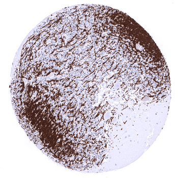
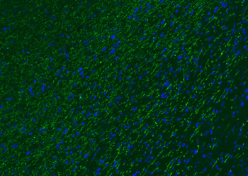
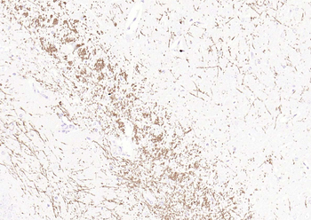
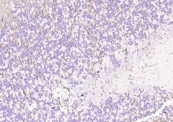
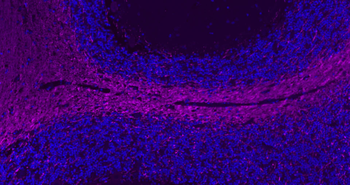
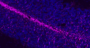
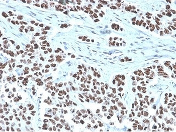
In the cerebellum a fibrillar MBP immunostaining occurs along intracerebral axons.
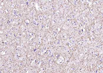
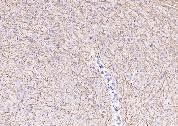
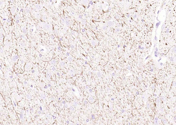
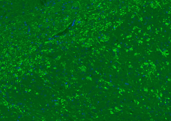
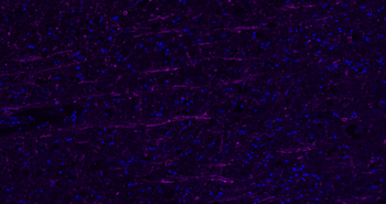
In the cerebrum a fibrillar MBP immunostaining occurs along intracerebral axons. Neurons are not stained.
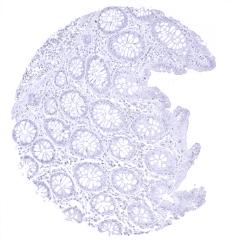
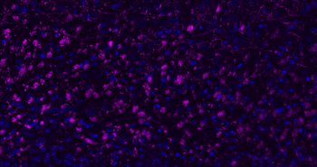
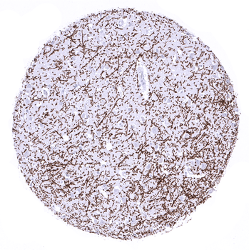
Complete absence of MBP immunostaining in epithelial and stromal cells of the appendix mucosa.
Quick Database Links
UniProt
UniProt Details
− No UniProt data available
Documents Download
Datasheet
Product Information
Request a Document
Myelin Basic Protein antibody (orb2276388)

- 0.0
Based on 0 reviews
Participating in our Biorbyt product reviews program enables you to support fellow scientists by sharing your firsthand experience with our products.
Login to Submit a ReviewAvailable Sizes
Select a size below