You have no items in your shopping cart.
Description
Research Area
Protein Biochemistry
Images & Validation
−Item 1 of 8
| Tested Applications | DOT, ELISA, SDS-PAGE, WB |
|---|---|
| Application Notes |
Key Properties
−| Source | Human |
|---|---|
| Biological Origin | Human |
| Isotype | IgG F(c) |
| Conjugation | Unconjugated |
| Purity | HUMAN IgG F(c) was prepared from normal serum by a multi-step process which includes delipidation, salt fractionation, ion exchange chromatography and papain digestion followed by chromatographic separation and extensive dialysis against the buffer stated above. Assay by immunoelectrophoresis resulted in a single precipitin arc against anti-Human Serum, anti-Human IgG and anti-Human IgG F(c). No reaction was observed against anti-Human IgG F(ab’)2 or anti-Papain. |
Storage & Handling
−| Storage | Store vial at 4° C prior to opening. This product is stable 4° C as an undiluted liquid. Dilute only prior to immediate use. For extended storage mix with an equal volume of glycerol, aliquot contents and freeze at -20° C or below. Avoid cycles of freezing and thawing. |
|---|---|
| Form/Appearance | Liquid (sterile filtered) |
| Buffer/Preservatives | Preservative: 0.01% (w/v) Sodium Azide. Stabilizer: None; Buffer: 0.02 M Potassium Phosphate, 0.15 M Sodium Chloride, pH 7.2 |
| Concentration | 1.029 mg/mL |
| Expiration Date | 12 months from date of receipt. |
| Disclaimer | For research use only |
Alternative Names
−Human Immunoglobulin G F(c) Fragment, IgG Fc
Similar Products
−- Item 1 of 9
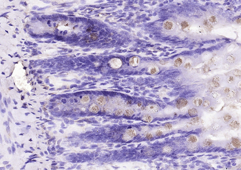
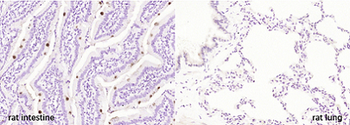
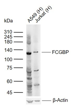
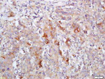
FCGBP Rabbit Polyclonal Antibody [orb156853]
IF, IHC-Fr, IHC-P, WB
Canine, Rabbit
Human, Mouse, Rat
Rabbit
Polyclonal
Unconjugated
50 μl, 100 μl, 200 μl - Item 1 of 11
Multi-rAb CoraLite Plus 750-Goat Mouse Recombinant Secondary Antibody (H+L) [orb2302689]
FC, IF, WB
Mouse
Goat
Recombinant
CoraLite® Plus 750
200 μl, 1 ml - Item 1 of 10
Multi-rAb CoraLite Plus 488-Goat Rabbit Recombinant Secondary Antibody (H+L) [orb2302686]
FC, IF
Rabbit
Goat
Recombinant
CoraLite® Plus 488
1 ml, 200 μl - Item 1 of 9
FCGR2A Rabbit Polyclonal Antibody [orb443179]
ELISA, IHC, WB
Human, Mouse, Rat
Rabbit
Polyclonal
Unconjugated
100 μg - Item 1 of 8
Multi-rAb CoraLite Plus 647-Goat Rabbit Recombinant Secondary Antibody (H+L) [orb2302683]
FC, IF
Rabbit
Goat
Recombinant
CoraLite® Plus 647
200 μl, 1 ml

Quality Guarantee
Explore bioreagents carefree to elevate your research. All our products are rigorously tested for performance. If a product does not perform as described on its datasheet, our scientific support team will provide expert troubleshooting, a prompt replacement, or a refund. For full details, please see our Terms & Conditions and Buying Guide. Contact us at [email protected].
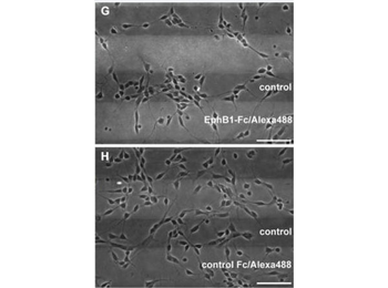
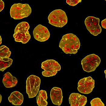
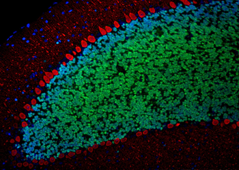
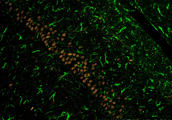
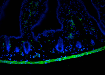
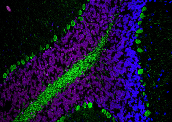
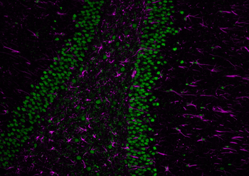
EphB1 acts repulsive on neurons of the superficial migratory stream via reverse signaling with stripe assay. (G) Dissociated neurons from the MGE clearly avoid EphB1-containing lanes in the stripe assay after 2 DIV. (H) On alternating stripes of labeled and unlabeled control protein the cells show no preferential growth. Scale bars: (G-H) 50 µm.

EphB1 acts repulsive on neurons of the superficial migratory stream via reverse signaling with stripe assay. (J) After down regulation of ephrin-B3 ligands by siRNA transfection (red) in MGE-derived neurons the repulsion induced by EphB1-Fc in the stripe assay is abolished after 2 DIV, while non-transfected interneurons still avoid the EphB1-Fc stripes. (K) Application of Alexa555 labeled control siRNA has no effect as most of the cells still avoid the EphB1-Fc containing stripes. (L) Addition of ephrin-B3 siRNA does not affect MGE-derived neurons growing on Fc/control stripes. Scale bars: (J–L) 50 µm.
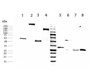
SDS-PAGE of Human IgG Fc (orb346220). Lane 1: Non-reduced Human IgG Fc. Lane 2: Non-reduced Human IgG Whole Molecule (orb346219). Lane 3: Non-reduced Human IgG Fab Fragment (orb346222). Lane 4: Non-reduced Human IgG F(ab')2 (orb346221). Middle Lane: 5 µl OPAL Pre-stained Marker. Lane 5: Reduced Human IgG Fc. Lane 6: Reduced Human IgG Whole Molecule (orb346219). Lane 7: Reduced Human IgG Fab Fragment (orb346222). Lane 8: Reduced Human IgG F(ab')2 (orb346221). Load: 1 µg per lane. Predicted/Observed size: Non-reduced at 50kDa, reduced at 25kDa/Non-reduced at 55-60kDa, reduced at 30-33kDa.
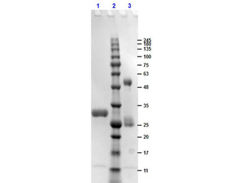
SDS-PAGE results of Human IgG F(c) Fragment. Lane 1: reduced Human IgG F(c) Fragment. Lane 2: Opal Prestained Molecular Weight Ladder. Lane 3: non-reduced Human IgG F(c) Fragment. Load: 1 µg. 4-20% Lonza SDS-PAGE; Coomassie Stained; BioRad ChemiDoc Imaged.
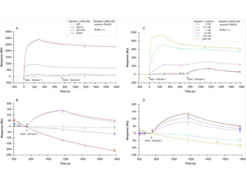
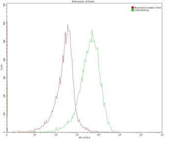
SPR interaction analyses regarding the aptamer binding site in Protein A.Biacore X100 / sensor chip CAP / ligand: biotinylated Protein A with immobilization level of ~560 RU / two-step analyte binding without regeneration in between, (A-B) analyte 1 = sample 1: human IgG, IgG-Fc fragment, IgG-Fab fragment with a concentration of 1000 nM each, or buffer, (C-D) analyte 1 = sample 1: concentration series of human IgG-Fc in the range of 0–1000 nM, (A-D) analyte 2 = sample 2: 2000 nM 5'-fluorescein-labeled aptamer PA#2/8 or buffer. Double-referenced sensorgrams are shown (blank reference surface without Protein A, buffer injection). Binding of sample 1 followed by sample 2 is shown in (A) and (C) with alignment to injection start of sample 1. In (B) and (D) only binding of sample 2 with alignment to injection start of sample 2 is shown.
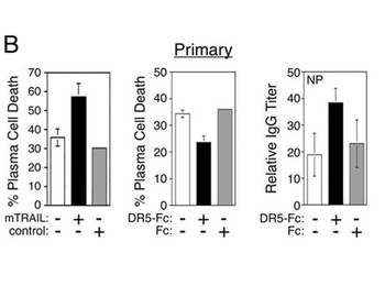
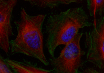
TRAIL induces apoptosis of primary plasma cells generated in a T-dependent immune response. (B) The in vivo-generated, syndecan-1-positive plasma cells were cultured alone (□) or with TRAIL-expressing 2PK3 cells (mTRAIL, ▪) or the parental 2PK3 (control, ▦) cells at a 10:1 ratio for 5 h before determination of cell death by trypan blue staining (left panel). In vivo-generated plasma cells were plated onto CD40L-expressing L cells at a 10:1 ratio in the absence (□) or presence (▪) of DR5-Fc or Fc (▦) (1 µg/ml). Cell viability was determined by trypan blue exclusion after a 5-h incubation (middle panel), and the relative amount of NP-specific IgG secreted into the medium for a total of 20 h was determined by ELISA (right panel). For each of the conditions, the values represent independent analysis of cells isolated from three mice.
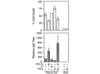
TRAIL-mediated apoptosis of human plasma cells. Top, Control CESS cells (blast) or IL-6-differentiated plasma cells were cultured for 24 h in the presence or absence of 1.0 µg/ml TRAIL, DR5-Fc, or the Fc portion of human IgG. Cell viability was determined by trypan blue exclusion, and the percent of plasma cell death was calculated by dividing the number of dead plasma cells by the total number of plasma cells in the culture. Bottom, ELISA of IgG secretion in the corresponding culture medium. Data represent the relative IgG titer required to obtain a single OD value that lies within the linear range. Data represent three independent cell populations from one experiment. Three independent experiments were performed with similar results.
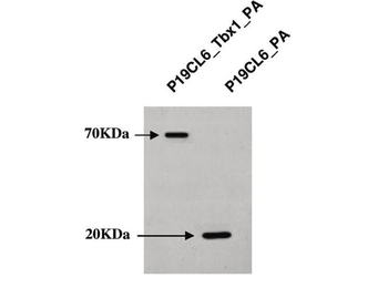
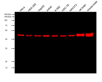
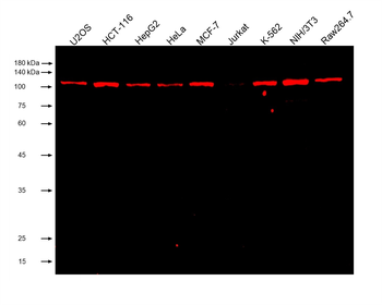
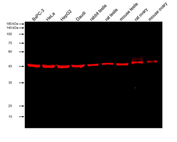
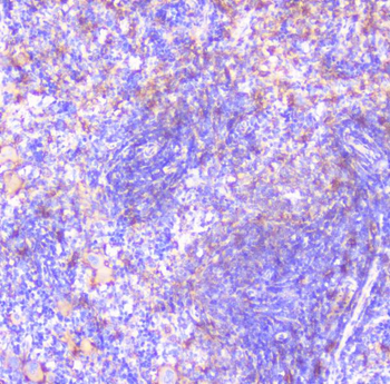
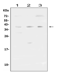
Western blot analysis of P19CL6_Tbx1-PA cells and P19CL6_PA control cells. Proteins were separated on 10% SDS-PAGE gel and immunoblotted with human IgG F(c).
Documents Download
Datasheet
Product Information
Request a Document
Protocol Information
Protein Handling and Storage Guide
Protein Handling Guide
WB
Western Blot (IB, immunoblot)
ELISA
Enzyme-linked Immunosorbent Assay (EIA)
DOT
Dot Blot
SDS-PAGE
Sodium Dodecyl Sulphate PolyAcrylamide Gel Electrophoresis
Human IgG Fc Antibody (orb346220)

- 0.0
Based on 0 reviews
Participating in our Biorbyt product reviews program enables you to support fellow scientists by sharing your firsthand experience with our products.
Login to Submit a ReviewAvailable Sizes
Select a size below