You have no items in your shopping cart.
Human HGF Protein
SKU: orb429387
Description
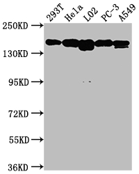
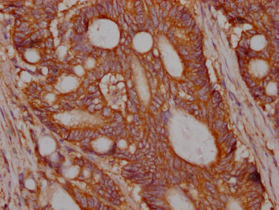
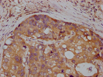
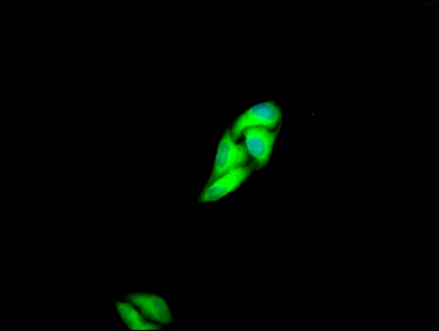
Images & Validation
−| Application Notes |
|---|
Key Properties
−| Source | Insect cells |
|---|---|
| Biological Activity | The activity was assayed for scattering activity in the MDCK cell assay. The ED50 for this effect is typically at 2.0-5.0 ng/ml. |
| Protein Sequence | QRKRRNTIHEFKKSAKTTLIKIDPALKIKTKKVNTADQCANRCTRNKGLPFTCKAFVFDKARKQCLWFPFNSMSSGVKKEFGHEFDLYENKDYIRNCIIGKGRSYKGTVSITKSGIKCQPWSSMIPHEHSYRGKDLQENYCRNPRGEEGGPWCFTSNPEVRYEVCDIPQCSEVECMTCNGESYRGLMDHTESGKICQRWDHQTPHRHKFLPERYPDKGFDDNYCRNPDGQPRPWCYTLDPHTRWEYCAIKTCADNTMNDTDVPLETTECIQGQGEGYRGTVNTIWNGIPCQRWDSQYPHEHDMTPENFKCKDLRENYCRNPDGSESPWCFTTDPNIRVGYCSQIPNCDMSHGQDCYRGNGKNYMGNLSQTRSGLTCSMWDKNMEDLHRHIFWEPDASKLNENYCRNPDDDAHGPWCYTGNPLIPWDYCPISRCEGDTTPTIVNLDHPVISCAKTKQLRVVNGIPTRTNIGWMVSLRYRNKHICGGSLIKESWVLTARQCFPSRDLKDYEAWLGIHDVHGRGDEKCKQVLNVSQLVYGPEGSDLVLMKLARPAVLDDFVSTIDLPNYGCTIPEKTSCSVYGWGYTGLINYDGLLRVAHLYIMGNEKCSQHHRGKVTLNESEICAGAEKIGSGPCEGDYGGPLVCEQHKMRMVLGVIVPGRGCAIPNRPGIFVRVAYYAKWIHKIILTYKVPQS |
| Purity | Greater than 95.0% as determined by SDS-PAGE. |
Storage & Handling
−| Storage | Stability: Lyophilized Hepatocyte Growth Factor although stable at room temperature for 3 weeks, should be stored desiccated below -18°C. Upon reconstitution HGF should be stored at 4°C between 2-7 days and for future use below -18°C. For long term storage it is recommended to add a carrier protein (0.1% HSA or BSA).Please prevent freeze-thaw cycles |
|---|---|
| Form/Appearance | Sterile Filtered White lyophilized (freeze-dried) powder. |
| Buffer/Preservatives | The sterile protein powder (1 mg/ml) is lyophilized from a solution containing 50mM acetic acid. |
| Expiration Date | 6 months from date of receipt. |
| Disclaimer | For research use only |
Alternative Names
−Scatter Factor (SF), Hepatopoietin (HPTA), HGF, HGFB, F-TCF.
Similar Products
−- Item 1 of 5
- Item 1 of 1
Human Hepatocyte Growth Factor Receptor (HGFR) ELISA Kit [orb777576]
Human
125-8000 pg/mL
48 pg/mL
48 T, 96 T - Item 1 of 1
- Item 1 of 1
- Item 1 of 1
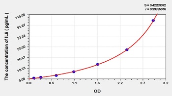
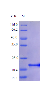
Human IL6 protein (Active) [orb358972]
> 96% as determined by SDS-PAGE and HPLC.
20.7 kDa
E.Coli
5 μg, 500 μg, 100 μg


Quality Guarantee
Explore bioreagents carefree to elevate your research. All our products are rigorously tested for performance. If a product does not perform as described on its datasheet, our scientific support team will provide expert troubleshooting, a prompt replacement, or a refund. For full details, please see our Terms & Conditions and Buying Guide. Contact us at [email protected].
Documents Download
Datasheet
Product Information
Request a Document
Protocol Information
Human HGF Protein (orb429387)

- 0.0
Based on 0 reviews
Participating in our Biorbyt product reviews program enables you to support fellow scientists by sharing your firsthand experience with our products.
Login to Submit a ReviewAvailable Sizes
Select a size below