You have no items in your shopping cart.
Description
Research Area
Infectious Disease & Virology
Images & Validation
−Item 1 of 4
| Tested Applications | IHC-P, WB |
|---|---|
| Dilution Range | WB: 1:1000, IHC: 1:50, IHC-P: 1:25, IHC-P: 1:25 |
| Reactivity | Human |
Key Properties
−| Host | Mouse |
|---|---|
| Clonality | Monoclonal |
| Isotype | IgG1 |
| Clone No. | 938CT5.1.1 |
| Target | This H2AFX antibody is generated from mice immunized with a KLH conjugated synthetic peptide between 115-143 amino acids from the C-terminal region of human H2AFX. |
| Molecular Weight | 15145 Da |
| Conjugation | Unconjugated |
Storage & Handling
−| Storage | Maintain refrigerated at 2-8°C for up to 2 weeks. For long term storage store at -20°C in small aliquots to prevent freeze-thaw cycles |
|---|---|
| Form/Appearance | Purified monoclonal antibody supplied in PBS with 0.09% (W/V) sodium azide. This antibody is purified through a protein G column, followed by dialysis against PBS. |
| Expiration Date | 12 months from date of receipt. |
| Disclaimer | For research use only |
Alternative Names
−Histone H2AX, H2a/x, Histone H2AX, H2AFX, H2AX
Similar Products
−- Item 1 of 2
- Item 1 of 1
Histone H2A Rabbit Polyclonal Antibody [orb1289766]
WB
Bovine, Gallus, Human, Mouse, Rat
Rabbit
Polyclonal
Unconjugated
30 μl, 100 μl, 200 μl, 50 μl
Quality Guarantee
Explore bioreagents carefree to elevate your research. All our products are rigorously tested for performance. If a product does not perform as described on its datasheet, our scientific support team will provide expert troubleshooting, a prompt replacement, or a refund. For full details, please see our Terms & Conditions and Buying Guide. Contact us at [email protected].
Immunohistochemical analysis of paraffin-embedded Human Thymus section using Pink1. Diluted at 1: 50 dilution. A undiluted biotinylated goat polyvalent antibody was used as the secondary, followed by DAB staining.
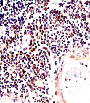
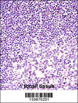
Immunohistochemical analysis of paraffin-embedded H. thymus section using H2AFX Antibody (C-term). Diluted at 1:25 dilution. A undiluted biotinylated goat polyvalent antibody was used as the secondary, followed by DAB staining.
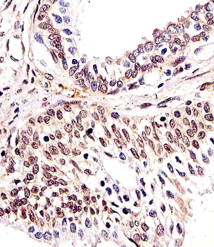
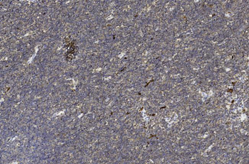
Immunohistochemical analysis of paraffin-embedded H. prostate section using H2AFX Antibody (C-term). Diluted at 1:25 dilution. A undiluted biotinylated goat polyvalent antibody was used as the secondary, followed by DAB staining.
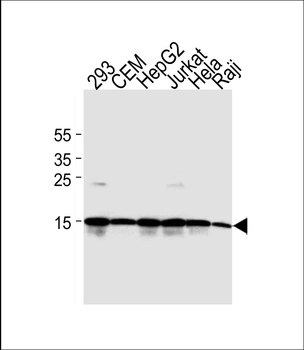
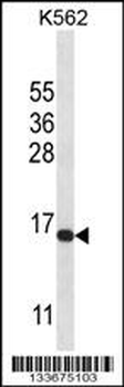
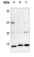
Western blot analysis of lysates from 293, CEM, HepG2, Jurkat, Hela, Raji cell line (from left to right). using H2AFX Antibody (C-term). Diluted at 1:2000 at each lane. A goat anti-mouse IgG H&L (HRP) at 1: 3000 dilution was used as the secondary antibody. Lysates at 35μg per lane.
Quick Database Links
Gene Symbol
This H2AFX antibody is generated from mice immunized with a KLH conjugated synthetic peptide between 115-143 amino acids from the C-terminal region of human H2AFX.
UniProt
RefSeq (Protein):NP_002096.1
UniProt Details
− No UniProt data available
NCBI Reference Sequences
−Associated Accession Numbers
Curated reference sequences for the gene transcript and protein product| Protein | NP_002096.1 |
|---|
Documents Download
Datasheet
Product Information
Request a Document
Protocol Information
WB
Western Blot (IB, immunoblot)
IHC-P
Immunohistochemistry Paraffin
H2AFX Antibody (C-term) (orb1927418)

- 0.0
Based on 0 reviews
Participating in our Biorbyt product reviews program enables you to support fellow scientists by sharing your firsthand experience with our products.
Login to Submit a ReviewAvailable Sizes
Select a size below