You have no items in your shopping cart.
GLS Antibody (C-term)
SKU: orb1928270
Description
Research Area
Cell Biology, Metabolism Research, Neuroscience, Signal Transduction
Images & Validation
−Item 1 of 7
| Tested Applications | FC, IF, IHC-P, WB |
|---|---|
| Dilution Range | WB: 1:1000, WB: 1:1000, IF: 1:25, IF: 1:25, IF: 1:25, WB: 1:1000, WB: 1:2000 |
| Reactivity | Human, Mouse, Rat |
Key Properties
−| Antibody Type | Primary Antibody |
|---|---|
| Host | Rabbit |
| Clonality | Polyclonal |
| Isotype | Rabbit IgG |
| Clone No. | RB22875 |
| Target | This GLS antibody is generated from rabbits immunized with a KLH conjugated synthetic peptide between 516-545 amino acids from the C-terminal region of human GLS. |
| Molecular Weight | 73461 Da |
| Conjugation | Unconjugated |
Storage & Handling
−| Storage | Maintain refrigerated at 2-8°C for up to 2 weeks. For long term storage store at -20°C in small aliquots to prevent freeze-thaw cycles |
|---|---|
| Form/Appearance | Purified polyclonal antibody supplied in PBS with 0.09% (W/V) sodium azide. This antibody is purified through a protein A column, followed by peptide affinity purification. |
| Expiration Date | 12 months from date of receipt. |
| Disclaimer | For research use only |
Alternative Names
−Glutaminase kidney isoform, mitochondrial, GLS, K-glutaminase, L-glutamine amidohydrolase, GLS, GLS1, KIAA0838
Similar Products
−- Item 1 of 1

GLS Antibody (C-term) [orb2995618]
IF, IHC-P, WB
Rat
Human, Mouse
Rabbit
Polyclonal
Unconjugated
50 μl, 100 μlGLS Antibody (C-term) [orb2996457]
FC, IF, IHC-P, WB
Human, Mouse, Rat
Rabbit
Polyclonal
Unconjugated
100 μl, 50 μl
Quality Guarantee
Explore bioreagents carefree to elevate your research. All our products are rigorously tested for performance. If a product does not perform as described on its datasheet, our scientific support team will provide expert troubleshooting, a prompt replacement, or a refund. For full details, please see our Terms & Conditions and Buying Guide. Contact us at [email protected].
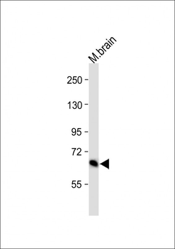
All lanes: Anti-GLS Antibody (C-term) at 1:1000 dilution. Lane 1: mouse brain lysate. Lysates/proteins at 20 µg per lane. Secondary Goat Anti-Rabbit IgG, (H+L), Peroxidase conjugated at 1/10000 dilution.Observed band size: 65 kDa. Blocking/Dilution buffer: 5% NFDM/TBST.
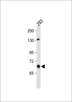
All lanes: Anti-GLS Antibody (C-term) at 1:1000 dilution. Lane 1: 293 whole cell lysate. Lysates/proteins at 20 µg per lane. Secondary Goat Anti-Rabbit IgG, (H+L), Peroxidase conjugated at 1/10000 dilution.Observed band size: 65 kDa. Blocking/Dilution buffer: 5% NFDM/TBST.
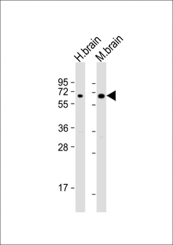
Western blot analysis of lysates from human brain, mouse brain ad rat kidney tissue lysate (from left to right), using GLS Antibody (C-term). Diluted at 1:1000 at each lane. A goat anti-rabbit IgG H&L (HRP) at 1:5000 dilution was used as the secondary antibody. Lysates at 35 ug per lane.
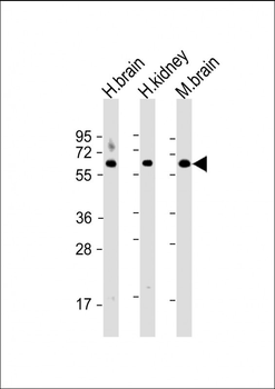
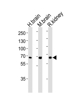
All lanes: Anti-GLS Antibody (C-term) at 1:2000 dilution. Lane 1: human brain lysate. Lane 2: human kidney lysate. Lane 3: mouse brain lysate. Lysates/proteins at 20 µg per lane. Secondary Goat Anti-Rabbit IgG, (H+L), Peroxidase conjugated at 1/10000 dilution. Predicted band size: 73 kDa. Blocking/Dilution buffer: 5% NFDM/TBST.
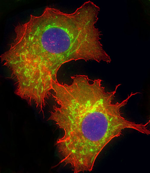
Immunofluorescent analysis of 4% paraformaldehyde-fixed, 0.1% Triton X-100 permeabilized U-251 MG cells labeling GLS at 1/25 dilution, followed by Dylight 488-conjugated goat anti-Rabbit IgG secondary antibody at 1/200 dilution (green). Immunofluorescence image showing Cytoplasm staining on U-251 MG cell line. Cytoplasmic actin is detected with Dylight 554 Phalloidin (red). The nuclear counter stain is DAPI (blue).
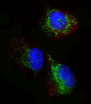
Immunofluorescent analysis of 4% paraformaldehyde-fixed, 0.1% Triton X-100 permeabilized HepG2 (human liver hepatocellular carcinoma cell line) cells labeling GLS at 1/25 dilution, followed by Dylight 488-conjugated goat anti-rabbit IgG (1583138) secondary antibody at 1/200 dilution (green). Immunofluorescence image showing mitochondrion staining on HepG2 cell line. Cytoplasmic actin is detected with Dylight 554 Phalloidin at 1/100 dilution (red). The nuclear counter stain is DAPI (blue).
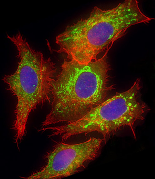
Immunofluorescent analysis of 4% paraformaldehyde-fixed, 0.1% Triton X-100 permeabilized HepG2 (human liver hepatocellular carcinoma cell line) cells labeling GLS at 1/25 dilution, followed by Dylight 488-conjugated goat anti-rabbit IgG (1583138) secondary antibody at 1/200 dilution (green). Immunofluorescence image showing mitochondrion staining on HepG2 cell line. Cytoplasmic actin is detected with Dylight 554 Phalloidin at 1/100 dilution (red). The nuclear counter stain is DAPI (blue).
Quick Database Links
Gene Symbol
This GLS antibody is generated from rabbits immunized with a KLH conjugated synthetic peptide between 516-545 amino acids from the C-terminal region of human GLS.
UniProt
RefSeq (Protein):NP_055720.3, NP_001243239.1
UniProt Details
− No UniProt data available
NCBI Reference Sequences
−Associated Accession Numbers
Curated reference sequences for the gene transcript and protein product| Protein | NP_055720.3, NP_001243239.1 |
|---|
Documents Download
Datasheet
Product Information
Request a Document
Protocol Information
WB
Western Blot (IB, immunoblot)
IHC-P
Immunohistochemistry Paraffin
FC
Flow Cytometry
IF
Immunofluorescence
GLS Antibody (C-term) (orb1928270)

- 0.0
Based on 0 reviews
Participating in our Biorbyt product reviews program enables you to support fellow scientists by sharing your firsthand experience with our products.
Login to Submit a ReviewAvailable Sizes
Select a size below