You have no items in your shopping cart.
FYN Antibody (N-term)
SKU: orb1928959
Description
Research Area
Signal Transduction
Images & Validation
−Item 1 of 4
| Tested Applications | FC, IHC-P, WB |
|---|---|
| Dilution Range | WB: 1:1000, WB: 1:1000, IHC-P: 1:50~100, FC: 1:10~50 |
| Reactivity | Human, Mouse |
| Predicted Reactivity | Rat |
Key Properties
−| Antibody Type | Primary Antibody |
|---|---|
| Host | Rabbit |
| Clonality | Polyclonal |
| Isotype | Rabbit IgG |
| Clone No. | RB03090 |
| Target | This FYN antibody is generated from rabbits immunized with a KLH conjugated synthetic peptide between 13-43 amino acids from the N-terminal region of human FYN. |
| Molecular Weight | 60762 Da |
| Conjugation | Unconjugated |
Storage & Handling
−| Storage | Maintain refrigerated at 2-8°C for up to 2 weeks. For long term storage store at -20°C in small aliquots to prevent freeze-thaw cycles |
|---|---|
| Form/Appearance | Purified polyclonal antibody supplied in PBS with 0.09% (W/V) sodium azide. This antibody is purified through a protein A column, followed by peptide affinity purification. |
| Expiration Date | 12 months from date of receipt. |
| Disclaimer | For research use only |
Alternative Names
−Tyrosine-protein kinase Fyn, Proto-oncogene Syn, Proto-oncogene c-Fyn, Src-like kinase, SLK, p59-Fyn, FYN
Similar Products
−- Item 1 of 3
- Item 1 of 3

FYN Antibody (N-term) [orb2996478]
FC, IHC-P, WB
Bovine, Gallus, Rat
Human, Mouse
Rabbit
Polyclonal
Unconjugated
100 μl, 50 μl


Quality Guarantee
Explore bioreagents carefree to elevate your research. All our products are rigorously tested for performance. If a product does not perform as described on its datasheet, our scientific support team will provide expert troubleshooting, a prompt replacement, or a refund. For full details, please see our Terms & Conditions and Buying Guide. Contact us at [email protected].

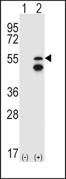
Western blot analysis of FYN (arrow) using rabbit polyclonal FYN Antibody (N-term). 293 cell lysates (2 ug/lane) either nontransfected (Lane 1) or transiently transfected (Lane 2) with the FYN gene.
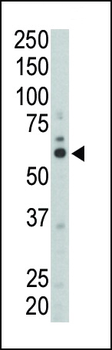
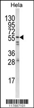
Western blot analysis of anti-FYN (N-term) Pab in mouse liver tissue lysate. FYN (arrow) was detected using purified Pab. Secondary HRP-anti-rabbit was used for signal visualization with chemiluminescence.
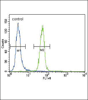
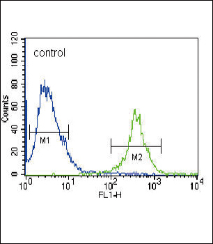
FYN Antibody (N-term) flow cytometric analysis of Hela cells (right histogram) compared to a negative control cell (left histogram). FITC-conjugated goat-anti-rabbit secondary antibodies were used for the analysis.
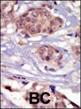
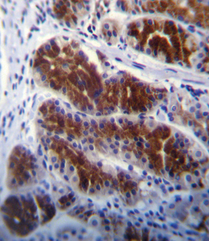
Formalin-fixed and paraffin-embedded human cancer tissue reacted with the primary antibody, which was peroxidase-conjugated to the secondary antibody, followed by DAB staining. This data demonstrates the use of this antibody for immunohistochemistry; clinical relevance has not been evaluated. BC = breast carcinoma; HC = hepatocarcinoma.
Quick Database Links
Gene Symbol
This FYN antibody is generated from rabbits immunized with a KLH conjugated synthetic peptide between 13-43 amino acids from the N-terminal region of human FYN.
UniProt
RefSeq (Protein):NP_694592.1, NP_002028.1, NP_694593.1
UniProt Details
− No UniProt data available
NCBI Reference Sequences
−Associated Accession Numbers
Curated reference sequences for the gene transcript and protein product| Protein | NP_694592.1, NP_002028.1, NP_694593.1 |
|---|
Documents Download
Datasheet
Product Information
Request a Document
Protocol Information
WB
Western Blot (IB, immunoblot)
IHC-P
Immunohistochemistry Paraffin
FC
Flow Cytometry
FYN Antibody (N-term) (orb1928959)

- 0.0
Based on 0 reviews
Participating in our Biorbyt product reviews program enables you to support fellow scientists by sharing your firsthand experience with our products.
Login to Submit a ReviewAvailable Sizes
Select a size below