You have no items in your shopping cart.
Beta Galactosidase Antibody Fluorescein Conjugated
SKU: orb344712
Description
Images & Validation
−Item 1 of 3
| Tested Applications | DOT, WB |
|---|---|
| Dilution Range | IF: User Optimized, WB: User Optimized |
| Application Notes |
Key Properties
−| Antibody Type | Primary Antibody |
|---|---|
| Host | Rabbit |
| Clonality | Polyclonal |
| Isotype | IgG |
| Immunogen | Beta Galactosidase (E. coli) |
| Purity | Anti-Beta Galactosidase is an IgG fraction antibody purified from monospecific antiserum by a multi-step process which includes delipidation, salt fractionation and ion exchange chromatography followed by extensive dialysis against the buffer stated above. Assay by immunoelectrophoresis resulted in a single precipitin arc against anti-fluorescein, anti-Rabbit Serum and purified and partially purified Beta Galactosidase (E. coli). |
| Conjugation | FITC |
Storage & Handling
−| Storage | Store vial antibody at 4° C prior to restoration. For extended storage aliquot contents and freeze at -20° C or below. Avoid cycles of freezing and thawing. Centrifuge product if not completely clear after standing at room temperature. This product is stable for several weeks at 4° C as an undiluted liquid. Dilute only prior to immediate use. |
|---|---|
| Form/Appearance | Lyophilized |
| Buffer/Preservatives | Preservative: 0.01% (w/v) Sodium Azide. Stabilizer: 10 mg/mL Bovine Serum Albumin (rAlbumin) - Immunoglobulin and Protease free; Buffer: 0.02 M Potassium Phosphate, 0.15 M Sodium Chloride, pH 7.2 |
| Concentration | 1.0 mg/mL |
| Expiration Date | 12 months from date of receipt. |
| Disclaimer | For research use only |
Alternative Names
−rabbit anti-Beta Galactosidase Antibody fluorescein Conjugation, FITC conjugated rabbit anti-Beta Galactosidase Antibody, rabbit anti-beta gal FITC conjugated antibody, ß-Gal Fluorescein, Anti-ß-Gal Fluorescein Antibody
Similar Products
−- Item 1 of 3
- Item 1 of 3


Quality Guarantee
Explore bioreagents carefree to elevate your research. All our products are rigorously tested for performance. If a product does not perform as described on its datasheet, our scientific support team will provide expert troubleshooting, a prompt replacement, or a refund. For full details, please see our Terms & Conditions and Buying Guide. Contact us at [email protected].

Western blot using Biorbyt's anti-b-Galactosidase antibody shows detection of a band at ~117 kDa (lane 1) corresponding to b-Gal present in a partially purified preparation (arrowhead). Approximately 1 µg of protein was resolved on a 4-20% Tris-Glycine gel by SDS-PAGE and transferred onto nitrocellulose. After blocking, the membrane was probed with the primary antibody diluted to 1:1000. Reaction occurred overnight at 4°C followed by washes and reaction with a 1:10000 dilution of IRDye® 800 conjugated Gt-a-Rabbit IgG (H&L) MX10 for 45 min at room temperature (800 nm channel, green). Molecular weight estimation was made by comparison to prestained MW markers in lane M (700 nm channel, red).

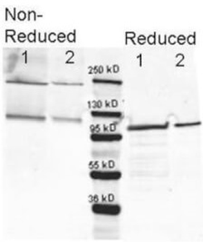
Western blotting using Biorbyt's anti-b-Galactosidase antibody. Lane 1 shows 80 ng and lane 2 shows 20 ng loaded onto gel. Results for non-reducing conditions of SDS-PAGE prior to transfer to nitrocellulose are shown on the left side of the figure; results obtainined under reducing conditions are shown on the right. Blots were blocked overnight at 4°C with Blocking Buffer for Fluorescent Western Blotting (p/n orb348637). The membrane was probed with anti-b-Galactosidase diluted to 1:10000. Reaction occurred overnight at 4°C. Dylight649™ conjugated Gt-a-anti-Rabbit IgG was used for detection. Molecular weight estimation was made by comparison to a prestained MW marker (center). in lane M.

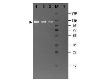
Western blotting using Biorbyt's Fluorescein conjugated anti-b-Galactosidase antibody shows a band at ~117 kDa (lanes 1 - 3) corresponding to 60 ng, 30 ng and 15 ng, respectively of b-Gal present in partially purified preparations (arrowhead). Lane 4 shows no cross reactivity with proteins present in a non-specific control E.coli lysate. Proteins were resolved on a 4-20% Tris-Glycine gel by SDS-PAGE and transferred to nitrocellulose and blocking using Blocking Buffer for Fluorescent Western Blotting (p/n orb348637). The membrane was probed with fluorescein conjugated anti-b-Galactosidase (p/n orb344848) diluted to 1:10000. Reaction occurred for 2 hours at room temperature. Molecular weight estimation was made by comparison to a prestained MW marker in lane M.
Quick Database Links
UniProt
RefSeq (Protein):NP_414878.1
UniProt Details
− No UniProt data available
NCBI Reference Sequences
−Associated Accession Numbers
Curated reference sequences for the gene transcript and protein product| Protein | NP_414878.1 |
|---|
Documents Download
Datasheet
Product Information
Request a Document
Protocol Information
WB
Western Blot (IB, immunoblot)
DOT
Dot Blot
Beta Galactosidase Antibody Fluorescein Conjugated (orb344712)

- 0.0
Based on 0 reviews
Participating in our Biorbyt product reviews program enables you to support fellow scientists by sharing your firsthand experience with our products.
Login to Submit a ReviewAvailable Sizes
Select a size below