You have no items in your shopping cart.
L1CAM Antibody
SKU: orb2650318
Description
Research Area
Neuroscience
Images & Validation
−Item 1 of 2
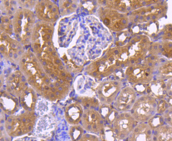
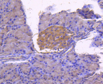
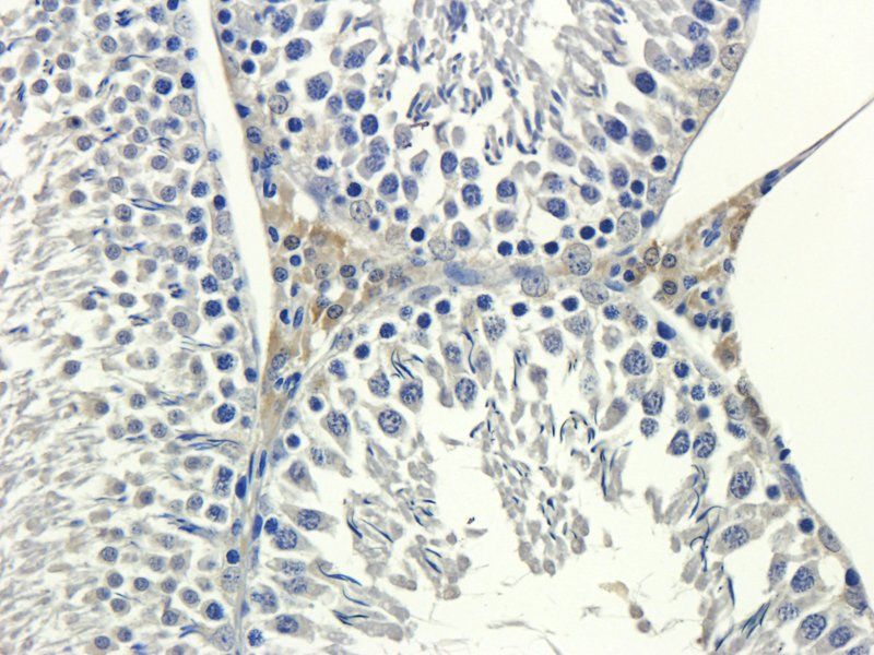
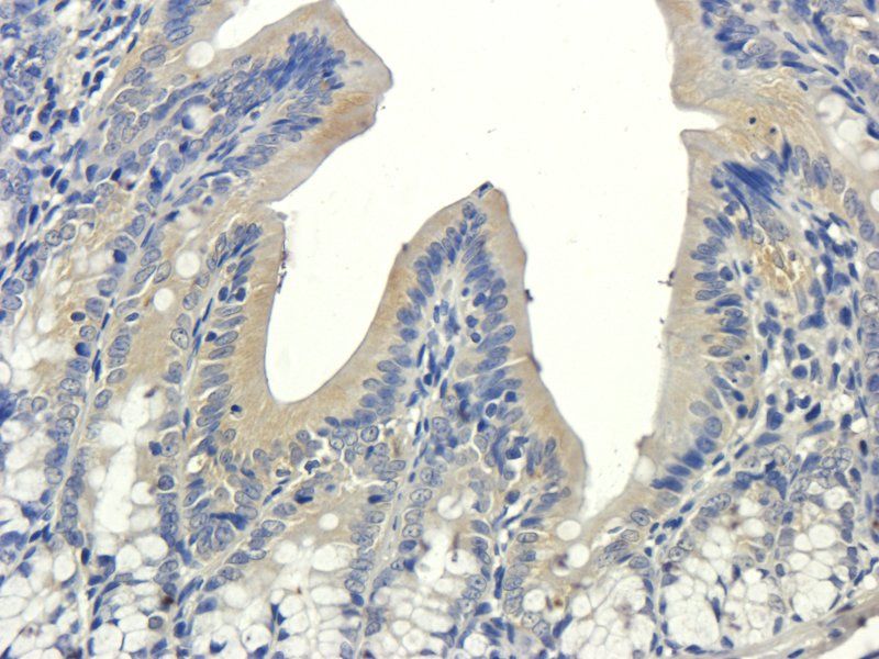
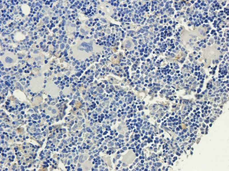
| Tested Applications | IF, WB |
|---|---|
| Dilution Range | WB: WB (1/500 - 1/1000), IF/IC (1/100 - 1/500), IF: WB (1/500 - 1/1000), IF/IC (1/100 - 1/500) |
| Reactivity | Human, Mouse, Rat, Zebrafish |
Key Properties
−| Host | Rabbit |
|---|---|
| Clonality | Polyclonal |
| Clone No. | L1CAM |
| Conjugation | Unconjugated |
Storage & Handling
−| Storage | Maintain refrigerated at 2-8°C for up to 2 weeks. For long term storage store at -20°C in small aliquots to prevent freeze-thaw cycles. |
|---|---|
| Expiration Date | 12 months from date of receipt. |
| Disclaimer | For research use only |
Alternative Names
−CAML1; MIC5; Neural cell adhesion molecule L1; N-CAM-L1; NCAM-L1; CD171
Similar Products
−- Item 1 of 11
Calnexin Recombinant Rabbit Monoclonal Antibody [orb704311]
IF, IHC-Fr, IHC-P, WB
Rat
Human
Rabbit
Recombinant
Unconjugated
50 μl, 100 μl, 25 μl - Item 1 of 6
VCAM1 Rabbit Polyclonal Antibody [orb348962]
IHC-P, WB
Human, Mouse, Rat
Rabbit
Polyclonal
Unconjugated
100 μg - Item 1 of 7
L1CAM Rabbit Polyclonal Antibody [orb402210]
IHC
Human, Mouse, Rat
Rabbit
Polyclonal
Unconjugated
100 μg - Item 1 of 1
Mouse L1-Cell Adhesion Molecule (L1CAM) ELISA Kit [orb777790]
Mouse
15.63-1000 pg/mL
7 pg/mL
48 T, 96 T - Item 1 of 1
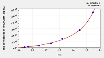
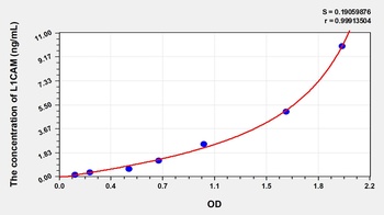
Human L1-Cell Adhesion Molecule (L1CAM) ELISA Kit [orb777665]
Human
0.16-10 ng/mL
0.055 ng/mL
96 T, 48 T
Quality Guarantee
Explore bioreagents carefree to elevate your research. All our products are rigorously tested for performance. If a product does not perform as described on its datasheet, our scientific support team will provide expert troubleshooting, a prompt replacement, or a refund. For full details, please see our Terms & Conditions and Buying Guide. Contact us at [email protected].
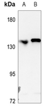
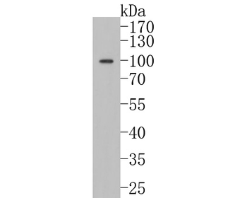
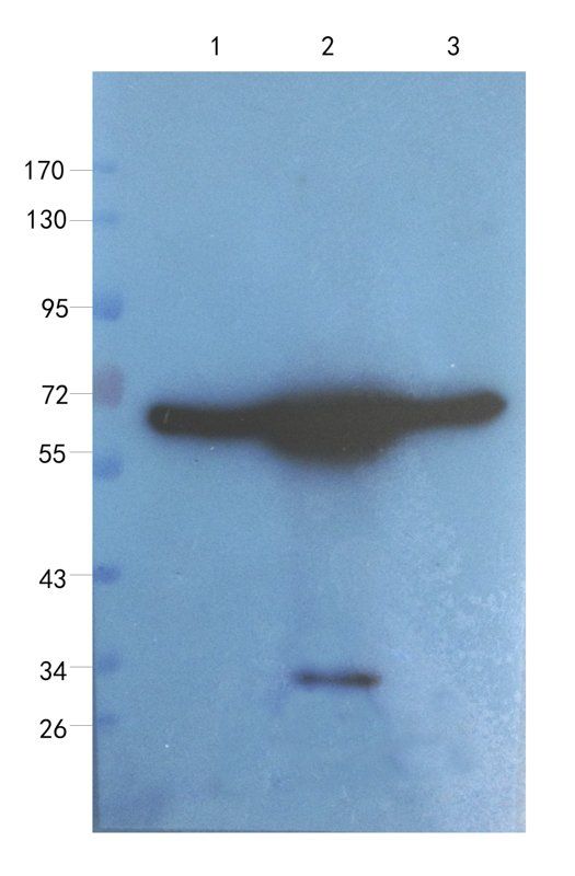
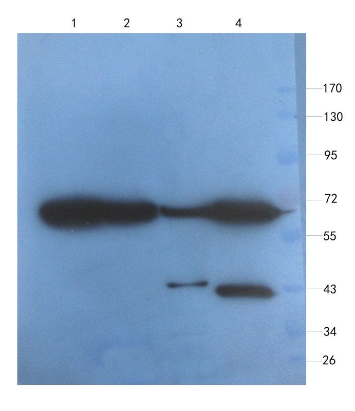
Western blot analysis of L1CAM expression in HEK293T (A), A549 (B) whole cell lysates.
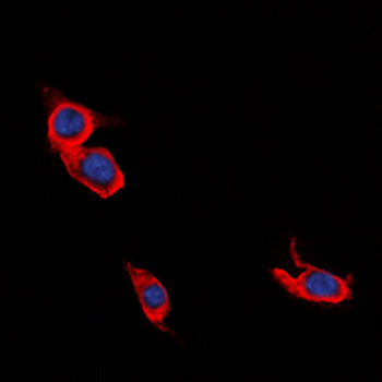
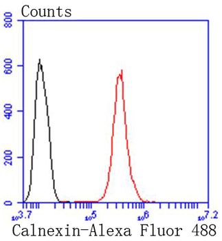
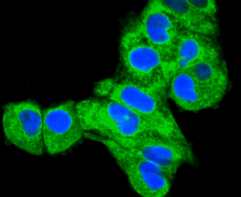
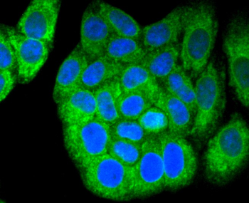
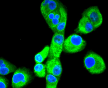
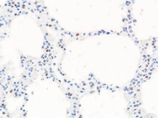
Immunofluorescent analysis of L1CAM staining in Jurkat cells. Formalin-fixed cells were permeabilized with 0.1% Triton X-100 in TBS for 5-10 minutes and blocked with 3% BSA-PBS for 30 minutes at room temperature. Cells were probed with the primary antibody in 3% BSA-PBS and incubated overnight at 4°C in a hidified chamber. Cells were washed with PBST and incubated with a DyLight 594-conjugated secondary antibody (red) in PBS at room temperature in the dark. DAPI was used to stain the cell nuclei (blue).
Quick Database Links
UniProt
UniProt Details
− No UniProt data available
Documents Download
Datasheet
Product Information
Request a Document
Protocol Information
WB
Western Blot (IB, immunoblot)
IF
Immunofluorescence
L1CAM Antibody (orb2650318)

- 0.0
Based on 0 reviews
Participating in our Biorbyt product reviews program enables you to support fellow scientists by sharing your firsthand experience with our products.
Login to Submit a ReviewAvailable Sizes
Select a size below