You have no items in your shopping cart.
Featured
Description
Research Area
Neuroscience
Images & Validation
−Item 1 of 7
| Tested Applications | In vitro, In vivo, SDS-PAGE, WB |
|---|---|
| Application Notes |
Key Properties
−| Source | Recombinant |
|---|---|
| Expression System | E. coli |
| Biological Origin | Human |
| Biological Activity | Does not induce Lewy body inclusion formation in Sprague-Dawley rat primary hippocampal neurons. Thioflavin T emission curve shows only a small increase in fluorescence (indicative of alpha synuclein aggregation) when Type 2 alpha synuclein PFFs are combined with alpha synuclein monomers. Certain biological activities in other neuronal cells cannot be ruled out. Researchers should test compatibility prior to use. |
| Target | Alpha Synuclein PFFs |
| Reactivity | Human |
| Tag | No tag |
| Protein Length | Full Length |
| Purification | Ion-exchange Purified |
| MW | ~14.46 kDa |
| Purity | >95% |
| Protein Sequence | MDVFMKGLSK AKEGVVAAAE KTKQGVAEAA GKTKEGVLYV GSKTKEGVVH GVATVAEKTK EQVTNVGGAV VTGVTAVAQK TVEGAGSIAA ATGFVKKDQL GKNEEGAPQE GILEDMPVDP DNEAYEMPSE EGYQDYEPEA |
Storage & Handling
−| Storage | -80°C |
|---|---|
| Buffer/Preservatives | PBS pH 7.4 |
| Concentration | 2 mg/ml |
| Expiration Date | 6 months from date of receipt. |
| Dry Ice Shipping | Please note: This product requires shipment on dry ice. A dry ice surcharge will apply. |
| Disclaimer | For research use only |
Alternative Names
−Alpha synuclein PFFs, Alpha synuclein PFF, Alpha synuclein aggregates, Alpha synuclein protein aggregates, Alpha synuclein aggregates, Alpha-synuclein protein, Non-A beta component of AD amyloid protein, Non-A4 component of amyloid precursor protein, NACP protein, SNCA protein, NACP protein, PARK1 protein, SYN protein, Parkinson disease familial 1 Protein
Similar Products
−- Item 1 of 9
Alpha Synuclein Pre-formed Fibrils [orb1822362]
In vitro, In vivo, SDS-PAGE, WB
>95%
~14.46 kDa
Recombinant
100 μg - Item 1 of 8
Alpha Synuclein A53T Mutant Pre-formed Fibrils [orb1822360]
In vitro, In vivo, SDS-PAGE, WB
>95%
~14.46 kDa
Recombinant
100 μg 
Alpha Synuclein Pre-formed Fibrils: ATTO 594 [orb3012996]
In vitro, In vivo, SDS-PAGE, WB
>95%
Recombinant
1 unitHuman Alpha-Synuclein (A53T) Pre-formed Fibrils Protein, Tag Free [orb3158027]
Unconjugated
90%
14.5 kDa
100 μg, 500 μg, 1 mgHuman Alpha-Synuclein Pre-formed Fibrils Protein, His Tag [orb3158057]
Unconjugated
90%
16.3 kDa
100 μg, 1 mg, 500 μg
Quality Guarantee
Explore bioreagents carefree to elevate your research. All our products are rigorously tested for performance. If a product does not perform as described on its datasheet, our scientific support team will provide expert troubleshooting, a prompt replacement, or a refund. For full details, please see our Terms & Conditions and Buying Guide. Contact us at [email protected].
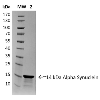
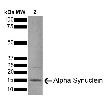
SDS-PAGE of ~14 kDa Human Recombinant Alpha Synuclein Protein Pre-formed Fibrils (Type 2). Lane 1: Molecular Weight Ladder (MW). Lane 2: Alpha Synuclein Protein Pre-formed Fibrils.
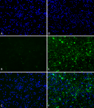
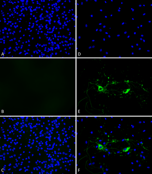
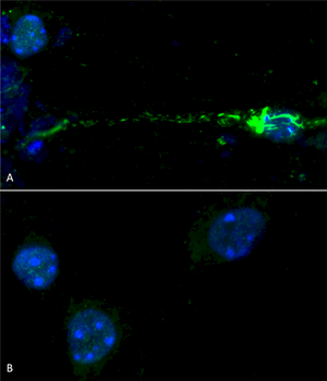
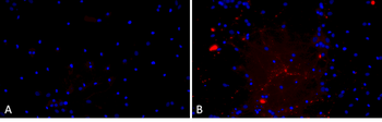
Primary rat hippocampal neurons show lewy body inclusion formation when treated with Type 1 Alpha Synuclein Pre-formed Fibrils at 4 μg/ml (D-F), but not when treated with Type 2 Alpha Synuclein Pre-formed Fibrils at 4 μg/ml (A-C). Tissue: Primary hippocampal neurons. Species: Sprague-Dawley rat. Fixation: 4% formaldehyde made from PFA. Primary Antibody: Mouse anti-pSer129 Antibody at 1:1000 24 hours at 4°C; Secondary Antibody: FITC Goat Anti-Mouse (green) at 1:700 for 1 hours at RT (B, E). Counterstain: Hoechst (blue) nuclear stain at 1:4000 for 1 hour at RT (A, D). Localization: Lewy body inclusions. Magnification: 20x. C is A and B merged and F is D and E merged.
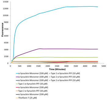
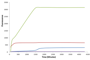
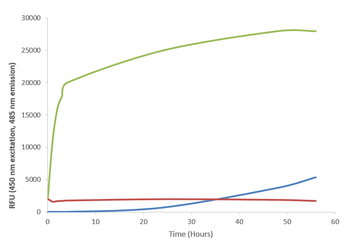
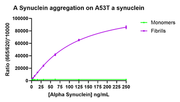
Type 1 alpha synuclein Pre-formed Fibrils seed the formation of new alpha synuclein fibrils from the pool of alpha synuclein monomers. Thioflavin T is a fluorescent dye that binds to beta sheet-rich structures, such as those in alpha synuclein fibrils. Upon binding, the emission spectrum of the dye experiences a red-shift and increased fluorescence intensity. Thioflavin T emission curves show increased fluorescence (correlated to alpha synuclein protein aggregation) over time when 10 μM of Type 1 alpha synuclein Pre-formed Fibrils is combined with 100 μM of alpha synuclein monomer, as compared to when 10 μM of Type 2 alpha synuclein Pre-formed Fibrils is combined with 100 μM of alpha synuclein monomer or 100 μM of alpha Synuclein monomer. Thioflavin T ex = 450 nm, em = 485 nm.
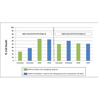
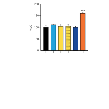
Toxicity results comparing Active Human Recombinant Alpha Synuclein Pre-formed Fibrils (Type 2) and Active Human Recombinant Alpha Synuclein Pre-formed Fibrils (Type 1). Data was graphed after live cell imaging results were obtained using the following procedure: After 8 days in vitro, primary rat mixed cortical neuron cells were washed with 1X PBS and treated with 500 μg/ml of Type 1 and Type 2 Alpha Synuclein Proteins for 20 hours at 37?C. Following treatements, cells were washed with 2X PBS and incubated with a staining solution (2.0 μM Cell Event + 2.5 μM Ethidium homodimer + 2.5 μg/ml Hoechst 33342 in sterile HBSS) for 30 minutes at 37?C. The addition of the Type 2 Alpha Synuclein Proteins resulted in a significant increase in cell death.

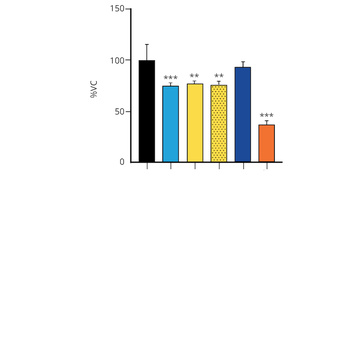
Evaluation of a-syn toxicity on primary mouse cortical neurons. Lactate dehydrogenase (LDH) is a soluble enzyme present in the cytosol that is released upon cell death. Toxicity was assessed with an LDH assay and displayed as % of vehicle control (VC). Data are presented as bar graphs and standard deviation.

Evaluation of a-syn toxicity on primary mouse cortical neurons. Mitochondrial dehydrogenase activity reduces yellow MTT to dark blue formazan crystals, a reaction catalyzed in living cells. Cell viability was assessed with an MTT assay and displayed as % of vehicle control (VC). Data are presented as bar graphs and standard deviation.
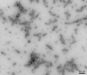
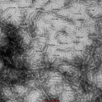
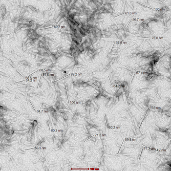
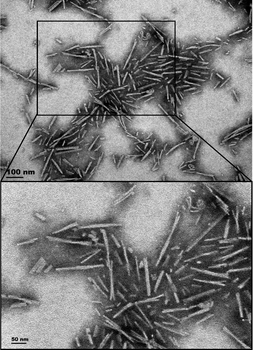
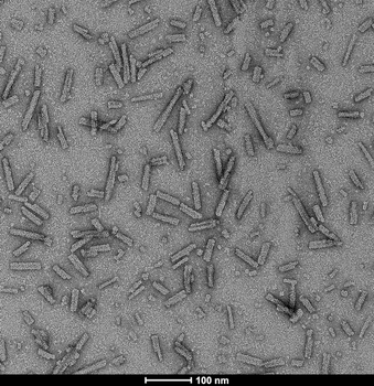
TEM of Type 2 Alpha Synuclein Pre-formed Fibrils (PFFs)
Quick Database Links
Gene Symbol
Alpha Synuclein PFFs
UniProt
UniProt Details
− No UniProt data available
Documents Download
Datasheet
Product Information
Request a Document
Protocol Information
Protein Handling and Storage Guide
Protein Handling Guide
WB
Western Blot (IB, immunoblot)
SDS-PAGE
Sodium Dodecyl Sulphate PolyAcrylamide Gel Electrophoresis
Alpha Synuclein Pre-formed Fibrils (orb1822369)

- 0.0
Based on 0 reviews
Participating in our Biorbyt product reviews program enables you to support fellow scientists by sharing your firsthand experience with our products.
Login to Submit a ReviewAvailable Sizes
Select a size below