You have no items in your shopping cart.
Featured
Description
Research Area
Neuroscience
Images & Validation
−Item 1 of 8
| Tested Applications | In vitro, In vivo, SDS-PAGE, WB |
|---|---|
| Application Notes |
Key Properties
−| Source | Recombinant |
|---|---|
| Expression System | E. coli |
| Biological Origin | Human |
| Biological Activity | 100 µM A53T alpha synuclein protein monomer seeded with 10 uM A53T alpha synuclein protein PFF in 25 µM Thioflavin T (PBS pH 7.4, 100 µl reaction volume) generated a fluorescence intensity of 28 000 Relative Fluorescence Units after incubation at 37°C with shaking at 600 rpm for 56 hours. Fluorescence was measured by excitation at 450 nm and emission at 485 nm on a Molecular Devices Gemini XPS microplate reader. |
| Target | Alpha Synuclein A53T PFFs |
| Reactivity | Human |
| Tag | No tag |
| Protein Length | Full Length |
| Purification | Ion-exchange Purified |
| MW | ~14.46 kDa |
| Purity | >95% |
| Protein Sequence | MDVFMKGLSK AKEGVVAAAE KTKQGVAEAA GKTKEGVLYV GSKTKEGVVH GVTTVAEKTK EQVTNVGGAV VTGVTAVAQK TVEGAGSIAA ATGFVKKDQL GKNEEGAPQE GILEDMPVDP DNEAYEMPSE EGYQDYEPEA |
Storage & Handling
−| Storage | -80°C |
|---|---|
| Buffer/Preservatives | PBS pH 7.4 |
| Concentration | 2 mg/ml |
| Expiration Date | 6 months from date of receipt. |
| Dry Ice Shipping | Please note: This product requires shipment on dry ice. A dry ice surcharge will apply. |
| Disclaimer | For research use only |
Alternative Names
−A53T mutant alpha synuclein, A53T mutated SNCA, A53T Alpha synuclein PFFs, Alpha synuclein PFF, Ala53thr mutant alpha synuclein, Alpha synuclein pre-formed fibrils, Alpha synuclein aggregates, Alpha synuclein protein aggregates, Alpha synuclein aggregates, Alpha-synuclein protein, Non-A beta component of AD amyloid protein, Non-A4 component of amyloid precursor protein, NACP protein, SNCA protein, NACP protein, PARK1 protein, SYN protein, Parkinson disease familial 1 Protein
Quality Guarantee
Explore bioreagents carefree to elevate your research. All our products are rigorously tested for performance. If a product does not perform as described on its datasheet, our scientific support team will provide expert troubleshooting, a prompt replacement, or a refund. For full details, please see our Terms & Conditions and Buying Guide. Contact us at [email protected].
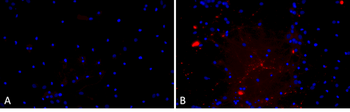
Primary rat hippocampal neurons show lewy body inclusion formation when treated with A53T mutant Alpha Synuclein Protein Pre-formed Fibrils (B) but not when treated with a media control (A). Tissue: Primary hippocampal neurons. Species: Sprague-Dawley rat. Primary Antibody: Rabbit anti-pSer129 Antibody. Fibrils were diluted to 1 ug/uL in neuronal media consisting of B27, Glutamax, penicillin/strip, and neurobasalA and sonicated for 1 hour in a water bath. The sonicated stock was then used to achieve the final concentration of 1 ug/mL in the wells.
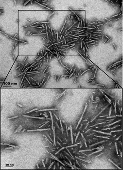
TEM of A53T alpha synuclein Pre-formed Fibrils
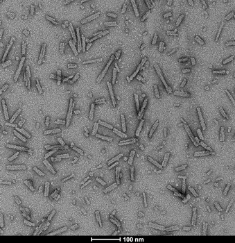
TEM of A53T alpha synuclein Pre-formed Fibrils
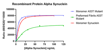
A53T alpha synuclein monomers and fibrils are well recognized by homogeneous time-resolved fluorescence (HTRF) total alpha synuclein assay.
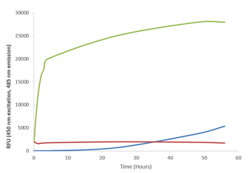
Thioflavin T is a fluorescent dye that binds to beta sheet-rich structures such as those in alpha synuclein fibrils. Upon binding, the emission spectrum of the dye experiences a red-shift and increased fluorescence intensity. Thioflavin T emission curves show a limited increase in fluorescence (correlated to alpha synuclein aggregation) over time in A53T alpha synuclein monomers. A much greater increase in fluorescence is seen when 100 μM monomer is combined with 10 μM of fibrils as the fibrils seed the formation of new fibrils from the pool of active monomers. Thioflavin T ex = 450 nm, em = 485 nm.
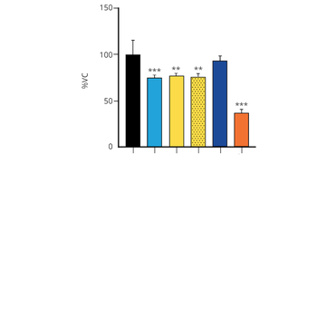
Evaluation of a-syn toxicity on primary mouse cortical neurons. Lactate dehydrogenase (LDH) is a soluble enzyme present in the cytosol that is released upon cell death. Toxicity was assessed with an LDH assay and displayed as % of vehicle control (VC). Data are presented as bar graphs and standard deviation.
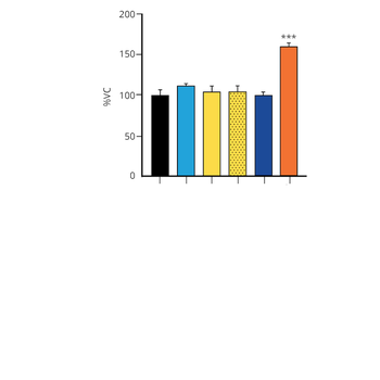
Evaluation of a-syn toxicity on primary mouse cortical neurons. Mitochondrial dehydrogenase activity reduces yellow MTT to dark blue formazan crystals, a reaction catalyzed in living cells. Cell viability was assessed with an MTT assay and displayed as % of vehicle control (VC). Data are presented as bar graphs and standard deviation.
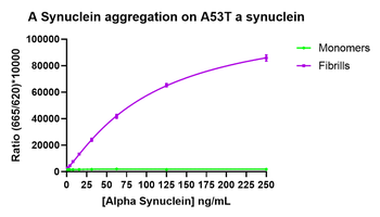
A53T alpha synuclein monomers and fibrils are well recognized by homogeneous time-resolved fluorescence (HTRF) alpha synuclein aggregation assay.
Quick Database Links
Gene Symbol
Alpha Synuclein A53T PFFs
UniProt
UniProt Details
− No UniProt data available
Documents Download
Datasheet
Product Information
Request a Document
Protocol Information
Protein Handling and Storage Guide
Protein Handling Guide
WB
Western Blot (IB, immunoblot)
SDS-PAGE
Sodium Dodecyl Sulphate PolyAcrylamide Gel Electrophoresis
Alpha Synuclein A53T Mutant Pre-formed Fibrils (orb1822360)

- 0.0
Based on 0 reviews
Participating in our Biorbyt product reviews program enables you to support fellow scientists by sharing your firsthand experience with our products.
Login to Submit a ReviewAvailable Sizes
Select a size below