You have no items in your shopping cart.
10X PBS pH 7.2
SKU: orb348532
Description
Images & Validation
−Item 1 of 2
| Application Notes |
|---|
Key Properties
−| Purity | 10X PBS buffer was aseptically filtered through a Millipore 0.22 micron filter into clean, pre-sterilized containers. The product was tested on trypticase soy agar for 24 hours, 48 hours and 72 hours and was found to be negative for bacteria. |
|---|---|
| Conjugation | Unconjugated |
Storage & Handling
−| Storage | Store container at room temperature (18° to 26° C) prior to opening. If desired, the solution may be stored at 4° C or less. Some salts may precipitate out of solution at lower temperature. Allow buffer to equilibrate to room temperature (18° to 26° C) to restore solubility of some salts. |
|---|---|
| Form/Appearance | Liquid |
| Buffer/Preservatives | Preservative: None. Stabilizer: None; Buffer: See application note. |
| Concentration | 10X |
| Expiration Date | 12 months from date of receipt. |
| Hazard Information | Non-Toxic |
| Disclaimer | For research use only |
Alternative Names
−Phosphate buffered saline, Phosphate buffered solution, PBS, 10X PBS
Similar Products
−- Item 1 of 2


Quality Guarantee
Explore bioreagents carefree to elevate your research. All our products are rigorously tested for performance. If a product does not perform as described on its datasheet, our scientific support team will provide expert troubleshooting, a prompt replacement, or a refund. For full details, please see our Terms & Conditions and Buying Guide. Contact us at [email protected].

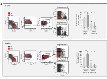
A subset of Tem-like cells sorted based on surface markers defining clusters 12 and 13 are highly susceptible to HIV infection. (A) Shown are the CyTOF datasets, with UI CD4+ T cells shown in gray and the HIV-susceptible PRE cells shown in red. Cells were pre-gated on live, singlet CD3 + CD19−CD8−CD4+ T cells. A sequential gating strategy was then implemented using surface markers characteristic of HIV-susceptible cells as defined by clusters 12 and 13. This strategy was used to characterize a final population of "population-1" cells (CD3 + CD4+ CD45RO + CD45RA−CCR7low/medCD29med/highCD69med/high CD62LlowCD57low/med), which were more abundant among PRE cells than among UI cells. For comparison, we characterized a "population-2" (CD3 + CD4+ CD45RO + CD45RA−CCR7low/medCD29lowCD69low and not CD62LlowCD57low/med) predicted to be much less susceptible to infection because it comprised a significantly lower proportion of PRE cells. The gating strategies are shown on the left, whereas the graphs on the right depict the frequencies of the population-1 and population-2 subsets within the UI and PRE cell populations. Note that the over-representation of population-1 cells among PRE cells suggest their preferential susceptibility to infection, whereas the under-representation of population-2 cells among PRE cells suggest their relative resistance to infection. *p < 0.05, ****p < 0.0001 as determined by a Student's paired t test. Error bars correspond to the standard deviation.
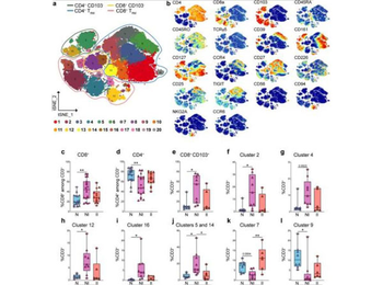
Quantification of CD8 and CD4 T cell clusters by CyTOF analysis in LP of control and CD patients. a Schematic t-SNE of CD4+ and CD8+ T cells from LP of all donors concatenated together (n = 18) controls (N), n = 8; CD, non-inflamed site (NI), n = 9; CD, inflamed site (II), n = 6. Total of 23 samples. b t-SNE of the indicated markers in CD4+ and CD8+ T cells. c, d Quantification of total CD8+ (c), total CD4+ (d) in LP of controls and CD patients by CyTOF (triangles = fresh samples) and FACS (circles = frozen samples). c, d Control (N), n = 17 (8 fresh, 9 frozen); CD, non-inflamed site (NI) n = 19 (9 fresh, 10 frozen); CD, inflamed site (II), n = 14 (6 fresh, 8 frozen). e–i Quantification of total CD8+ TRM (e) and CD8+ clusters 2 (f), 4 (g), 12 (h), and 16 (i) in LP of controls and CD patients by CyTOF. j–l Quantification of the CD4+ clusters 5 and 14 (j), 7 (k), and 9 (l) in LP of controls and CD patients by CyTOF. e–l Controls (N), n = 8; CD, non-inflamed site (NI), n = 9; CD, inflamed site (II), n = 6. Circles and triangles on the boxplots show data collected for each individual donor. Data were median and interquartile range. Significance was calculated using an ordinary, one-way ANOVA, multiple comparisons test with Prism v8 software. c **P = 0.0014; d **P = 0.028; e *P = 0.0139; f *P = 0.0178; h *P = 0.0178; i *P = 0.0219; j N vs. NI *P = 0.0156, NI vs. II *P = 0.0465; k **P = 0.0014; l **P = 0.0283. TRM tissue-resident memory T cell.
Documents Download
Datasheet
Product Information
Request a Document
Protocol Information
WB
Western Blot (IB, immunoblot)
ELISA
Enzyme-linked Immunosorbent Assay (EIA)
10X PBS pH 7.2 (orb348532)

- 0.0
Based on 0 reviews
Participating in our Biorbyt product reviews program enables you to support fellow scientists by sharing your firsthand experience with our products.
Login to Submit a ReviewAvailable Sizes
Select a size below