
You have no items in your shopping cart.
Cancer Biology Research
Tumor Suppressor Proteins: Pathways, Functions, and Detection
Explore the critical roles of tumor suppressors like p53, Rb, and PTEN in maintaining genomic stability, compare their mechanisms against oncogenes, and validate expression using specialized reagents like Biorbyt p53 (WT-p53) Antibody orb13631.
1. What Are Tumor Suppressor Proteins?
Tumor suppressor proteins are critical cellular regulators that inhibit excessive proliferation, prevent abnormal cell growth, and maintain genomic stability. They operate at the core of the cellular machinery to control cell division, promote DNA repair, and trigger apoptosis (programmed cell death) when genetic damage is beyond repair.
The functional loss of these protective proteins is a fundamental driver in the multi-step process of carcinogenesis. According to Alfred G. Knudson's seminal 1971 "Two-Hit Hypothesis", tumor suppressor genes generally behave recessively at the cellular level. Because humans inherit two alleles for most genes, both copies typically must be inactivated through somatic mutation, inherited germline mutation, or epigenetic silencing before the protective tumor-suppressive effect is fully lost.
Restoring or mimicking the function of these pathways represents a major focus of modern cancer therapeutics, highlighting the importance of accurately studying tumor suppressor expression and mutation status in the laboratory.
Key point: While oncogenes drive cancer through "gain-of-function" mutations that act like a stuck accelerator pedal, tumor suppressor genes cause cancer through "loss-of-function" mutations, effectively acting as cellular brakes that have failed.
2. Tumor Suppressor Genes vs. Oncogenes
To understand cancer progression, it is essential to distinguish between the two major classes of cancer-associated genes. Both play roles in tumor development, but they do so through fundamentally different molecular mechanisms.
| Feature | Tumor Suppressor Genes | Oncogenes |
|---|---|---|
| Normal physiological role | Inhibit cell cycle progression, repair DNA, and induce apoptosis | Promote cell growth, division, and proliferation |
| Effect of cancer mutation | Loss of function (inactivation) | Gain of function (hyperactivation) |
| Nature of mutation | Recessive (typically requires "two hits" to lose function) | Dominant (one mutated allele is often sufficient to drive cancer) |
| Key Examples | TP53, RB1, PTEN, BRCA1/2, APC | RAS, MYC, HER2, EGFR |
Selection guidance: When developing targeted therapies or studying cellular signaling, verifying whether a cancer model has lost a tumor suppressor (e.g., via knockout) or acquired an overactive oncogene determines the experimental strategy and appropriate antibody detection methods.
3. Featured Product: p53 (WT-p53) Antibody
Often referred to as the "Guardian of the Genome," p53 is the most frequently mutated tumor suppressor across all human cancers. Evaluating p53 expression and cellular localization is an essential component of cancer research.
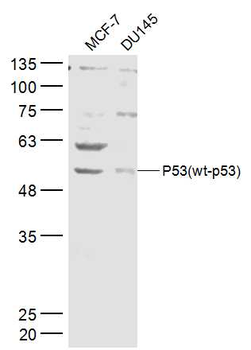
Representative validation image for p53 (WT-p53) Antibody orb13631 .
Primary Tumor Suppressor Detection
View orb13631
p53 (WT-p53) Antibody — orb13631
orb13631 is a highly validated primary antibody designed to detect the p53 tumor suppressor protein. It provides robust performance across multiple essential laboratory techniques, enabling researchers to monitor p53 accumulation, degradation, and tissue localization in normal and neoplastic samples.
Product:
p53 (WT-p53) Antibody
SKU:
orb13631
Tested applications:
WB, IHC, IF, ELISA
Reactivity:
Human, Mouse, Rat
Available size:
100 µg
Protocol:
Validated protocols available
4. Key Tumor Suppressor Proteins
p53 (TP53)
The p53 protein acts as an upstream regulator of cell cycle arrest and apoptosis. In response to cellular stress or DNA damage, p53 induces the expression of targets like p21 to halt the cell cycle, allowing for DNA repair or triggering programmed cell death if the damage is unrepairable.
Retinoblastoma Protein (Rb)
Rb serves as a crucial cell cycle gatekeeper. It blocks the progression from the G1 phase to the S phase by binding to and inactivating E2F transcription factors. When hyperphosphorylated, Rb releases E2F, allowing DNA replication to proceed.
PTEN
PTEN acts as a negative regulator of the PI3K/AKT signaling pathway. By dephosphorylating PIP3 back to PIP2, PTEN prevents the unchecked cellular growth, survival, and proliferation signals normally driven by PI3K activation.
5. Extensive Cancer Research Reagents
Beyond primary antibodies, a robust cancer biology workflow requires high-quality biochemicals, assay kits, and recombinant proteins. Explore our expanded catalog below to support your tumor suppressor and oncogenic pathway assays.
Small Molecules & Biochemicals
| SKU | Product |
|---|---|
| orb1307109 | Capivasertib Small Molecule |
| orb1310103 | 5-Fluorouracil Small Molecule |
| orb1309994 | Docetaxel Small Molecule |
| orb1306206 | Kaempferol Small Molecule |
| orb1307179 | BAY 11-7082 Small Molecule |
Recombinant Proteins & VLPs
| SKU | Product |
|---|---|
| orb1095883 | Recombinant Human Leukemia Inhibitory Factor Protein |
| orb1095892 | Recombinant Human Tumor Necrosis Factor Protein |
| orb1095885 | Recombinant Human Tumor Necrosis Factor Protein |
| orb1478125 | Human CLDN6 VLPs Protein VLP |
| orb1478133 | Human MS4A1 VLPs Protein VLP |
ELISA & Assay Kits
| SKU | Product |
|---|---|
| orb775055 | Human Tumor Necrosis Factor alpha ELISA Kit ELISA Kit |
| orb775642 | Mouse Aspartate Aminotransferase ELISA Kit ELISA Kit |
| orb777584 | Mouse Melanoma Cell Adhesion Marker ELISA Kit ELISA Kit |
| orb775909 | Human PCX ELISA Kit ELISA Kit |
| orb774971 | Human B Lymphocyte Chemoattractant ELISA Kit ELISA Kit |
| orb775234 | Human Renal Tumor Antigen RAGE ELISA Kit ELISA Kit |
| orb774889 | Human Growth Regulated Oncogene ELISA Kit ELISA Kit |
6. Companion Resources
Loading Control
GAPDH Antibody — orb500826
A highly reliable loading control essential for the accurate normalization of tumor suppressor protein expression in Western blot experiments.
Proliferation Marker
Ki67 Antibody — orb389335
Evaluate the downstream effects of tumor suppressor loss by staining for Ki67, a classic marker of active cellular proliferation.
Primary Antibodies
Browse all Cancer Portfolio
Explore thousands of primary antibodies targeting key cell cycle regulators, oncogenes, and DNA repair mechanisms.
Western Blot Detection
ECL Plus Substrate — orb527071
Chemiluminescent HRP substrate designed for the sensitive Western blot detection of low-abundance tumor suppressor proteins.
7. Experimental Design Considerations
Loss of Heterozygosity (LOH)
When studying tumor suppressors, consider the status of both alleles. Even if one allele is mutated, the remaining wild-type allele may produce enough functional protein. Evaluating LOH is critical for proving full inactivation.
Epigenetic Silencing
A lack of detectable protein via Western Blot does not always mean the gene is physically deleted or mutated. Promoter hypermethylation frequently silences tumor suppressor genes in cancer, requiring epigenetic investigation.
Appropriate Controls
Always validate antibody specificity using appropriate biological controls. Use genetically modified knockout (KO) or siRNA-mediated knockdown (KD) cell lines as negative controls to ensure the observed signal is specific to the target protein.
8. Why Choose Biorbyt for Cancer Research?
Extensive Validation
Biorbyt commits to rigorous validation strategies, including KO/KD testing where applicable, ensuring high specificity for crucial targets like p53 and PTEN.
Application Diversity
Our antibodies are formulated and tested to support a broad array of applications, from standard Western blotting to advanced multiplex immunofluorescence.
Comprehensive Portfolio
Beyond primary tumor suppressor antibodies, Biorbyt supplies the essential loading controls, secondary antibodies, and assay kits required to complete your workflow.
Expert Support
Backed by thousands of peer-reviewed citations, our scientific support team is available to help troubleshoot protocols and optimize your assay conditions.
9. Frequently Asked Questions
Why does mutant p53 often accumulate in cancer cells?
Unlike the wild-type p53 protein, which has a very short half-life and is rapidly degraded via the ubiquitin-proteasome pathway, mutated p53 proteins frequently misfold and evade degradation. This leads to a massive accumulation of the mutant protein in the nucleus, which can be readily detected via IHC.
Can one mutated allele of a tumor suppressor gene cause cancer?
Generally, both alleles must be inactivated (the "two-hit" hypothesis) for complete loss of tumor suppression. However, certain mutations can exert a "dominant-negative" effect (where the mutant protein interferes with the remaining wild-type protein) or display "haploinsufficiency" (where a 50% reduction in protein levels is enough to promote tumorigenesis).
How do I verify if my antibody is detecting mutant or wild-type protein?
Most standard primary antibodies, including broad-spectrum p53 antibodies, detect both mutant and wild-type forms because they bind to conserved epitopes. To distinguish between the two, researchers typically sequence the gene, use mutation-specific antibodies (if available), or analyze functional downstream targets (like p21 expression).
10. Scientific References
- Vogelstein, B., & Kinzler, K. W. (2004). Cancer genes and the pathways they control. Nature Medicine, 10(8), 789–799. https://doi.org/10.1038/nm1087
- Knudson A. G. (1971). Mutation and cancer: statistical study of retinoblastoma. Proceedings of the National Academy of Sciences of the United States of America, 68(4), 820–823. (See related retrospective: The two-hit theory hits 50. https://pmc.ncbi.nlm.nih.gov/articles/PMC8694077/)
- Pylayeva-Gupta, Y., Grabocka, E. & Bar-Sagi, D. (2011). RAS oncogenes: weaving a tumorigenic web. Nature Reviews Cancer, 11, 761–774. https://doi.org/10.1038/nrc3106
- Niu, G., et al. (2026). Clinical metastatic cancer from initiation to colonization: emerging challenges and breakthroughs. Oncogene. https://doi.org/10.1038/s41388-026-03944-0
- Xiao, Q., Liu, Y., Li, T. et al. (2025). Viral oncogenesis in cancer: From mechanisms to therapeutics. Signal Transduction and Targeted Therapy, 10(151). https://doi.org/10.1038/s41392-025-02197-9