You have no items in your shopping cart.
CD74 Antibody (RPE)
SKU: orb396386
Featured
Description
Research Area
Immunology & Inflammation
Images & Validation
−Item 1 of 2
| Tested Applications | ICC, IF, WB |
|---|---|
| Dilution Range | WB (1:1000); ICC/IF (1:100) |
| Reactivity | Human |
| Application Notes |
Key Properties
−| Host | Mouse |
|---|---|
| Clonality | Monoclonal |
| Isotype | IgG1 |
| Clone No. | 6D9 |
| Immunogen | Synthetic peptide corresponding to the N-terminal (1-100 aa) of human CD74 conjugated to KLH. Overlaps 100% with all 3 isoforms |
| Target | CD74 |
| Molecular Weight | 33 kDa |
| Purification | Protein G Purified |
| Conjugation | RPE |
Storage & Handling
−| Storage | Conjugated antibodies should be stored according to the product label |
|---|---|
| Buffer/Preservatives | 95.46mM Phosphate, 2.48mM MES and 2mM EDTA |
| Concentration | 1 mg/ml |
| Expiration Date | 12 months from date of receipt. |
| Disclaimer | For research use only |
Alternative Names
−CD74, DHLAG, HLA DR gamma, HLADG, p33, p35, Protein 41, CD74 antigen (invariant polypeptide of major histocompatibility complex, class II antigen-associated), CD74 antigen, CD74 molecule, CD74 molecule, major histocompatibility complex, class II invariant chain, CLIP, Gamma chain of class II antigens, HG2A_HUMAN, HLA class II histocompatibility antigen gamma chain, HLA DR antigens associated invariant chain, HLA-DR antigens-associated invariant chain, HLA-DR-gamma, HLADR antigens associated invariant chain, Ia antigen associated invariant chain, Ia antigen-associated invariant chain, Ia associated invariant chain, Ia gamma, Ii, Invariant polypeptide of major histocompatibility complex class II antigen associated, la-gamma, Major histocompatibility complex class II invariant chain, MHC HLA DR gamma chain, MHC HLA-DR gamma chain
Similar Products
−- Item 1 of 4
- Item 1 of 2
- Item 1 of 2
Quality Guarantee
Explore bioreagents carefree to elevate your research. All our products are rigorously tested for performance. If a product does not perform as described on its datasheet, our scientific support team will provide expert troubleshooting, a prompt replacement, or a refund. For full details, please see our Terms & Conditions and Buying Guide. Contact us at [email protected].

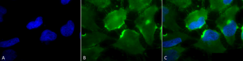
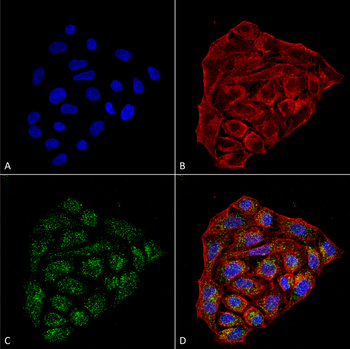
Immunocytochemistry/Immunofluorescence analysis using Mouse Anti-CD74 Monoclonal Antibody, Clone 6D9. Tissue: Cervical cancer cell line (HeLa). Species: Human. Fixation: 4% Formaldehyde for 15 min at RT. Primary Antibody: Mouse Anti-CD74 Monoclonal Antibody at 1:100 for 60 min at RT. Secondary Antibody: Goat Anti-Mouse ATTO 488 at 1:200 for 60 min at RT. Counterstain: Phalloidin Texas Red F-Actin stain; DAPI (blue) nuclear stain at 1:1000, 1:5000 for 60 min at RT, 5 min at RT. Localization: Cell membrane, Endoplasmic Reticulum, Golgi apparatus. Magnification: 60X. (A) DAPI (blue) nuclear stain. (B) Phalloidin Texas Red F-Actin stain. (C) CD74 Antibody. (D) Composite.
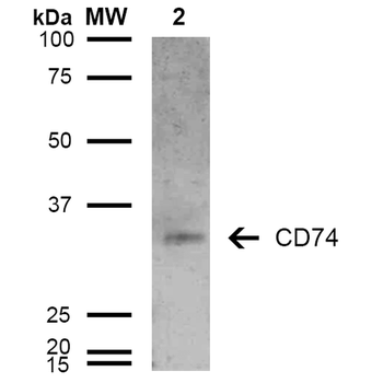
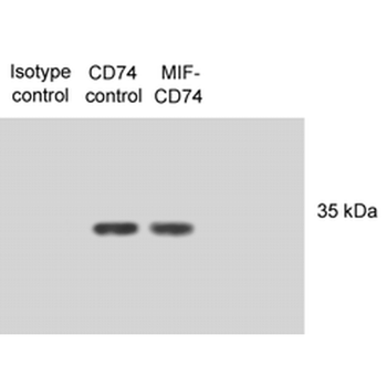
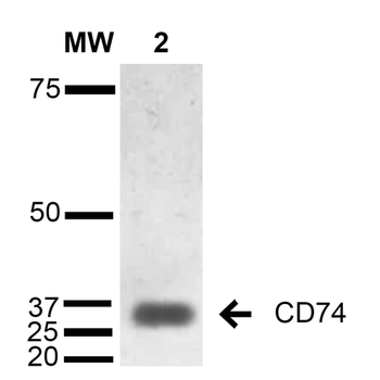
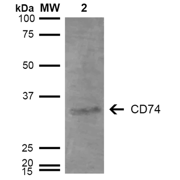
Western Blot analysis of Human Lymphoblastoid cell line (Raji) showing detection of 33-35 kDa CD74 protein using Mouse Anti-CD74 Monoclonal Antibody, Clone 6D9. Lane 1: Molecular Weight Ladder (MW). Lane 2: Raji cell lysate. Load: 15 μg. Block: 5% Skim Milk in TBST. Primary Antibody: Mouse Anti-CD74 Monoclonal Antibody at 1:1000 for 2 hours at RT. Secondary Antibody: Goat Anti-Mouse IgG: HRP at 1:1000 for 60 min at RT. Color Development: ECL solution for 5 min in RT. Predicted/Observed Size: 33-35 kDa.
Quick Database Links
UniProt Details
− No UniProt data available
NCBI Gene Details
− No NCBI Gene data available
NCBI Reference Sequences
−Associated Accession Numbers
Curated reference sequences for the gene transcript and protein product| Protein | NP_001020329.1 |
|---|
Documents Download
Datasheet
Product Information
Request a Document
Protocol Information
WB
Western Blot (IB, immunoblot)
IF
Immunofluorescence
ICC
Immunocytochemistry
CD74 Antibody (RPE) (orb396386)

- 0.0
Based on 0 reviews
Participating in our Biorbyt product reviews program enables you to support fellow scientists by sharing your firsthand experience with our products.
Login to Submit a ReviewAvailable Sizes
Select a size below