You have no items in your shopping cart.
Synapsin I rabbit pAb Antibody
SKU: orb766406
Featured
Description
Images & Validation
−Item 1 of 4
| Tested Applications | ELISA, IF, IHC, WB |
|---|---|
| Dilution range | Western Blot: 1/500 - 1/2000. Immunohistochemistry: 1/100 - 1/300. Immunofluorescence: 1/200 - 1/1000. ELISA: 1/20000. Not yet tested in other applications. |
| Reactivity | Human, Mouse, Rat |
Key Properties
−| Clonality | Polyclonal |
|---|---|
| Isotype | IgG |
| Immunogen | The antiserum was produced against synthesized peptide derived from human Synapsin. AA range:3-52 |
| Molecular Weight | 74 |
| Purification | The antibody was affinity-purified from rabbit antiserum by affinity-chromatography using epitope-specific immunogen. |
| Conjugation | Unconjugated |
Storage & Handling
−| Storage | Maintain refrigerated at 2-8°C for up to 2 weeks. For long term storage store at -20°C in small aliquots to prevent freeze-thaw cycles. |
|---|---|
| Buffer/Preservatives | PBS with 0.02% sodium azide and 50% glycerol pH 7.4. |
| Concentration | 1 mg/ml |
| Disclaimer | For research use only |
Alternative Names
−Anti-SYN1 antibody, anti-Synapsin-1 antibody, anti-Brain protein 4.1 antibody, anti-Synapsin I antibody
Similar Products
−- Item 1 of 4
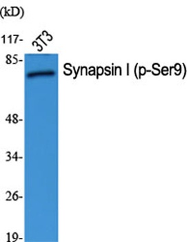
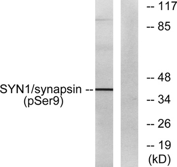
Synapsin I (phospho Ser9) rabbit pAb Antibody [orb764285]
ELISA, IF, IHC, WB
Human, Mouse, Rat
Polyclonal
Unconjugated
100 μl, 50 μl - Item 1 of 4
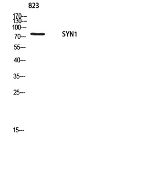
Synapsin-1 rabbit pAb Antibody [orb771286]
ELISA, IHC, WB
Human, Mouse, Rat
Polyclonal
Unconjugated
100 μl, 50 μl - Item 1 of 4
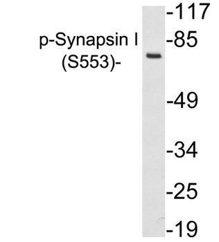
Synapsin-1 (phospho Ser553) rabbit pAb Antibody [orb771314]
IHC, WB
Human, Mouse, Rat
Polyclonal
Unconjugated
50 μl, 100 μl - Item 1 of 3
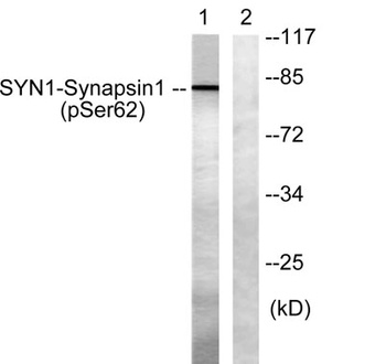
Synapsin I (phospho Ser62) rabbit pAb Antibody [orb770199]
ELISA, IF, IHC, WB
Human, Mouse, Rat
Polyclonal
Unconjugated
100 μl, 50 μl - Item 1 of 3
Synapsin I (phospho Ser605) rabbit pAb Antibody [orb770200]
ELISA, IF, IHC
Human, Mouse, Rat
Polyclonal
Unconjugated
100 μl, 50 μl


Quality Guarantee
Explore bioreagents carefree to elevate your research. All our products are rigorously tested for performance. If a product does not perform as described on its datasheet, our scientific support team will provide expert troubleshooting, a prompt replacement, or a refund. For full details, please see our Terms & Conditions and Buying Guide. Contact us at [email protected].

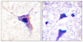
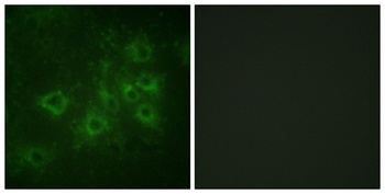
Immunofluorescence analysis of Hela cell. 1, Synapsin I Polyclonal Antibody (green) was diluted at 1:200 (4°C overnight). 2, Goat Anti Rabbit Alexa Fluor 488 was diluted at 1:1000 (room temperature, 50min). 3 DAPI (blue) 10min.
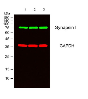
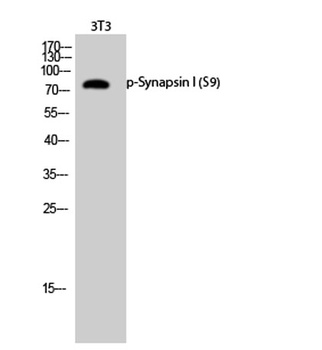
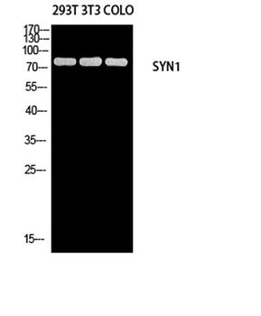
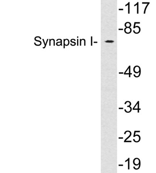
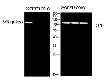
Western blot analysis of lysates from 1) Hela, 2) 293, 3) NIH-3T3 cells, (Green) primary antibody was diluted at 1:1000, 4°C over night, secondary antibody was diluted at 1:10000, 37°C 1 hour. (Red) GAPDH Monoclonal Antibody (2B8) antibody was diluted at 1:5000 as loading control, 4°C over night, secondary antibody was diluted at 1:10000, 37°C 1 hour.
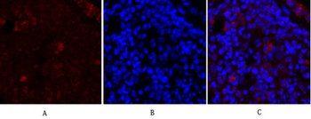
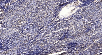
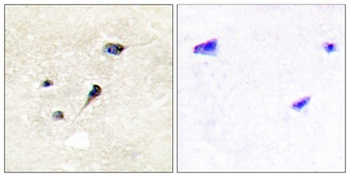
Immunofluorescence analysis of rat-lung tissue. 1, Synapsin I Polyclonal Antibody (red) was diluted at 1:200 (4°C, overnight). 2, Cy3 labled Secondary antibody was diluted at 1:300 (room temperature, 50min). 3, Picture B: DAPI (blue) 10min. Picture A:Target. Picture B: DAPI. Picture C: merge of A + B.
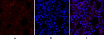
Immunofluorescence analysis of rat-lung tissue. 1, Synapsin I Polyclonal Antibody (red) was diluted at 1:200 (4°C, overnight). 2, Cy3 labled Secondary antibody was diluted at 1:300 (room temperature, 50min). 3, Picture B: DAPI (blue) 10min. Picture A:Target. Picture B: DAPI. Picture C: merge of A + B.
Quick Database Links
Documents Download
Datasheet
Product Information
Request a Document
Protocol Information
WB
Western Blot (IB, immunoblot)
IHC
Immunohistochemistry
IF
Immunofluorescence
ELISA
Enzyme-linked Immunosorbent Assay (EIA)
Synapsin I rabbit pAb Antibody (orb766406)

- 0.0
Based on 0 reviews
Participating in our Biorbyt product reviews program enables you to support fellow scientists by sharing your firsthand experience with our products.
Login to Submit a ReviewAvailable Sizes
Select a size below