You have no items in your shopping cart.
Rabbit anti-Phospho KAP-1 (S824) Recombinant Monoclonal Antibody
SKU: orb1519970
Description
Images & Validation
−Item 1 of 8
| Tested Applications | FC, ICC, IHC, IP, SW-Size, WB |
|---|---|
| Dilution range | WB - 1:1000; IP - 20 µl/mg lysate; IHC - 1:100 - 1:500. Epitope retrieval with citrate buffer pH 6.0 is recommended for FFPE tissue sections.; ICC-IF - 1:100 - 1:500. Formaldehyde fixation is recommended. Permeabilization with Triton-X 100 is recommended for formaldehyde-fixed cells. |
| Reactivity | Human |
| Application Notes |
Key Properties
−| Antibody Type | Primary Antibody |
|---|---|
| Host | Rabbit |
| Clonality | Recombinant |
| Clone No. | BL-246-7B5 |
| Immunogen | Surrounding serine 824 |
| Target | KAP-1, Phospho (S824) |
Storage & Handling
−| Storage | 2 - 8°C |
|---|---|
| Form/Appearance | Whole IgG |
| Buffer/Preservatives | Borate Buffered Saline (BBS) pH 8.2 with 0.1% rAlbumin and 0.09% Sodium Azide |
| Concentration | 100 µg/ml |
| Disclaimer | For research use only |
Alternative Names
−KRAB [Kruppel-associated box domain]-associated protein 1; RING finger protein 96; Tripartite motif-containing protein 28; transcriptional intermediary factor 1-beta; transcription intermediary factor 1-beta; TIF1-beta; TIF1B; TF1B; RNF96; RING-type E3 ubiquitin transferase TIF1-beta; protein phosphatase 1, regulatory subunit 157; PPP1R157; nuclear corepressor KAP-1; KRIP-1; KRAB-associated protein 1; KAP-1; KAP1; E3 SUMO-protein ligase TRIM28; KRAB-interacting protein 1
Similar Products
−- Item 1 of 8
Rabbit anti-Phospho KAP-1 (S824) Recombinant Monoclonal Antibody [orb1519972]
FC, ICC, IHC, IP, SW-Size, WB
Human, Mouse
Rabbit
Recombinant
Unconjugated
100 μl 
Rabbit anti-Phospho KAP-1 (S824) Recombinant Monoclonal Antibody [orb1519971]
FC, ICC, IHC, IP, SW-Size, WB
Human, Mouse
Rabbit
Recombinant
Unconjugated
100 μg


Quality Guarantee
Explore bioreagents carefree to elevate your research. All our products are rigorously tested for performance. If a product does not perform as described on its datasheet, our scientific support team will provide expert troubleshooting, a prompt replacement, or a refund. For full details, please see our Terms & Conditions and Buying Guide. Contact us at [email protected].

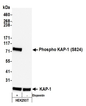
Detection of human Phospho KAP-1 (S824) by western blot. Samples: Whole cell lysate (25 µg) from HEK293T cells treated with 100 µM etoposide (+) or mock treated (-) prepared using NETN lysis buffer. Antibody: Rabbit anti-Phospho KAP-1 (S824) recombinant monoclonal antibody [BL-246-7B5] (orb1519970) used at 1:1000. Secondary: HRP-conjugated goat anti-rabbit IgG. Chemiluminescence with an exposure time of 3 seconds. Lower Panel: Rabbit anti-KAP1 recombinant monoclonal antibody [BL-248-2G6].

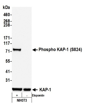
Detection of mouse Phospho KAP-1 (S824) by western blot. Samples: Whole cell lysate (50 µg) from NIH3T3 cells treated with 100 µM etoposide (+) or mock treated (-) prepared using NETN lysis buffer. Antibody: Rabbit anti-Phospho KAP-1 (S824) recombinant monoclonal antibody [BL-246-7B5] (orb1519970) used at 1:1000. Secondary: HRP-conjugated goat anti-rabbit IgG. Chemiluminescence with an exposure time of 3 seconds. Lower Panel: Rabbit anti-KAP1 recombinant monoclonal antibody [BL-248-2G6].

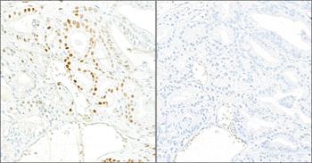
Detection of human Phospho KAP-1 (S824) in FFPE prostate carcinoma by IHC. Mock phosphatase treated section (left) and calf intestinal phosphatase-treated section (right). Antibody: Rabbit anti-Phospho KAP-1 (S824) recombinant monoclonal [BL-246-7B5] (orb1519970). Secondary: HRP-conjugated goat anti-rabbit IgG. Substrate: DAB.

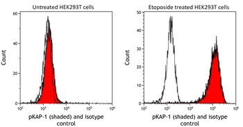
Detection of human phospho KAP-1 (shaded) in etoposide treated HEK293T cells (right) and untreated HEK293T cells (left) by flow cytometry. Antibody: Rabbit anti-phospho KAP-1 recombinant monoclonal [BL-246-7B5] (orb1519970) or isotype control (unshaded). Secondary: DyLight® 488-conjugated goat anti-rabbit IgG.
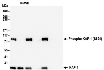
Detection of human Phospho KAP-1 (S824) by western blot of immunoprecipitates. Samples: Whole cell lysate (1.0 mg per IP reaction; 5% of IP loaded) from HEK293T cells prepared using NETN lysis buffer that were treated with 100 µM etoposide (+) or mock treated (-). Antibodies: Rabbit anti-Phospho KAP-1 (S824) recombinant monoclonal antibody [BL-246-7B5] (orb1519970) used for IP at 20 µl/mg lysate.

Detection of human Phospho KAP-1 (S824) in FFPE etoposide treated HeLa cells by ICC. Mock phosphatase treated section (left) and calf intestinal phosphatase-treated section (right). Antibody: Rabbit anti-Phospho KAP-1 (S824) recombinant monoclonal [BL-246-7B5] (orb1519970). Secondary: HRP-conjugated goat anti-rabbit IgG. Substrate: DAB.

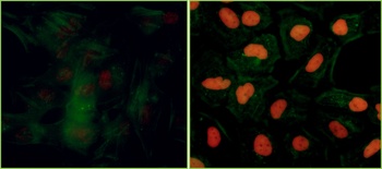
Detection of human Phospho KAP-1 (S824) by immunocytochemistry. Samples: Formaldehyde-fixed asynchronous HeLa cells grown in chambered microscope slides and treated with etoposide (right) or untreated (left). Antibody: Rabbit anti-Phospho KAP-1 (S824) recombinant monoclonal antibody [BL-246-7B5] (orb1519970) used at of 1:100. Secondary: DyLight® 594-conjugated goat anti-rabbit IgG. Counterstain: Phalloidin conjugated Alexa Fluor® 488 (green).

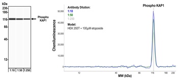
Detection of human Phospho KAP-1 (S824) by Simple Western™. Samples: Whole cell lysate (0.4 mg/mL) from HEK293T cells treated with 100 µM etoposide prepared using NETN lysis buffer. Antibody: Rabbit anti-Phospho KAP-1 (S824) recombinant monoclonal antibody [BL-246-7B5] (orb1519970) used at 1:10, 1:50, and 1:250. Separation and Detection: SallySue ProteinSimple instrument with the 12-230 kDa separation module and anti-Rabbit detection module. Left Panel: Virtual Lane View. Right Panel: Electropherogram.
Quick Database Links
UniProt Details
− No UniProt data available
NCBI Reference Sequences
−Associated Accession Numbers
Curated reference sequences for the gene transcript and protein product| Protein | NP_005753.1 |
|---|
Documents Download
Datasheet
Product Information
Request a Document
Protocol Information
WB
Western Blot (IB, immunoblot)
IHC
Immunohistochemistry
FC
Flow Cytometry
ICC
Immunocytochemistry
IP
Immunoprecipitation
Rabbit anti-Phospho KAP-1 (S824) Recombinant Monoclonal Antibody (orb1519970)

- 0.0
Based on 0 reviews
Participating in our Biorbyt product reviews program enables you to support fellow scientists by sharing your firsthand experience with our products.
Login to Submit a ReviewAvailable Sizes
Select a size below