You have no items in your shopping cart.
Cytokeratin 19 Antibody
SKU: orb248454
Description
Images & Validation
−Item 1 of 5
| Tested Applications | IF, IHC-P, WB |
|---|---|
| Dilution range | Western blot: 1-2ug/ml,Immunofluorescence: 1-2ug/ml,Immunohistochemistry (FFPE): 1-2ug/ml for 30 min at RT |
| Reactivity | Human |
| Application Notes |
Key Properties
−| Antibody Type | Primary Antibody |
|---|---|
| Host | Mouse |
| Clonality | Monoclonal |
| Isotype | Mouse IgG2a, lambda |
| Clone No. | A53-B/A2.26 |
| Immunogen | Human breast cancer MCF-7 cells were used as the immunogen for this Cytokeratin 19 antibody. |
| Purification | Protein G affinity chromatography |
| Conjugation | Unconjugated |
Storage & Handling
−| Storage | Maintain refrigerated at 2-8°C for up to 2 weeks. For long term storage store at -20°C in small aliquots to prevent freeze-thaw cycles. |
|---|---|
| Buffer/Preservatives | 1X PBS, pH 7.4 |
| Disclaimer | For research use only |
Similar Products
−- Item 1 of 22
Cytokeratin 19/KRT19 Antibody [orb316578]
FC, ICC, IF, IHC, WB
Human, Mouse, Rat
Rabbit
Polyclonal
Unconjugated
100 μg - Item 1 of 13
Cytokeratin 19 Mouse Monoclonal Antibody [orb500700]
ICC, IF, IHC-Fr, IHC-P, WB
Mouse
Human, Mouse, Rat
Mouse
Monoclonal
Unconjugated
200 μg, 200 μl, 50 μl, 100 μl - Item 1 of 8
Cytokeratin 19 Rabbit Polyclonal Antibody [orb5872]
FC, IF, IHC-Fr, IHC-P, WB
Bovine, Canine, Equine, Gallus, Mouse, Porcine, Rabbit, Rat
Human
Rabbit
Polyclonal
Unconjugated
200 μl, 50 μl, 100 μl - Item 1 of 7
Mouse Cytokeratin 19 / Keratin K19 Antibody [orb98265]
FC, ICC, IHC-Fr, IHC-P, WB
Human, Zebrafish
Mouse
Monoclonal
Unconjugated
0.1 mg - Item 1 of 10
CK19 Antibody / Cytokeratin 19 [orb749642]
IF, IHC-P, WB
Human, Rat
Mouse
Monoclonal
Unconjugated
100 μg, 20 μg
Quality Guarantee
Explore bioreagents carefree to elevate your research. All our products are rigorously tested for performance. If a product does not perform as described on its datasheet, our scientific support team will provide expert troubleshooting, a prompt replacement, or a refund. For full details, please see our Terms & Conditions and Buying Guide. Contact us at [email protected].
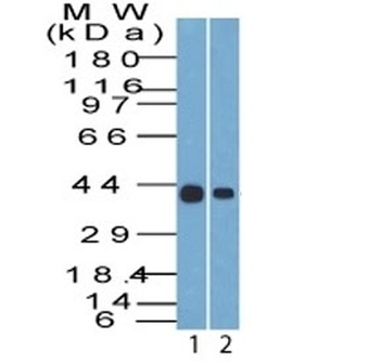
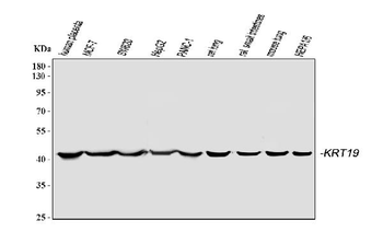
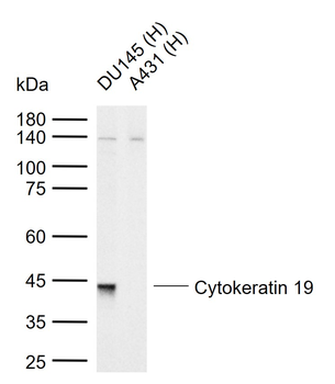
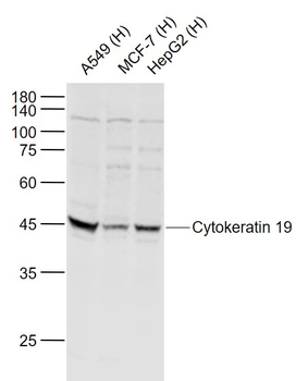
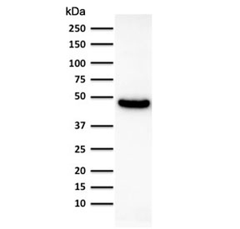
Western blot testing of human 1) HepG2 and 2) MCF7 cell lysate with Cytokeratin 19 antibody (clone A53-B/A2.26). Predicted molecular weight ~43 kDa.
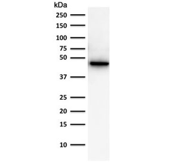
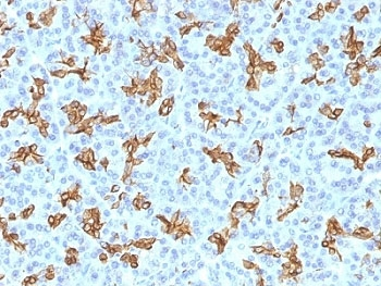
Western blot testing of human PC3 cell lysate using of Cytokeratin 19 antibody (clone A53-B/A2.26). Predicted molecular weight ~43 kDa.
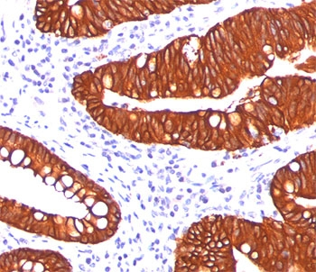
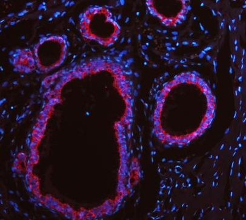
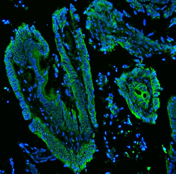
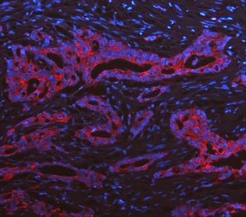
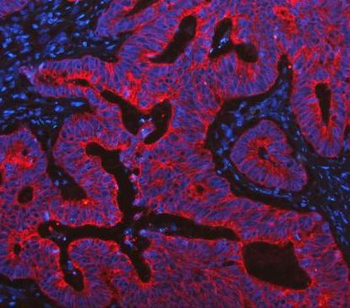
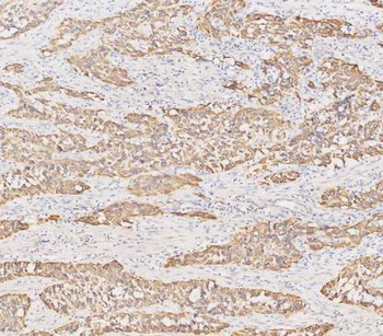
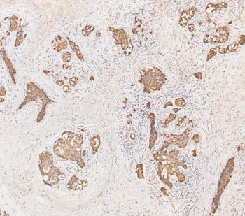
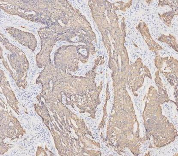
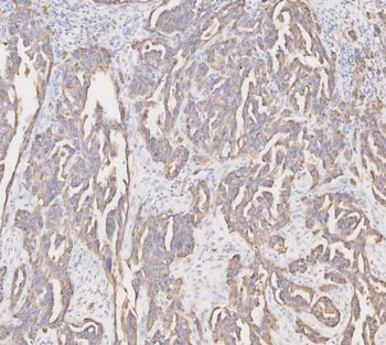
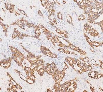
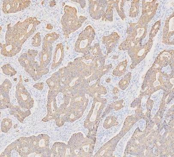
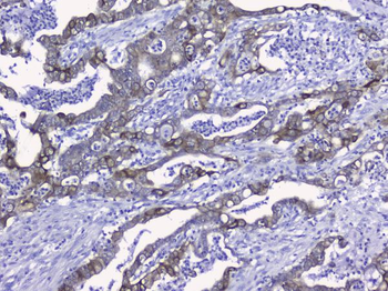
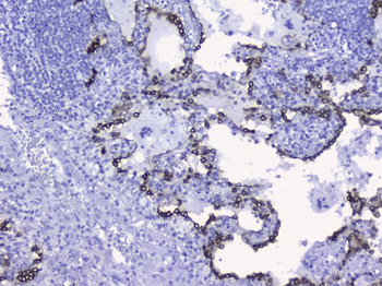
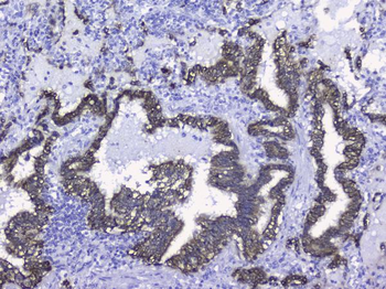
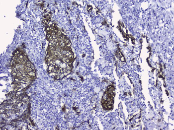
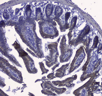
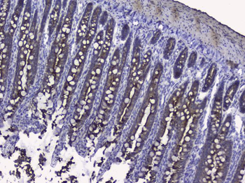
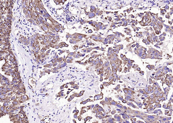
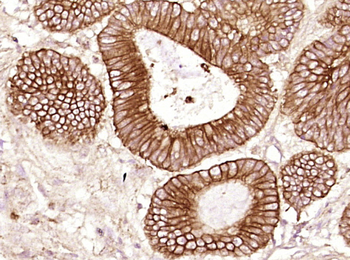
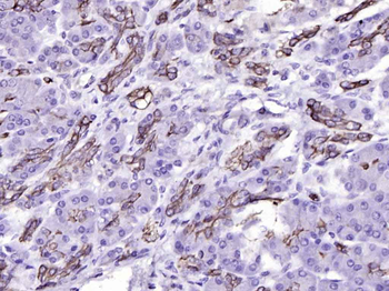
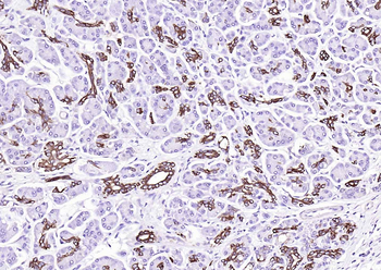
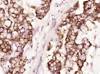
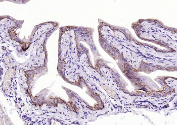
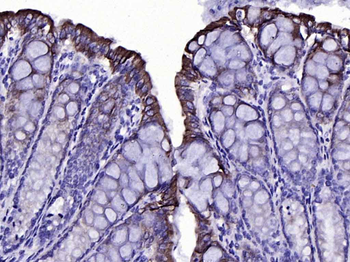
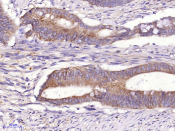
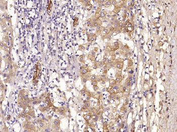
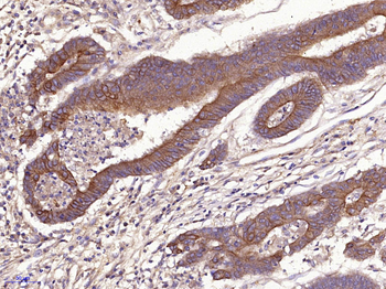
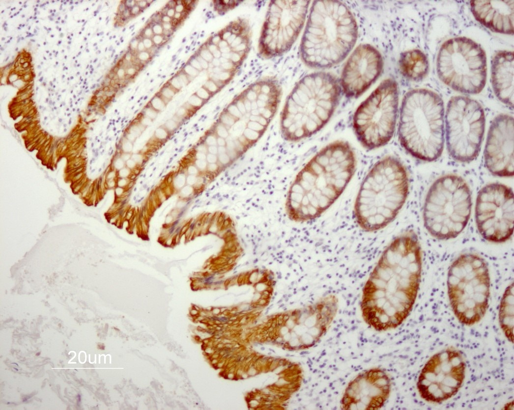
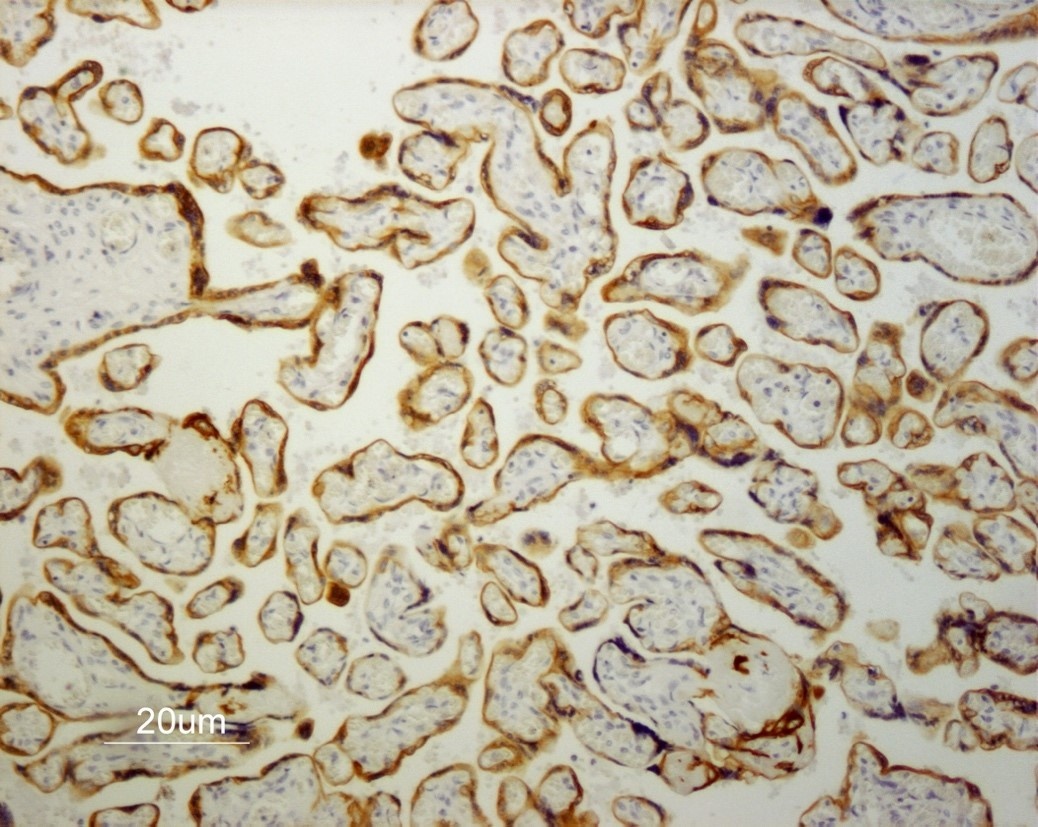
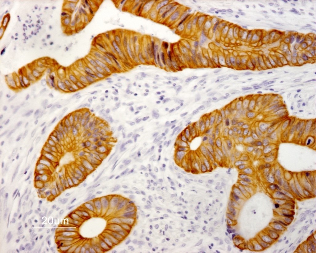
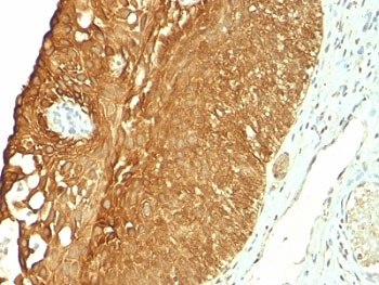
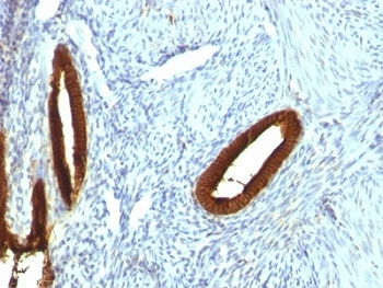
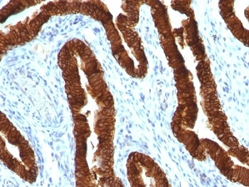
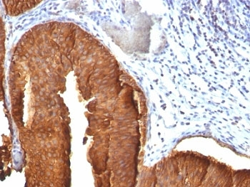
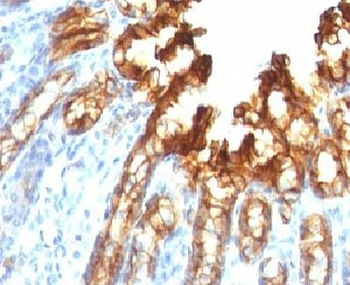
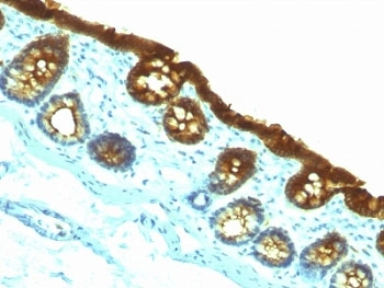
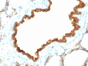
IHC testing of FFPE human colon carcinoma stained with Cytokeratin 19 antibody (clone A53-B/A2.26).
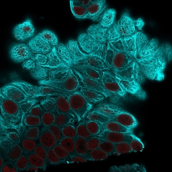
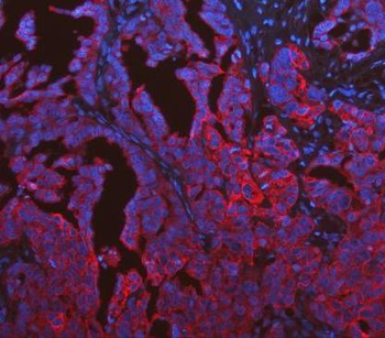
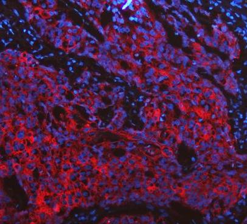
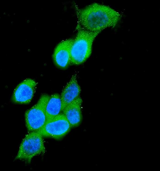
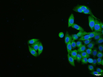
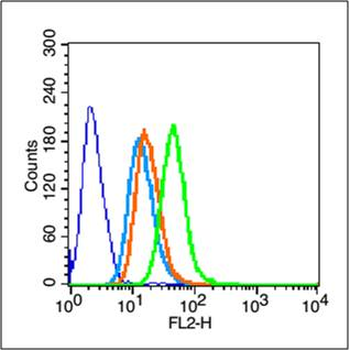
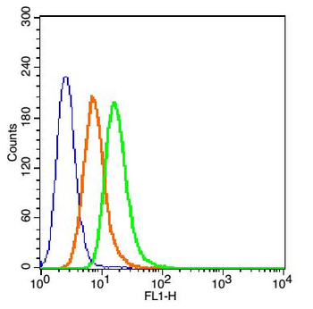
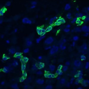
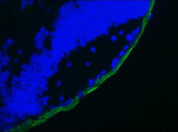
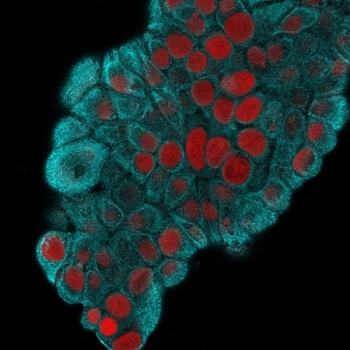
Immunofluorescent staining of MeOH fixed human MCF7 cells with Cytokeratin 19 antibody (clone A53-B/A2.26, blue) and Reddot nuclear stain (red).
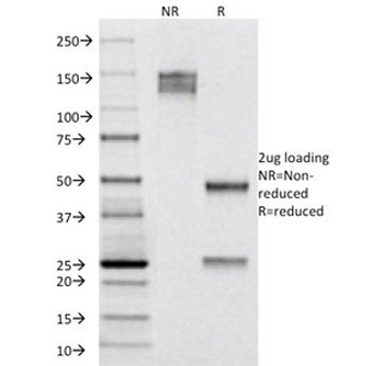
SDS-PAGE analysis of purified, BSA-free Cytokeratin 19 antibody (clone A53-B/A2.26) as confirmation of integrity and purity.
Quick Database Links
Documents Download
Datasheet
Product Information
Request a Document
Protocol Information
WB
Western Blot (IB, immunoblot)
IHC-P
Immunohistochemistry Paraffin
IF
Immunofluorescence
Cytokeratin 19 Antibody (orb248454)

- 0.0
Based on 0 reviews
Participating in our Biorbyt product reviews program enables you to support fellow scientists by sharing your firsthand experience with our products.
Login to Submit a ReviewAvailable Sizes
Select a size below