You have no items in your shopping cart.
SOX2 Antibody
SKU: orb1939275
Description
Research Area
Neuroscience, Signal Transduction, Stem Cell & Developmental Biology
Images & Validation
−Item 1 of 7
| Tested Applications | FC, IF, IHC-P, WB |
|---|---|
| Dilution Range | WB - 1:200-2000, IF - 1:100, IHC-P - 1:100-500, FC - 1:10-50, IHC - 1:100 |
| Reactivity | Human, Mouse |
Key Properties
−| Host | Mouse |
|---|---|
| Clonality | Monoclonal |
| Isotype | IgG1 |
| Molecular Weight | 34310 Da |
| Conjugation | Unconjugated |
Storage & Handling
−| Storage | Maintain refrigerated at 2-8°C for up to 2 weeks. For long term storage store at -20°C in small aliquots to prevent freeze-thaw cycles |
|---|---|
| Form/Appearance | Purified monoclonal antibody supplied in PBS with 0.09% (W/V) sodium azide. This antibody is purified through a protein G column, followed by dialysis against PBS. |
| Expiration Date | 12 months from date of receipt. |
| Disclaimer | For research use only |
Similar Products
−- Item 1 of 9
SOX2 Rabbit Polyclonal Antibody [orb11398]
IHC-P, WB
Human, Mouse, Rat
Rabbit
Polyclonal
Unconjugated
100 μg - Item 1 of 9
SOX2 Rabbit Polyclonal Antibody [orb33646]
IHC-P, WB
Human, Mouse, Rat
Rabbit
Polyclonal
Unconjugated
100 μg - Item 1 of 7
SOX2 Rabbit Polyclonal Antibody [orb500791]
IF, IHC-Fr, IHC-P
Bovine, Canine, Equine, Gallus, Human, Sheep
Mouse, Rat
Rabbit
Polyclonal
Unconjugated
50 μl, 100 μl, 200 μl - Item 1 of 9
SOX2 Rabbit Polyclonal Antibody [orb573909]
IHC, WB
Bovine, Canine, Equine, Goat, Porcine, Rabbit, Rat, Sheep, Zebrafish
Frog, Human, Mouse
Rabbit
Polyclonal
Unconjugated
100 μl - Item 1 of 9

Quality Guarantee
Explore bioreagents carefree to elevate your research. All our products are rigorously tested for performance. If a product does not perform as described on its datasheet, our scientific support team will provide expert troubleshooting, a prompt replacement, or a refund. For full details, please see our Terms & Conditions and Buying Guide. Contact us at [email protected].
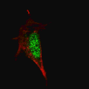
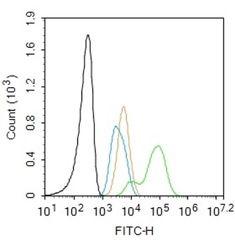
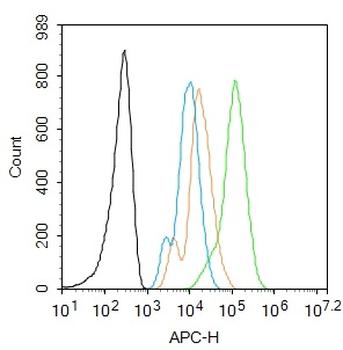
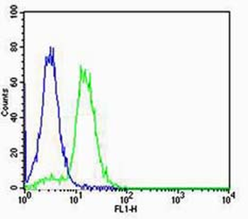
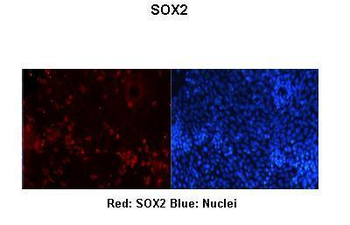
Fluorescent confocal image of SY5Y cells stained with SOX2 Antibody. SY5Y cells were fixed with 4% PFA (20 min), permeabilized with Triton X-100 (0.2%, 30 min). Cells were then incubated with SOX2 primary antibody (1:100, 2 h at room temperature). For secondary antibody, Alexa Fluor 488 conjugated donkey anti-mouse antibody (green) was used (1:1000, 1h).
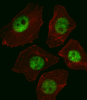
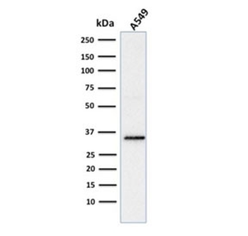
Fluorescent image of A549 cell stained with SOX2 Antibody. A549 cells were fixed with 4% PFA (20 min), permeabilized with Triton X-100 (0.1%, 10 min), then incubated with SOX2 primary antibody (1:25, 1 h at 37°C. For secondary antibody, Alexa Fluor 488 conjugated donkey anti-mouse antibody (green) was used (1:400, 50 min at 37°C.Cytoplasmic actin was counterstained with Alexa Fluor 555 (red) conjugated Phalloidin (7units/ml, 1 h at 37°C.SOX2 immunoreactivity is localized to Nucleus significantly.
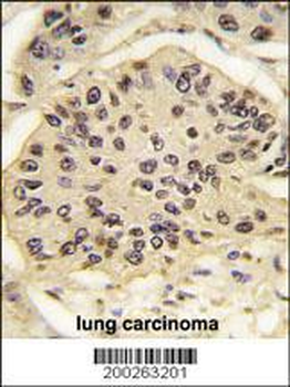
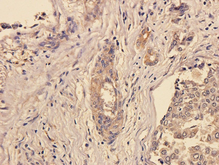
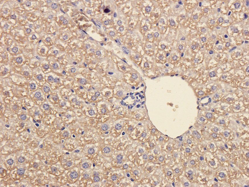
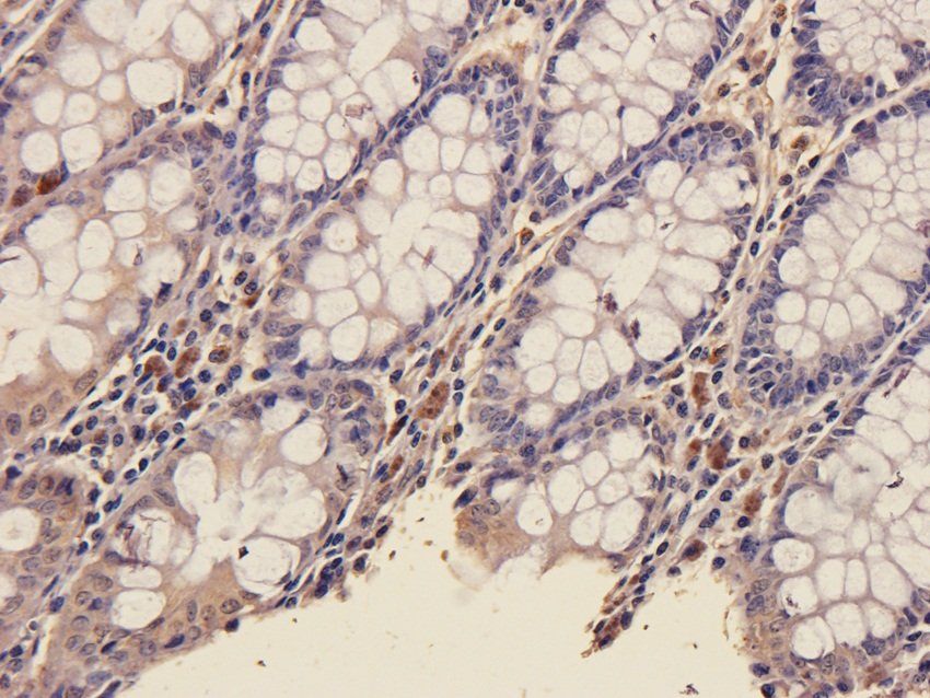
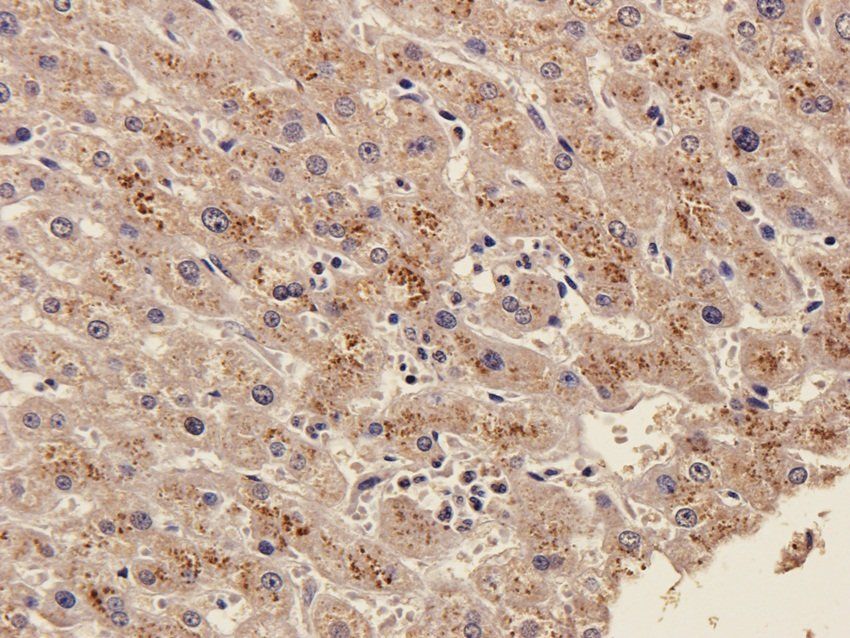
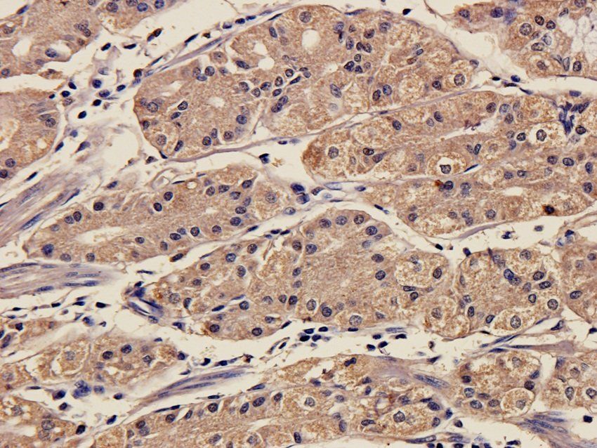
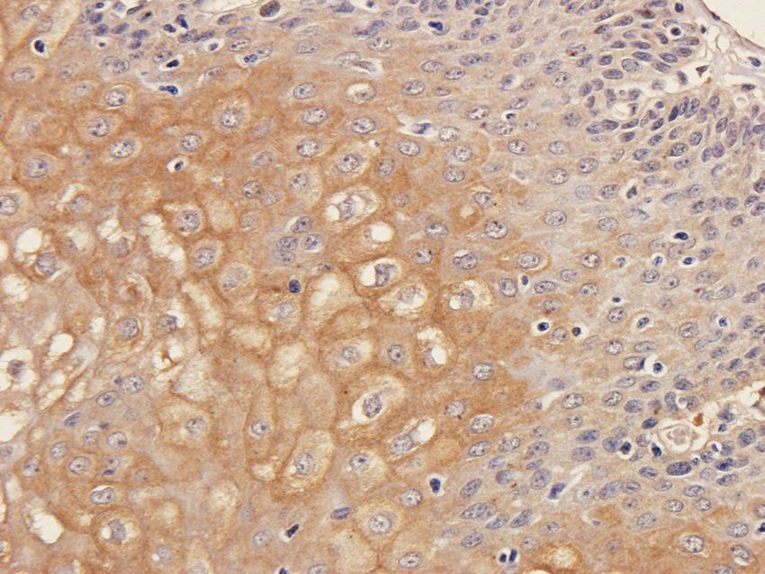
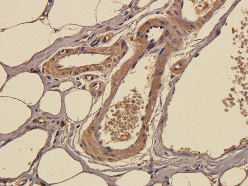
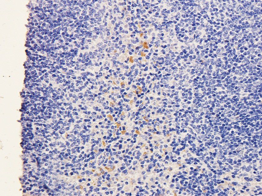
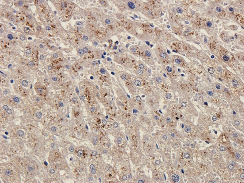
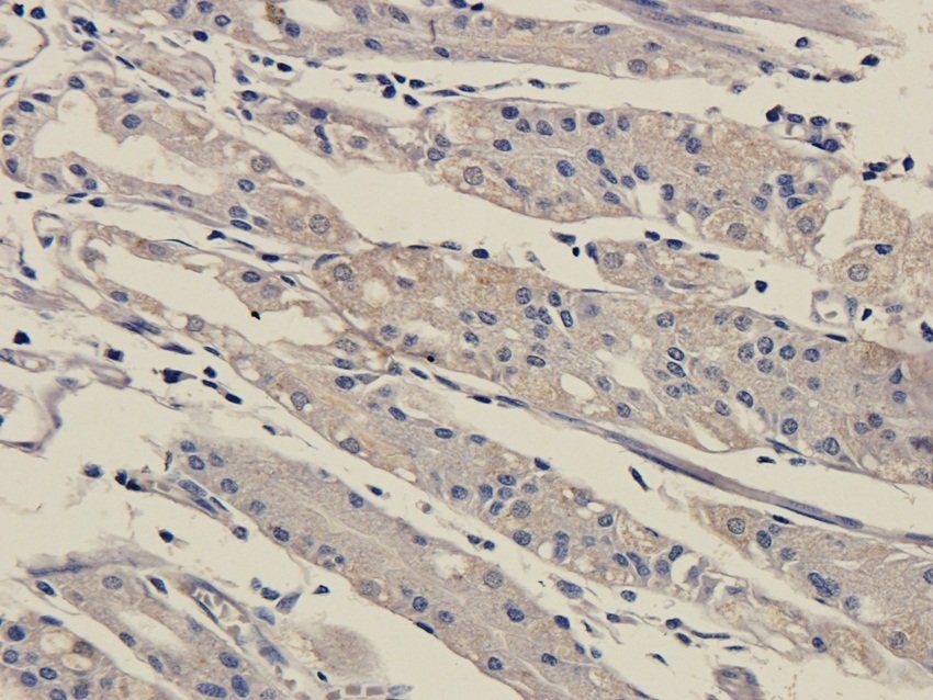
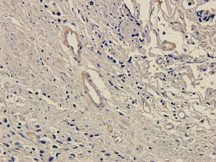
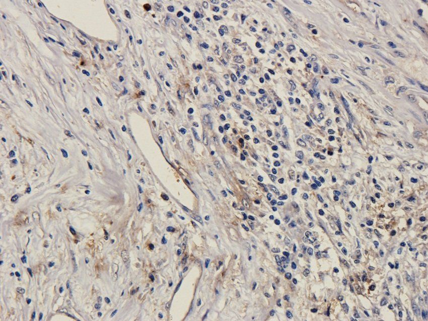
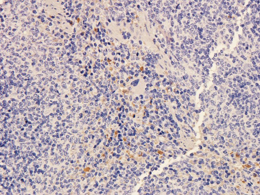
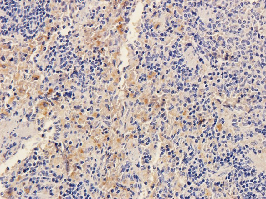
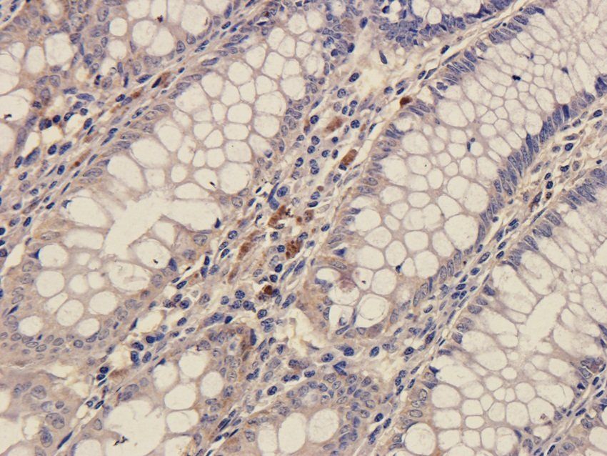
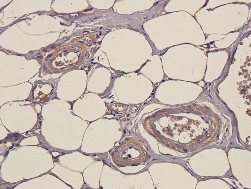
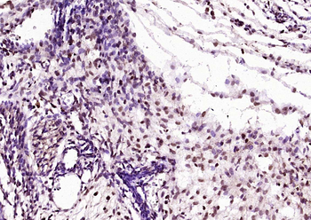
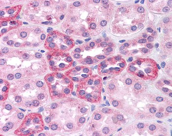
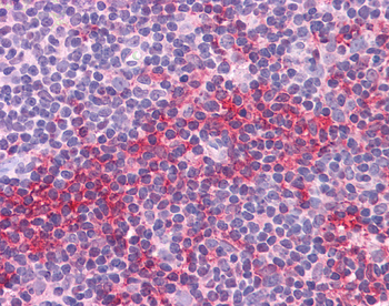
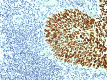
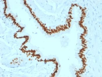
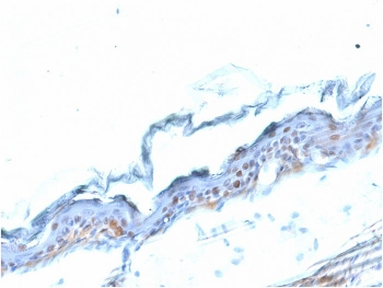
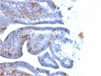
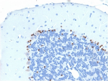
Formalin-fixed and paraffin-embedded human lung carcinoma tissue reacted with SOX2 Antibody, which was peroxidase-conjugated to the secondary antibody, followed by DAB staining. This data demonstrates the use of this antibody for immunohistochemistry; clinical relevance has not been evaluated.
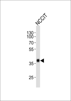
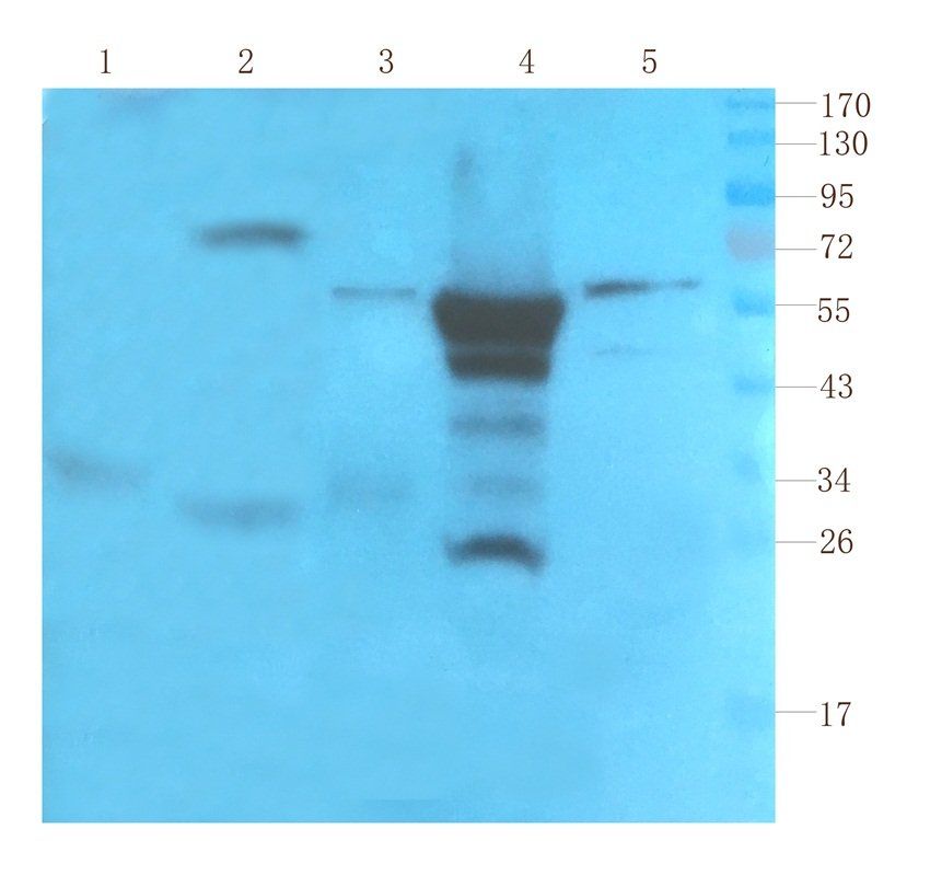
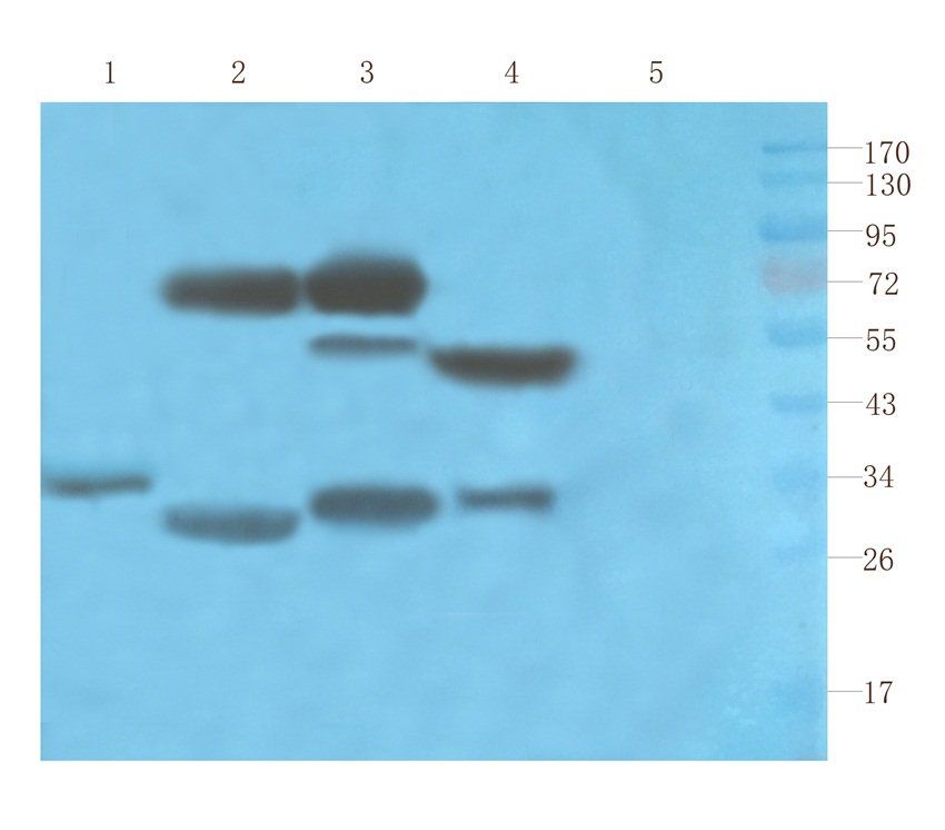
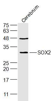
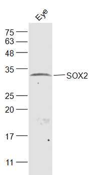
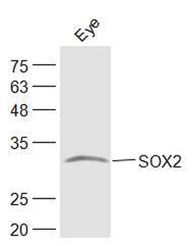
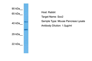
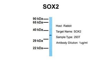
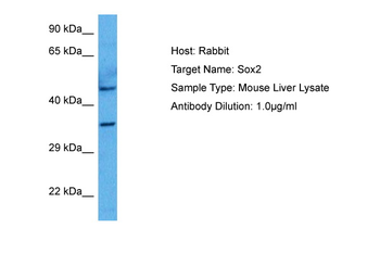
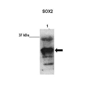
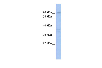
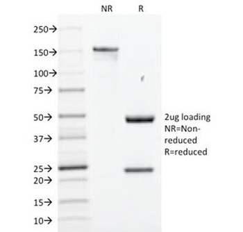
Western blot analysis of lysate from NCCIT cell line, using SOX2 Antibody. diluted at 1:1000. A goat anti-mouse IgG H&L (HRP) at 1:3000 dilution was used as the secondary Antibody. Lysate at 20 μg.
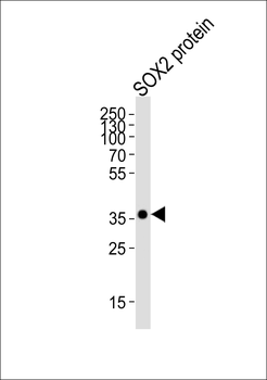
Western blot analysis of lysate from SOX2 protein, using SOX2 Antibody. diluted at 1:4000. A goat anti-mouse IgG H&L (HRP) at 1:3000 dilution was used as the secondary Antibody. Lysate at 20 μg.
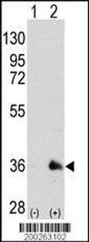
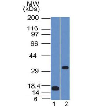
Western blot analysis of SOX2 (arrow) using mouse monoclonal SOX2 Antibody. 293 cell lysates (2 μg/lane) either nontransfected (Lane 1) or transiently transfected with the SOX2 gene (Lane 2)
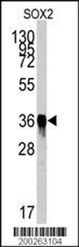
Western blot analysis of SOX2 Antibody by SOX2 recombinant protein. SOX2 (arrow) was detected using the purified Mab.
Quick Database Links
UniProt
UniProt Details
− No UniProt data available
Documents Download
Datasheet
Product Information
Request a Document
Protocol Information
WB
Western Blot (IB, immunoblot)
IHC-P
Immunohistochemistry Paraffin
FC
Flow Cytometry
IF
Immunofluorescence
SOX2 Antibody (orb1939275)

- 0.0
Based on 0 reviews
Participating in our Biorbyt product reviews program enables you to support fellow scientists by sharing your firsthand experience with our products.
Login to Submit a ReviewAvailable Sizes
Select a size below