You have no items in your shopping cart.
Featured
Description
Research Area
Cancer Research, Signal Transduction, Tumor Biomarkers
Images & Validation
−Item 1 of 8
| Tested Applications | FC, ICC, IF, IHC-Fr, IHC-P, WB |
|---|---|
| Dilution Range | WB=1:1000-5000, IHC-P=1:200-1000, IHC-F=1:200-1000, ICC/IF=1:100-500, IF=1:200-1000, Flow-Cyt=1ug/Test |
| Reactivity | Human, Mouse, Rat |
| Predicted Reactivity | A. thaliana, Bovine, Gallus, Porcine, Rabbit, Sheep |
Related Conjugates & Formulations
−Key Properties
−| Antibody Type | Primary Antibody |
|---|---|
| Host | Rabbit |
| Clonality | Polyclonal |
| Isotype | IgG |
| Immunogen | KLH conjugated synthetic peptide derived from human HSP70 (500-600/641aa) |
| Target | HSPA1A |
| Molecular Weight | 68 kDa |
| Purification | Affinity purified by Protein A |
| Conjugation | Unconjugated |
Storage & Handling
−| Storage | Maintain refrigerated at 2-8°C for up to 2 weeks. For long term storage store at -20°C in small aliquots to prevent freeze-thaw cycles. |
|---|---|
| Form/Appearance | Liquid |
| Buffer/Preservatives | 0.01M TBS (pH7.4) with 1% rAlbumin, 0.02% Proclin300 and 50% Glycerol. |
| Concentration | 1mg/ml |
| Expiration Date | 12 months from date of receipt. |
| Disclaimer | For research use only |
Alternative Names
−HSP70-1; HSPA1; HEL-S-103; HSP70; HSP70-1A; HSP70-2; HSP70.1; HSP70.2; HSP70I; HSP72; Hsp70-3; Hsp70.3; hsp68; hsp70A1; Hspa1a; Hspa2; HS71A_BOVIN; Heat shock 70 kDa protein 1 (HSP70.1); HS71A_HUMAN; Heat shock 70 kDa protein 1 (HSP70-1 | HSP70.1); Heat shock protein family A member 1A; HSX70; HS71B_HUMAN; HSPA1B; Heat shock 70 kDa protein 2 (HSP70-2 | HSP70.2); Heat shock protein family A member 1B; HS71B_MOUSE; Hcp70.1; HS71A_MOUSE; Heat shock 70 kDa protein 3 (HSP70.3); HSP71_RAT; heat shock protein family A (Hsp70) member 1A; heat shock 70kD protein 1A; heat shock 70kDa protein 1A
Similar Products
−- Item 1 of 12
Phospho-SRF (Ser77) Rabbit Polyclonal Antibody [orb1497]
IF, IHC-Fr, IHC-P
Bovine, Canine, Porcine
Human, Mouse, Rat
Rabbit
Polyclonal
Unconjugated
50 μl, 100 μl, 200 μl - Item 1 of 7
GRP75 Rabbit Polyclonal Antibody [orb10746]
IF, IHC-Fr, IHC-P, WB
Bovine, Canine, Equine, Porcine, Rabbit
Human, Mouse, Rat
Rabbit
Polyclonal
Unconjugated
50 μl, 100 μl, 200 μl - Item 1 of 4
HSP70 Rabbit Polyclonal Antibody [orb10848]
FC, ICC, IF, IHC-Fr, IHC-P, WB
Bovine, Gallus, Rabbit, Sheep
Human, Mouse, Rat
Rabbit
Polyclonal
Unconjugated
50 μl, 100 μl, 200 μl - Item 1 of 8
Phospho-HSP70 (Tyr611) Rabbit Polyclonal Antibody [orb157590]
FC, IF, IHC-Fr, IHC-P
Canine, Guinea pig, Porcine, Rabbit
Human, Mouse, Rat
Rabbit
Polyclonal
Unconjugated
50 μl, 100 μl, 200 μl - Item 1 of 8
Hsp70 Rabbit Polyclonal Antibody [orb556728]
ICC, IHC-P, IP, WB
Human, Monkey, Mouse, Other, Rat
Rabbit
Polyclonal
Unconjugated
100 μl

Quality Guarantee
Explore bioreagents carefree to elevate your research. All our products are rigorously tested for performance. If a product does not perform as described on its datasheet, our scientific support team will provide expert troubleshooting, a prompt replacement, or a refund. For full details, please see our Terms & Conditions and Buying Guide. Contact us at [email protected].
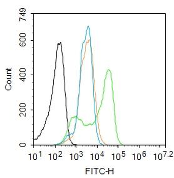
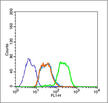
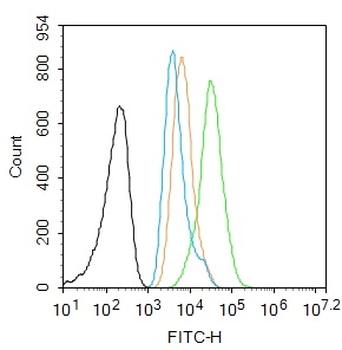
Blank control: 293T. Primary Antibody (green line): Rabbit Anti-HSP70 antibody (orb157591), Dilution: 2 µg/10^6 cells, Isotype Control Antibody (orange line): Rabbit IgG. Secondary Antibody: Goat anti-rabbit IgG-FITC, Dilution: 1 µg/Test. Protocol, The cells were fixed with 4% PFA (10 min at room temperature) and then permeabilized with 0.1% PBST for 20 min at room temperature. The cells were then incubated in 5% BSA to block non-specific protein-protein interactions for 30 min at room temperature. Cells stained with Primary Antibody for 30 min at room temperature. The secondary antibody used for 40 min at room temperature. Acquisition of 20000 events was performed.
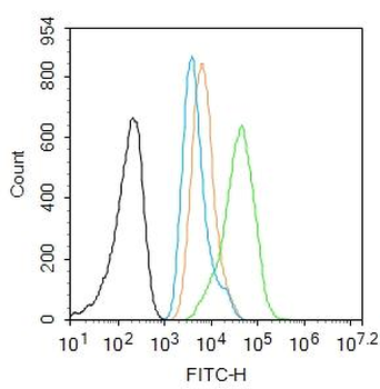
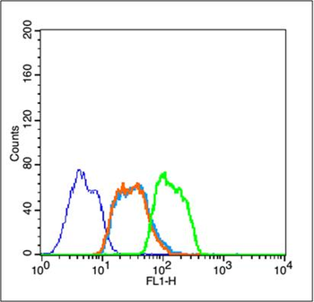
Blank control: A549. Primary Antibody (green line): Rabbit Anti-HSP70 antibody (orb157591), Dilution: 1 µg/10^6 cells, Isotype Control Antibody (orange line): Rabbit IgG. Secondary Antibody: Goat anti-rabbit IgG-AF488, Dilution: 1 µg/Test. Protocol, The cells were fixed with 4% PFA (10 min at room temperature) and then permeabilized with 90% ice-cold methanol for 20 min at -20°C. The cells were then incubated in 5% BSA to block non-specific protein-protein interactions for 30 min at room temperature. Cells stained with Primary Antibody for 30 min at room temperature. The secondary antibody used for 40 min at room temperature. Acquisition of 20000 events was performed.
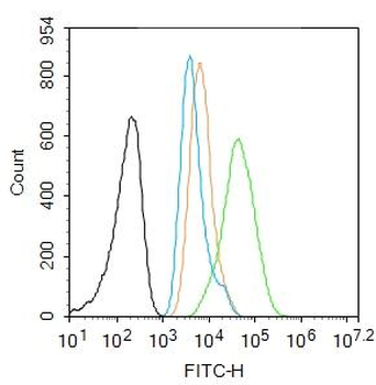
Blank control: A549. Primary Antibody (green line): Rabbit Anti-HSP70 antibody (orb157591), Dilution: 1 µg/10^6 cells, Isotype Control Antibody (orange line): Rabbit IgG. Secondary Antibody: Goat anti-rabbit IgG-AF488, Dilution: 1 µg/Test. Protocol, The cells were fixed with 4% PFA (10 min at room temperature) and then permeabilized with 90% ice-cold methanol for 20 min at -20°C. The cells were then incubated in 5% BSA to block non-specific protein-protein interactions for 30 min at room temperature. Cells stained with Primary Antibody for 30 min at room temperature. The secondary antibody used for 40 min at room temperature. Acquisition of 20000 events was performed.
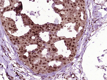
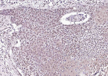
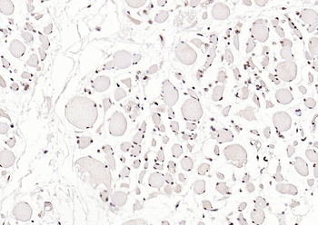
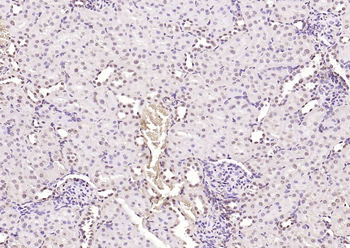
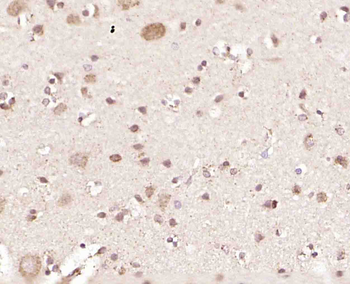
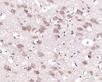
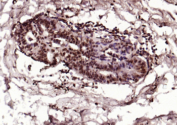
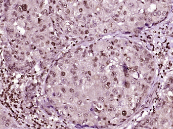
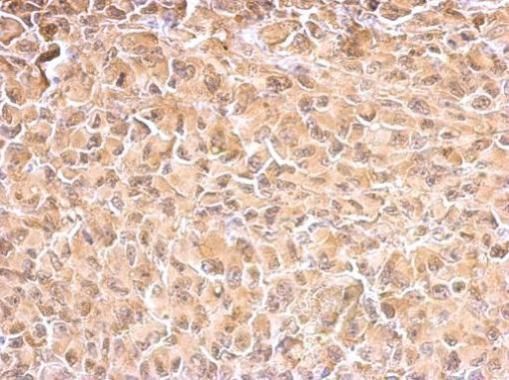
Paraformaldehyde-fixed, paraffin embedded (Human breast carcinoma), Antigen retrieval by boiling in sodium citrate buffer (pH6.0) for 15 min, Block endogenous peroxidase by 3% hydrogen peroxide for 20 minutes, Blocking buffer (normal goat serum) at 37°C for 30 min, Antibody incubation with (HSP70) Polyclonal Antibody, Unconjugated (orb157591) at 1:400 overnight at 4°C, followed by operating according to SP Kit (Rabbit) instructionsand DAB staining.
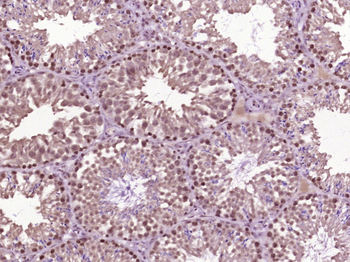
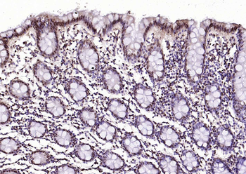
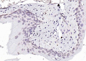
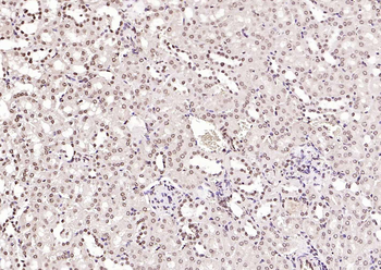
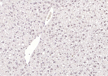
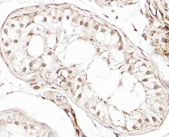
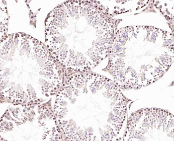
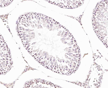
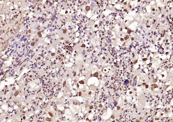
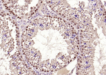
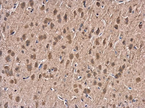
Paraformaldehyde-fixed, paraffin embedded (Mouse testis), Antigen retrieval by boiling in sodium citrate buffer (pH6.0) for 15 min, Block endogenous peroxidase by 3% hydrogen peroxide for 20 minutes, Blocking buffer (normal goat serum) at 37°C for 30 min, Antibody incubation with (HSP70) Polyclonal Antibody, Unconjugated (orb157591) at 1:400 overnight at 4°C, followed by operating according to SP Kit (Rabbit) instructionsand DAB staining.
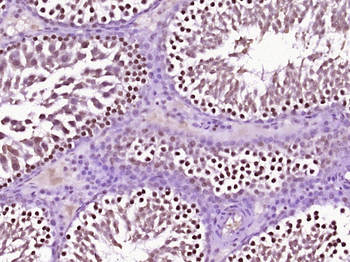
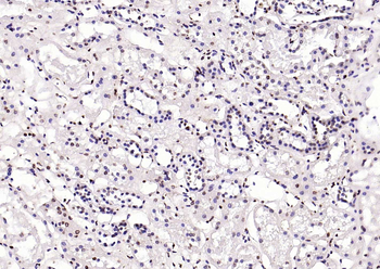
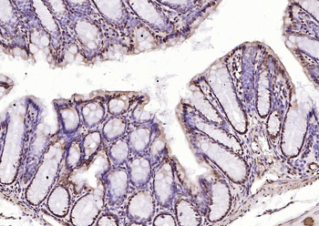
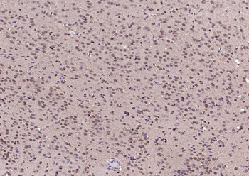
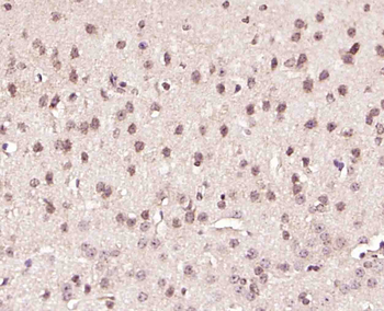
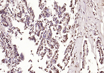
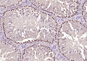
Paraformaldehyde-fixed, paraffin embedded (Rat testis), Antigen retrieval by boiling in sodium citrate buffer (pH6.0) for 15 min, Block endogenous peroxidase by 3% hydrogen peroxide for 20 minutes, Blocking buffer (normal goat serum) at 37°C for 30 min, Antibody incubation with (HSP70) Polyclonal Antibody, Unconjugated (orb157591) at 1:400 overnight at 4°C, followed by operating according to SP Kit (Rabbit) instructionsand DAB staining.

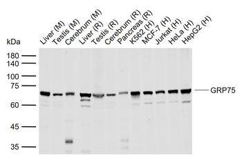
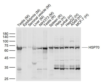
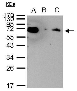
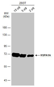
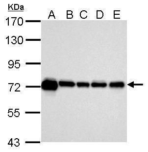
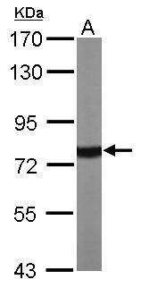
Sample: Lane 1: Testis (Mouse) Lysate at 40 ug, Lane 2: Epididymis (Mouse) Lysate at 40 ug, Lane 3: Seminal vesicle (Mouse) Lysate at 40 ug, Lane 4: Liver (Mouse) Lysate at 40 ug, Lane 5: NIH/3T3 (Mouse) Cell Lysate at 30 ug, Lane 6: Testis (Rat) Lysate at 40 ug, Lane 7: Spleen (Rat) Lysate at 40 ug, Lane 8: Hela (Human) Cell Lysate at 30 ug, Lane 9: Jurkat (Human) Cell Lysate at 30 ug, Lane 10: HepG2 (Human) Cell Lysate at 30 ug, Lane 11: A549 (Human) Cell Lysate at 30 ug, Lane 12: MCF-7 (Human) Cell Lysate at 30 ug, Primary: Anti-HSP70 (orb157591) at 1/1000 dilution, Secondary: IRDye800CW Goat Anti-Rabbit IgG at 1/20000 dilution, Predicted band size: 70 kD, Observed band size: 68 kD.
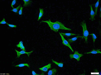
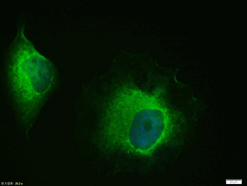
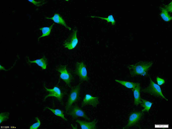
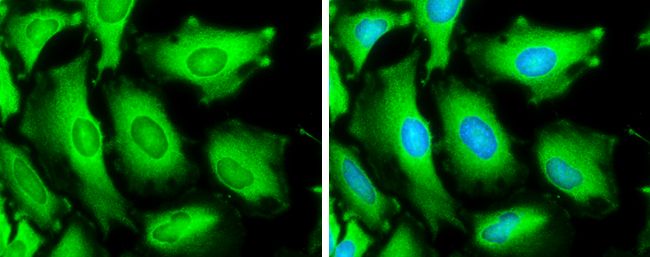
Tissue/Cell: U-87MG cell, 4% Paraformaldehyde-fixed, Triton X-100 at room temperature for 20 min, Blocking buffer (normal goat serum) at 37°C for 20 min, Antibody incubation with (HSP70) polyclonal Antibody, Unconjugated (orb157591) 1:100, 90 minutes at 37°C, followed by a FITC conjugated Goat Anti-Rabbit IgG antibody at 37°C for 90 minutes, DAPI (blue) was used to stain the cell nuclei.
Quick Database Links
Gene Symbol
HSPA1A
UniProt
UniProt Details
− No UniProt data available
Documents Download
Datasheet
Product Information
Request a Document
Protocol Information
WB
Western Blot (IB, immunoblot)
IHC-P
Immunohistochemistry Paraffin
IHC-Fr
Immunohistochemistry Frozen
FC
Flow Cytometry
IF
Immunofluorescence
ICC
Immunocytochemistry
Filter by Applications
Filter by Species
Suna Karadeniz Saygili et al. The effect of salubrinal on the endoplasmic reticulum stress pathway in heat-stressed spermatogonial cells in vitro FEBS Open Bio, (2025)
Applications
IF
Reactivity
Mouse
HSP70 Rabbit Polyclonal Antibody (orb157591)

- 0.0
Based on 0 reviews
Participating in our Biorbyt product reviews program enables you to support fellow scientists by sharing your firsthand experience with our products.
Login to Submit a ReviewAvailable Sizes
Select a size below
Free Secondary Antibody (20 ul)0/0
Please add an antibody product to your cart first.