You have no items in your shopping cart.
Description
Research Area
Neuroscience
Images & Validation
−Item 1 of 8
| Tested Applications | IF, IHC-Fr, IHC-P, WB |
|---|---|
| Dilution Range | Western Blot 1:1000-1:2000, Immunofluorescence 1:200 – 1:500, Immunohistochemistry (Frozen) 1:100 – 1:300, Immunohistochemistry (Paraffin) 1:100 – 1:300 |
| Reactivity | Human, Mouse, Rat |
Key Properties
−| Antibody Type | Primary Antibody |
|---|---|
| Host | Rabbit |
| Clonality | Polyclonal |
| Isotype | Rabbit-IgG |
| Immunogen | Synthetic peptide / encompassing a sequence within the C-terminus region. |
| Target | GFAP |
| Conjugation | Unconjugated |
Storage & Handling
−| Storage | Maintain refrigerated at 2-8°C for up to 2 weeks. For long term storage store at -20°C in small aliquots to prevent freeze-thaw cycles. |
|---|---|
| Buffer/Preservatives | 100mM Tris Glycine, 1% rAlbumin, 20% Glycerol (pH7). 0.025% ProClin 300 was added as a preservative |
| Expiration Date | 12 months from date of receipt. |
| Disclaimer | For research use only |
Similar Products
−- Item 1 of 15
GFAP Rabbit Polyclonal Antibody [orb10706]
ELISA, ICC, IF, IHC-P, WB
Human, Mouse, Rat
Rabbit
Polyclonal
Unconjugated
100 μg - Item 1 of 12
GFAP Rabbit Polyclonal Antibody [orb500829]
FC, IF, IHC-Fr, IHC-P
Bovine, Canine, Porcine, Rabbit, Sheep
Human, Mouse, Rat
Rabbit
Polyclonal
Unconjugated
50 μl, 100 μl, 200 μl - Item 1 of 5
GFAP Rabbit Polyclonal Antibody [orb221420]
FC, WB
Bovine, Gallus, Porcine, Rabbit, Sheep
Human, Mouse, Rat
Rabbit
Polyclonal
Unconjugated
- Item 1 of 4
GFAP Rabbit Polyclonal Antibody (Cy5) [orb913879]
FC, IF
Bovine, Canine, Porcine, Rabbit, Sheep
Human, Mouse, Rat
Rabbit
Polyclonal
Cy5
100 μl - Item 1 of 7
GFAP Rabbit pAb [orb539616]
ELISA, IF, IHC-P, WB
Human, Mouse, Rat
Rabbit
Polyclonal
Unconjugated


Quality Guarantee
Explore bioreagents carefree to elevate your research. All our products are rigorously tested for performance. If a product does not perform as described on its datasheet, our scientific support team will provide expert troubleshooting, a prompt replacement, or a refund. For full details, please see our Terms & Conditions and Buying Guide. Contact us at [email protected].
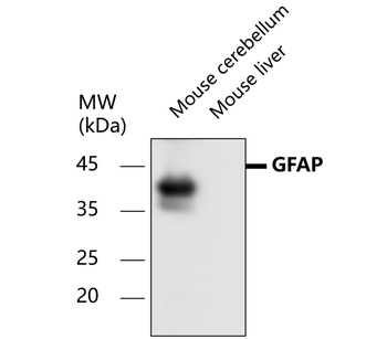
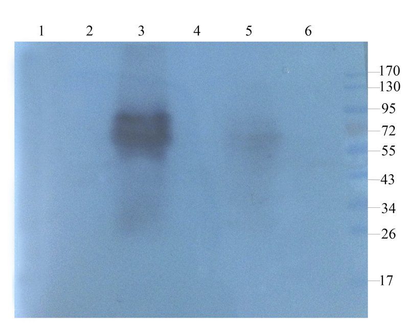
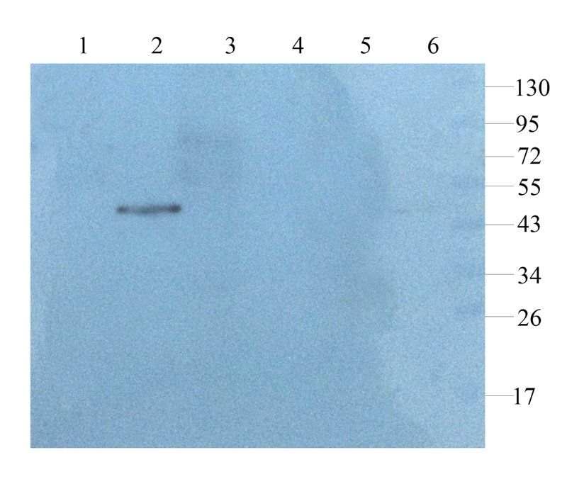
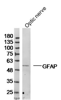
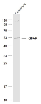
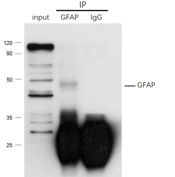
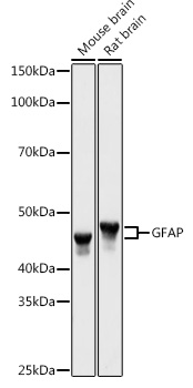
All lanes: Anti-GFAP antibody at 1/1000 dilution, Lysates/proteins at 60 µg per lane. This blot was produced using a 12% SDS-PAGE. Nitrocellulose was then blocked for an hour before being incubated with orb1294403 overnight at 4°C.
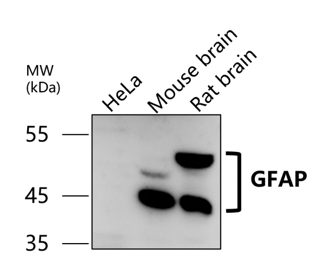
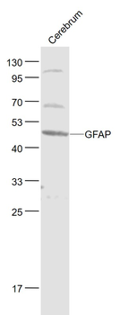
All lanes: Anti-GFAP antibody at 1/1000 dilution, Lysates/proteins at 60 µg per lane. This blot was produced using a 12% SDS-PAGE. Nitrocellulose was then blocked for an hour before being incubated with orb1294403 overnight at 4°C.
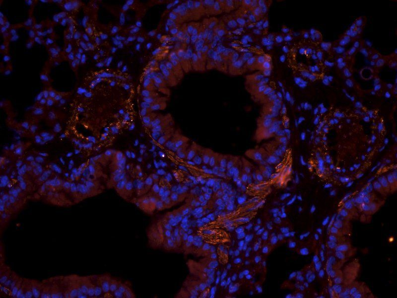
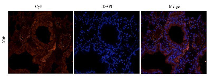
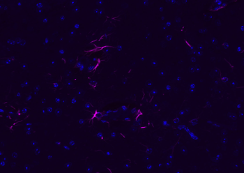
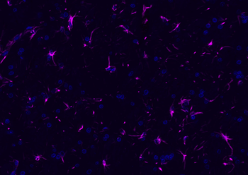
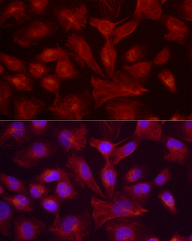
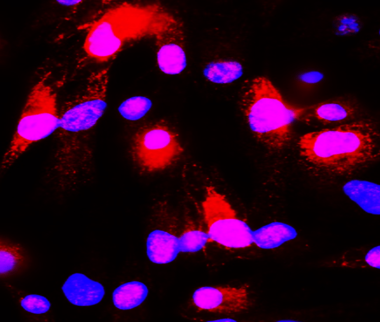
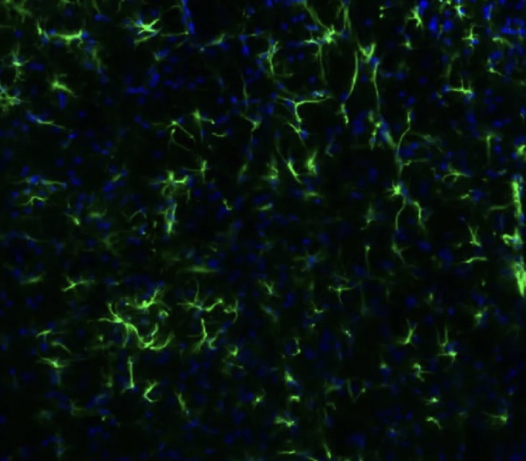
Immunofluorescence: cells were fixed with 4% paraformaldehyde for 10 min at RT, permeabilized with 0.1% NP-40 for 10 min at RT then blocked with 5% BSA for 30 min at room temperature. Cells were stained with orb1294403 anti-GFAP antibody (red) at 1:200 and 4°C. DAPI (blue) was used as the nuclear counter stain.
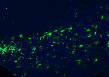
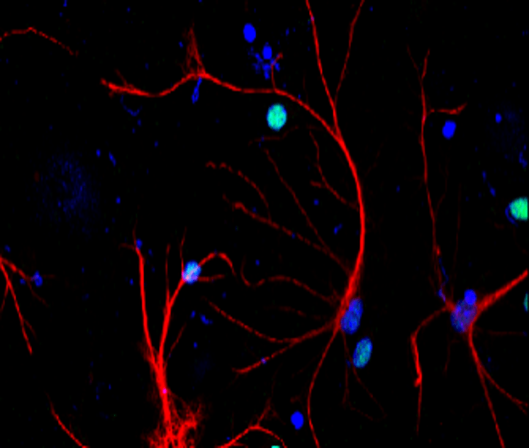
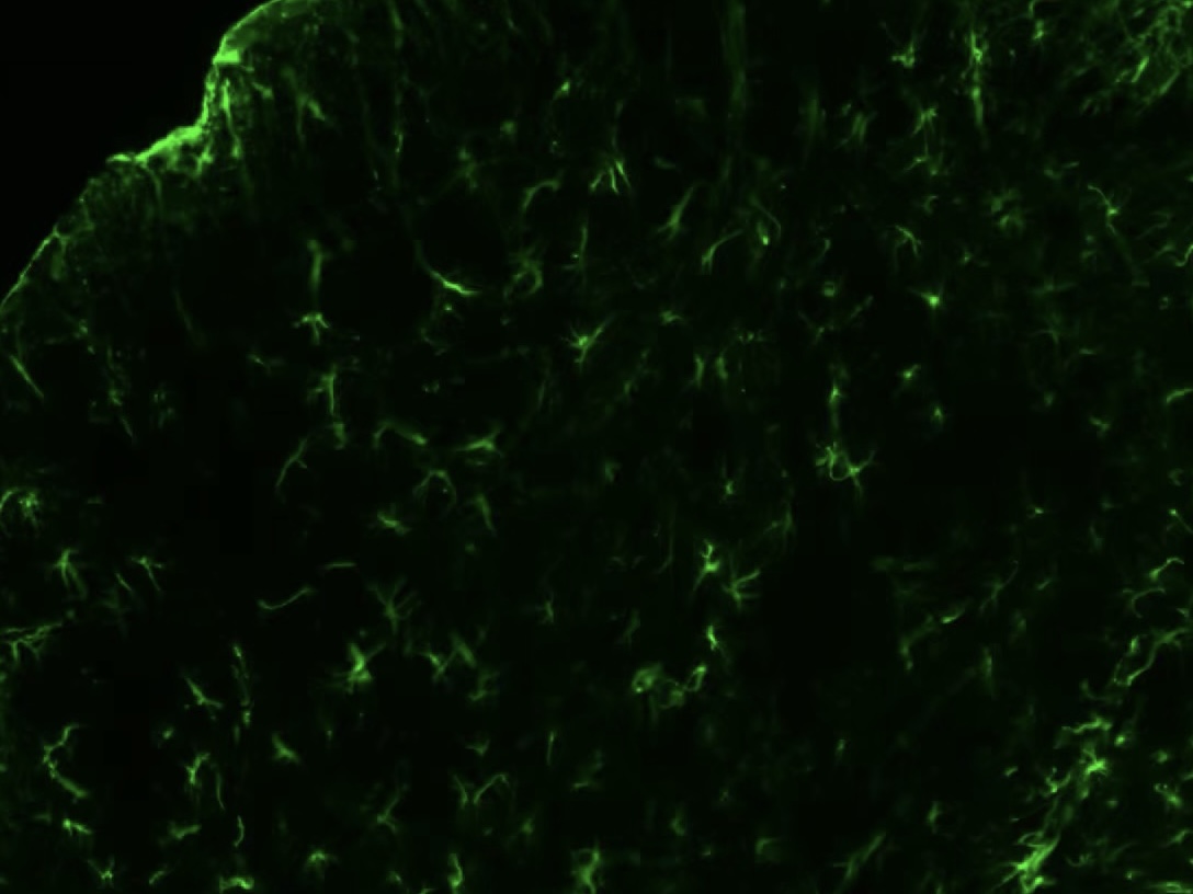
Immunofluorescent analysis. Sample: primary cortical neurons, Red: GFAP (orb1294403): 1-200, Blue: DAPI was used as the nuclear counter stain. Fixed: 4% paraformaldehyde at RT for 20 min.
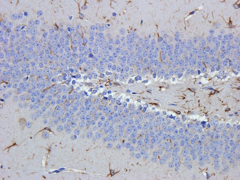
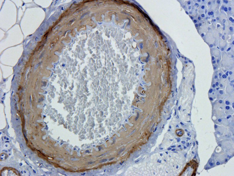
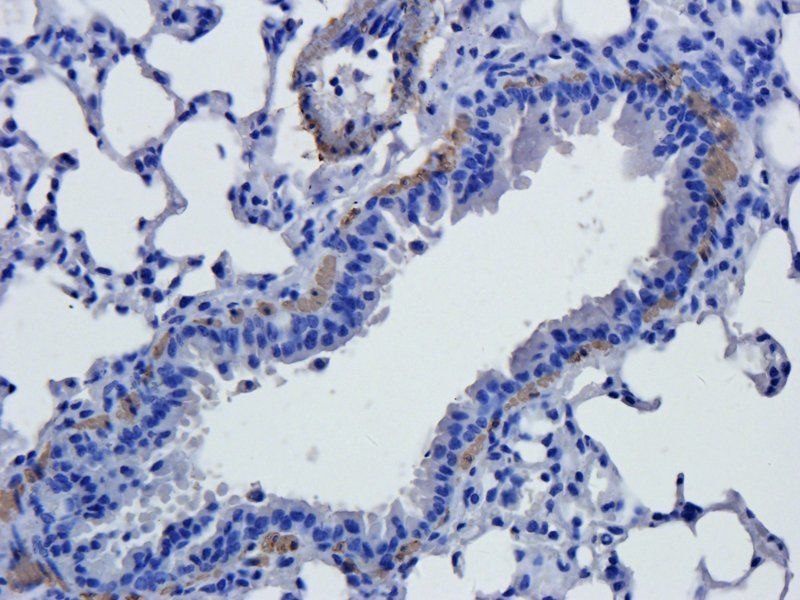
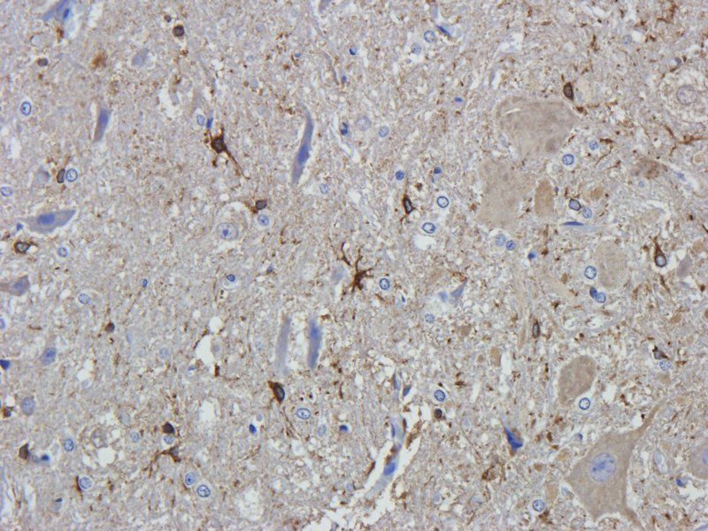
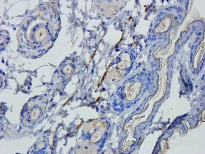
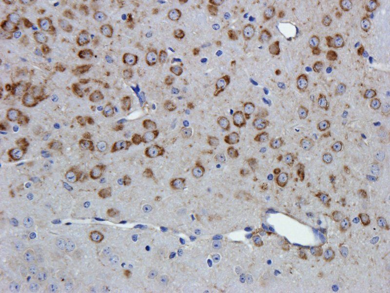
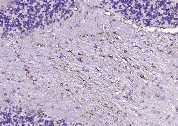
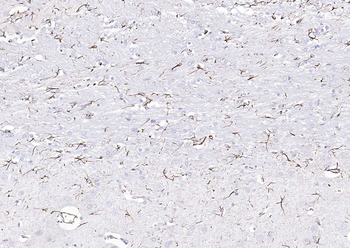
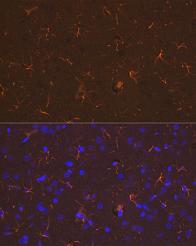
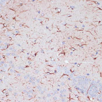
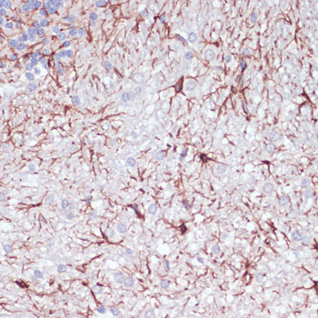
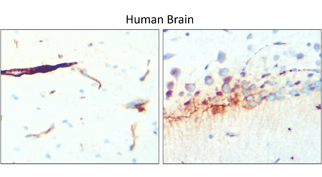
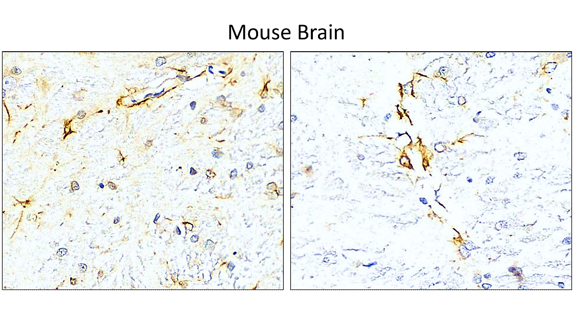
Immunohistochemical analysis of paraffin embedded Human brain tissue labeling GFAP antibody with orb1294403 at 1/100.
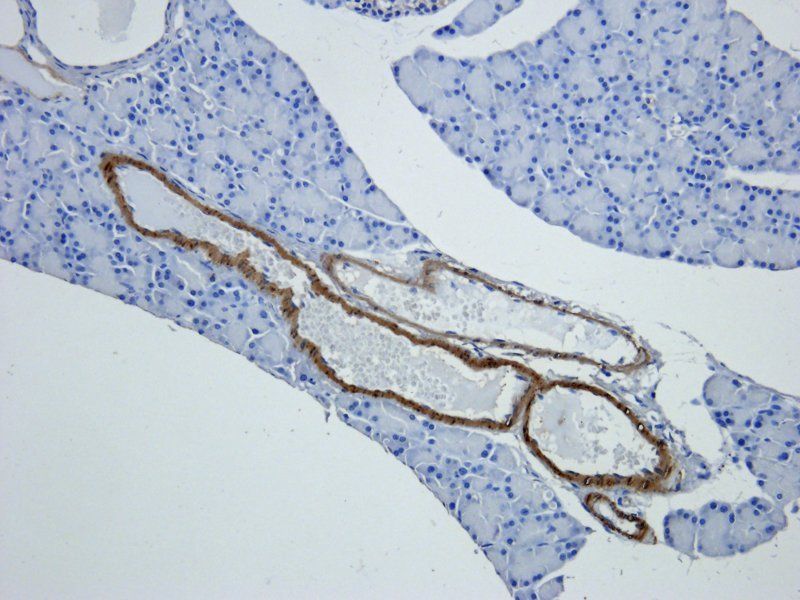
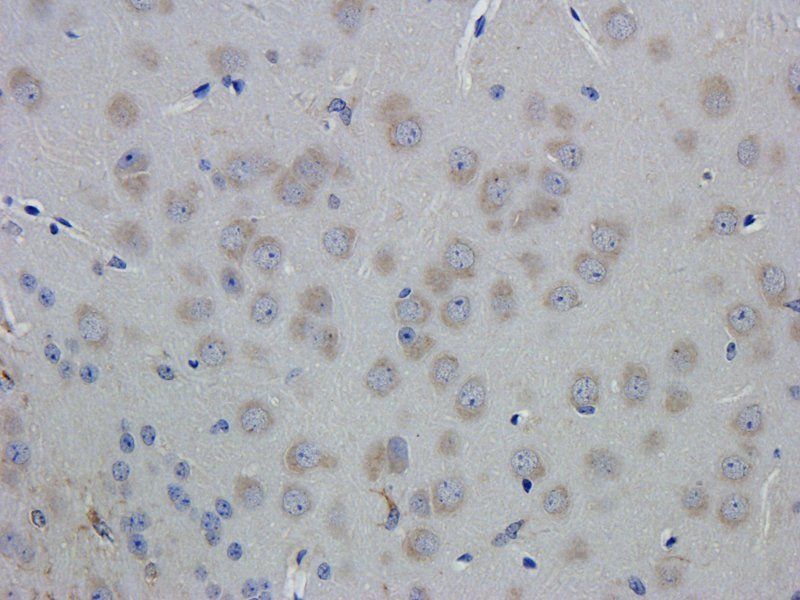
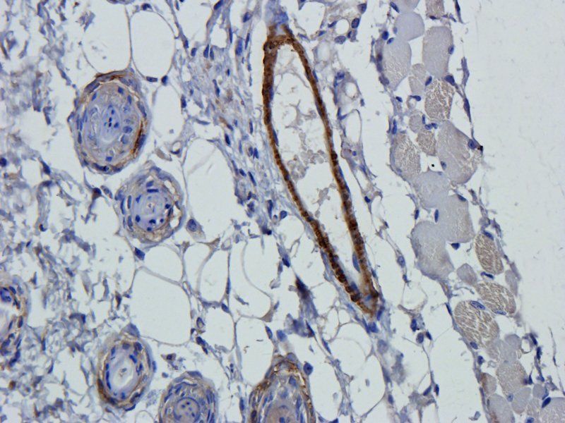
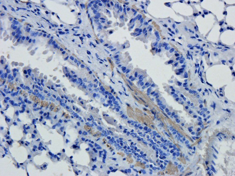
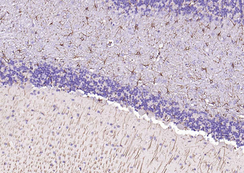
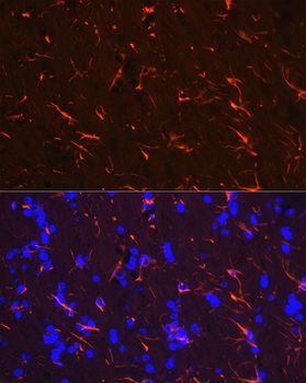
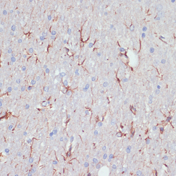
Immunohistochemical analysis of paraffin embedded Mouse brain tissue labeling GFAP antibody with orb1294403 at 1/100.
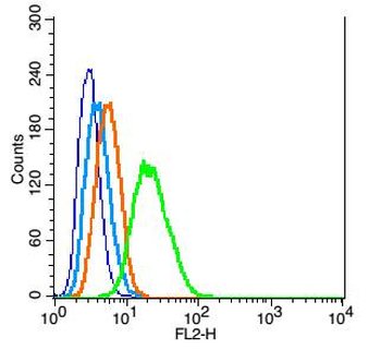
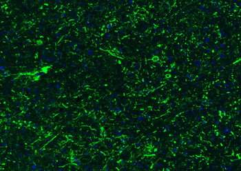
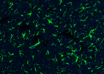
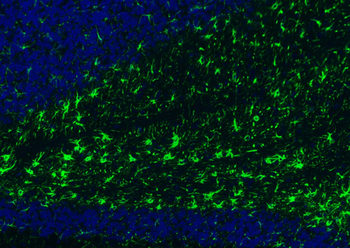
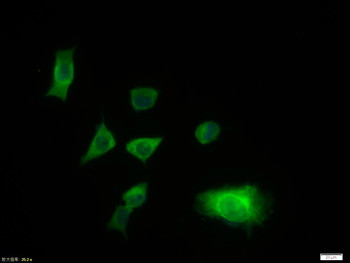
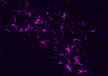
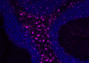
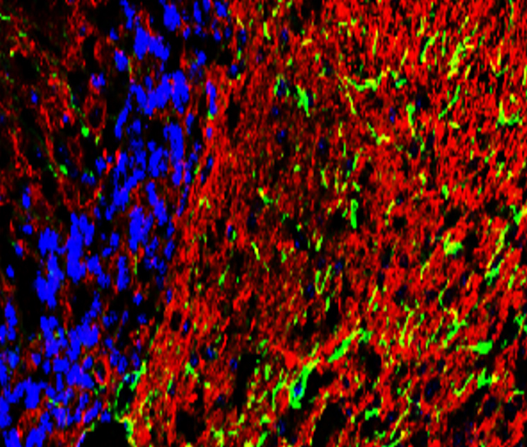
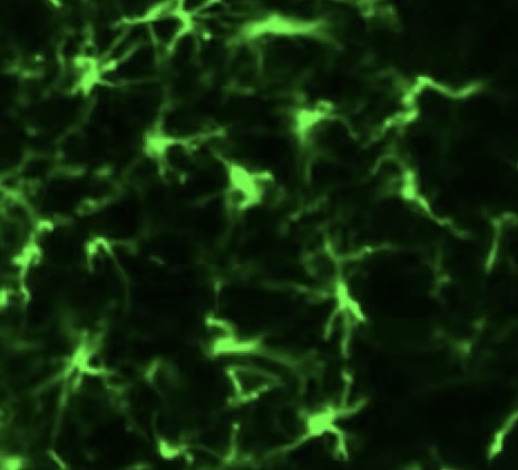
Immunohistochemical of frozen sections. Sample: mouse cerebellum. Green: GFAP (orb1294403): 1-200, Anti-rabbit 488: 1-500.
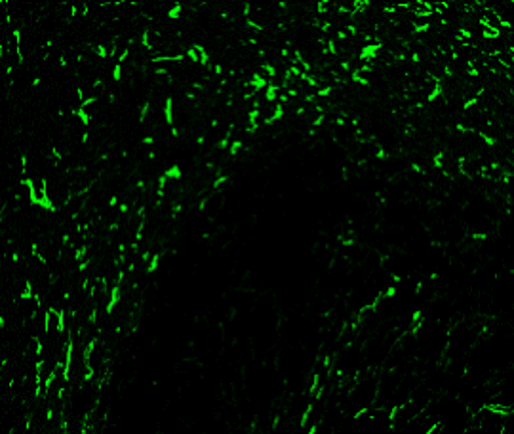
Immunohistochemical of frozen sections. Sample: mouse cerebellum. Green: GFAP (orb1294403): 1-200, Anti-rabbit 488: 1-500.
Quick Database Links
Gene Symbol
GFAP
Documents Download
Datasheet
Product Information
Request a Document
Protocol Information
WB
Western Blot (IB, immunoblot)
IHC-P
Immunohistochemistry Paraffin
IHC-Fr
Immunohistochemistry Frozen
IF
Immunofluorescence
GFAP Rabbit Polyclonal Antibody (orb1294403)

- 5.0
Based on 1 review
Participating in our Biorbyt product reviews program enables you to support fellow scientists by sharing your firsthand experience with our products.
Login to Submit a ReviewFilter by Rating
- 5 stars

- 4 stars
- 3 stars
- 2 stars
- 1 stars
Filter by Applications
Filter by Species
- 5 stars
GFAP antibody works great on mouse spinal frozen sections! Our immunofluorescence signal is specific, astrocytes show distinct morphology, and the staining effect is very beautiful!
Available Sizes
Select a size below
Choose Conjugation or Carrier Free Version
Free Secondary Antibody (20 ul)0/0
Please add an antibody product to your cart first.