You have no items in your shopping cart.
Description
Research Area
Cell Biology, Disease Biomarkers, Neuroscience, Stem Cell & Developmental Biology
Images & Validation
−Item 1 of 4
| Tested Applications | FC, IHC, WB |
|---|---|
| Dilution Range | Western blot, 0.1-0.5μg/ml, Human Immunohistochemistry (Paraffin-embedded Section), 0.5-1μg/ml, Human Flow Cytometry (Fixed), 1-3μg/1x10^6 cells, Human |
| Reactivity | Human |
Related Conjugates & Formulations
−Key Properties
−| Antibody Type | Primary Antibody |
|---|---|
| Host | Rabbit |
| Clonality | Polyclonal |
| Isotype | Rabbit IgG |
| Immunogen | E.coli-derived human PROM1 recombinant protein (Position: P531-H865). Human PROM1 shares 61% amino acid (aa) sequence identity with mouse PROM1. |
| Target | Prominin-1 |
| Molecular Weight | 120 kDa |
| Purification | Immunogen affinity purified. |
| Conjugation | Unconjugated |
Storage & Handling
−| Storage | Maintain refrigerated at 2-8°C for up to 2 weeks. For long term storage store at -20°C in small aliquots to prevent freeze-thaw cycles. |
|---|---|
| Form/Appearance | Lyophilized |
| Buffer/Preservatives | Each vial contains antibody formulated with stabilizing components, 0.9 mg NaCl, 0.2 mg Na2HPO4, and 0.05 mg NaN3. *This antibody is supplied in a stabilized formulation. Compatibility with conjugation reactions depends on the chemistry of the conjugation method used. For conjugation methods that are not compatible with the stabilizing components present in this formulation, a carrier-free antibody format is required. |
| Concentration | Adding 0.2 ml of distilled water will yield a concentration of 500 μg/ml. |
| Expiration Date | 12 months from date of receipt. |
| Disclaimer | For research use only |
Alternative Names
−Prominin-1; Antigen AC133; Prominin-like protein 1; CD133; PROM1; PROML1; MSTP061
Similar Products
−- Item 1 of 30
CD133 Rabbit Polyclonal Antibody [orb99113]
ELISA, ICC, IF, IHC-P, WB
Human, Mouse, Rat
Rabbit
Polyclonal
Unconjugated
100 μg - Item 1 of 9
- Item 1 of 5
CD133 Rabbit Polyclonal Antibody [orb526485]
FC, WB
Rat
Human, Mouse
Rabbit
Polyclonal
Unconjugated
50 μl, 100 μl, 200 μl - Item 1 of 5
- Item 1 of 4


Quality Guarantee
Explore bioreagents carefree to elevate your research. All our products are rigorously tested for performance. If a product does not perform as described on its datasheet, our scientific support team will provide expert troubleshooting, a prompt replacement, or a refund. For full details, please see our Terms & Conditions and Buying Guide. Contact us at [email protected].
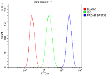
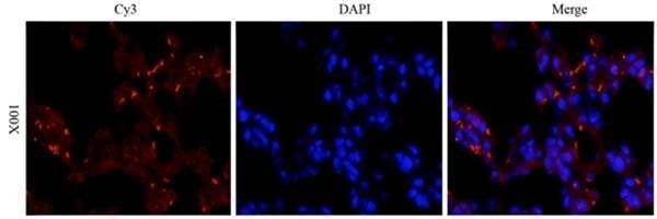
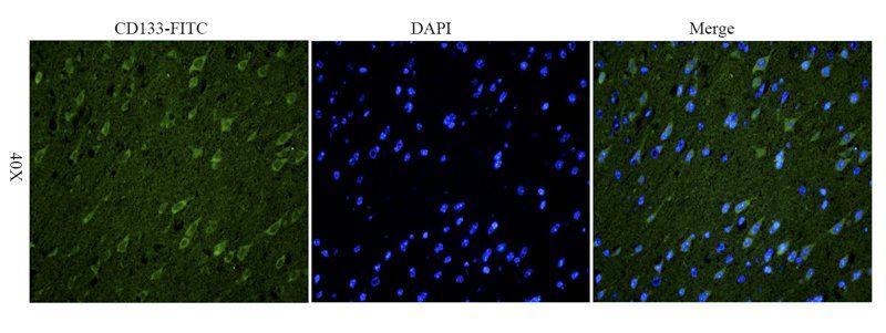
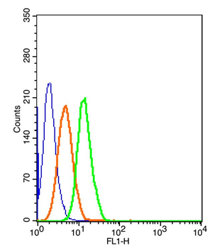
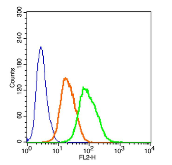
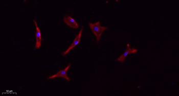
Flow Cytometry analysis of CACO-2 cells using anti-PROM1 antibody. Overlay histogram showing CACO-2 cells (Blue line). The cells were fixed with 4% paraformaldehyde and blocked with 10% normal goat serum. And then incubated with rabbit anti-PROM1 Antibody (1 µg/1x10^6 cells) for 30 min at 20°C. DyLight®488 conjugated goat anti-rabbit IgG (5-10 µg/1x10^6 cells) was used as secondary antibody for 30 minutes at 20°C. Isotype control antibody (Green line) was rabbit IgG (1 µg/1x10^6) used under the same conditions. Unlabelled sample without incubation with primary antibody and secondary antibody (Red line) was used as a blank control.
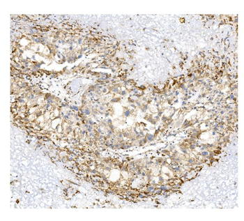
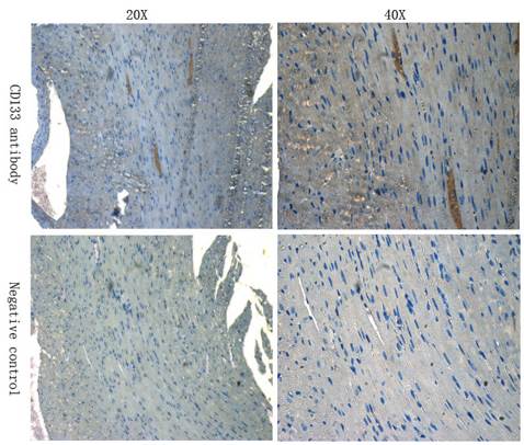
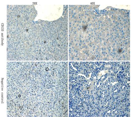
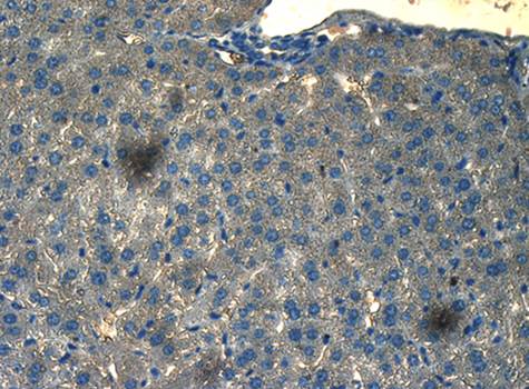
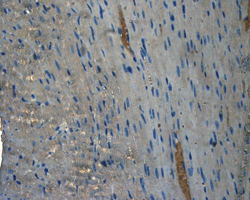
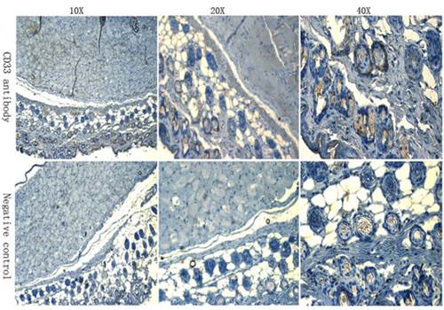
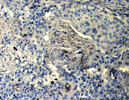
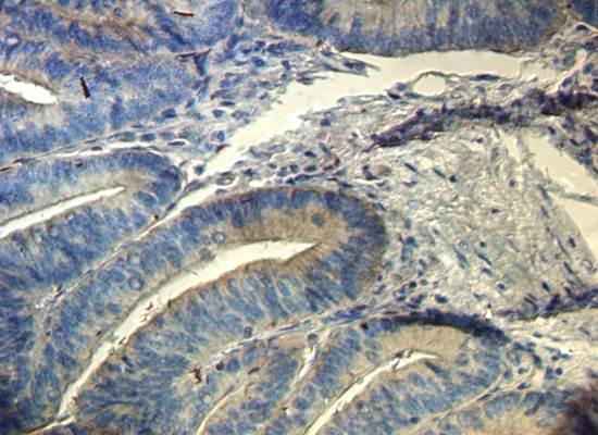
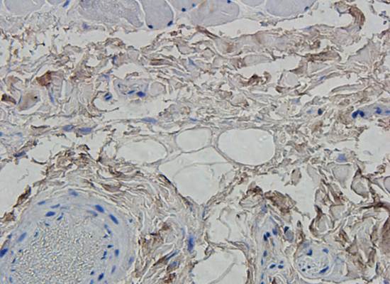
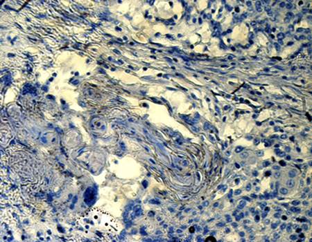
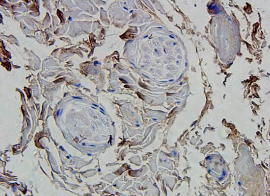
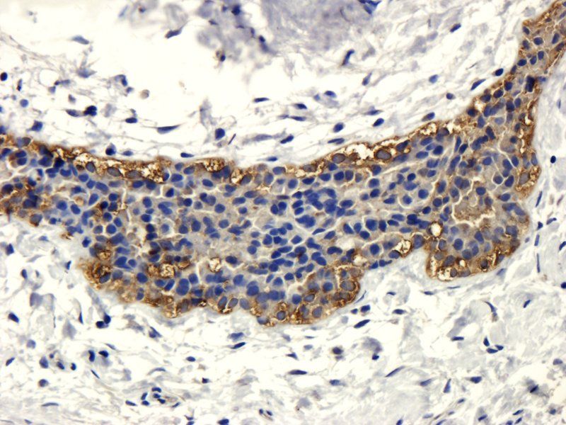
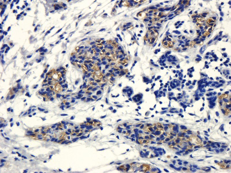
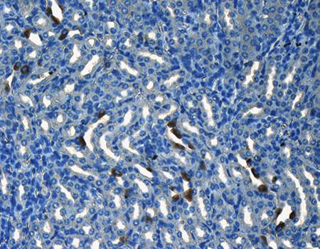
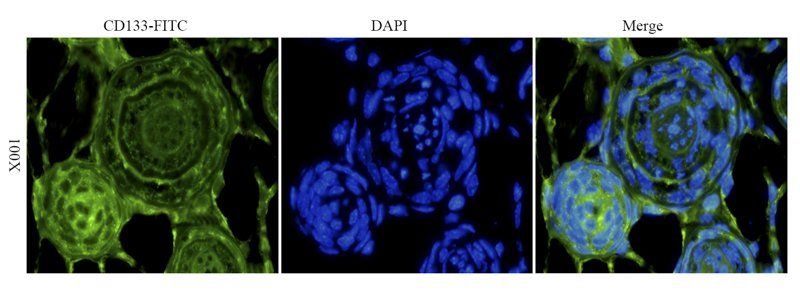
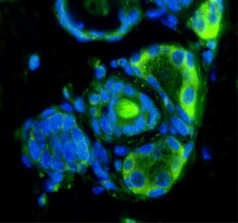
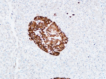
IHC analysis of PROM1 using anti-PROM1 antibody. PROM1 was detected in paraffin-embedded section of human liver cancer tissue. Heat mediated antigen retrieval was performed in EDTA buffer (pH8.0, epitope retrieval solution). The tissue section was blocked with 10% goat serum. The tissue section was then incubated with 1 µg/ml rabbit anti-PROM1 Antibody overnight at 4°C. Biotinylated goat anti-rabbit IgG was used as secondary antibody and incubated for 30 minutes at 37°C. The tissue section was developed using Strepavidin-Biotin-Complex (SABC) with DAB as the chromogen.
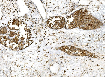
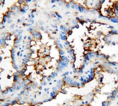
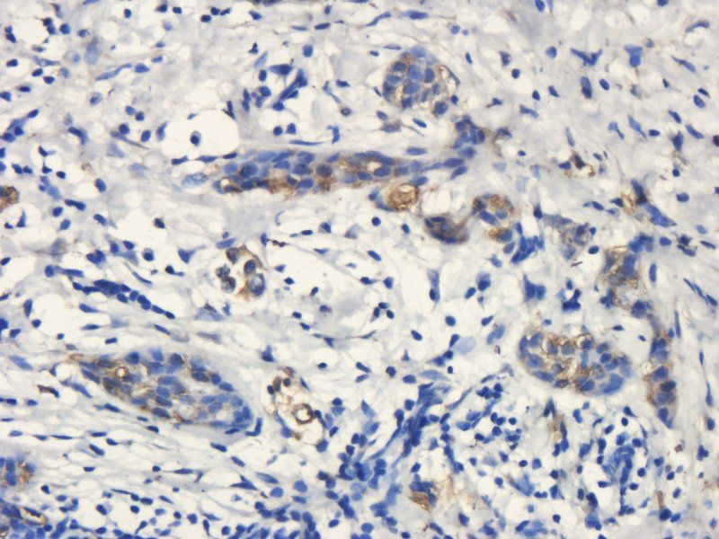
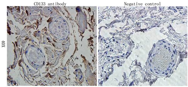
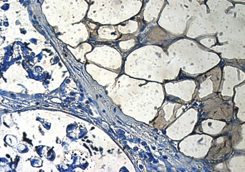
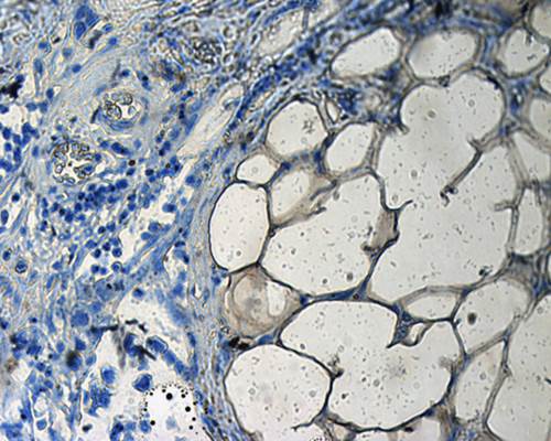
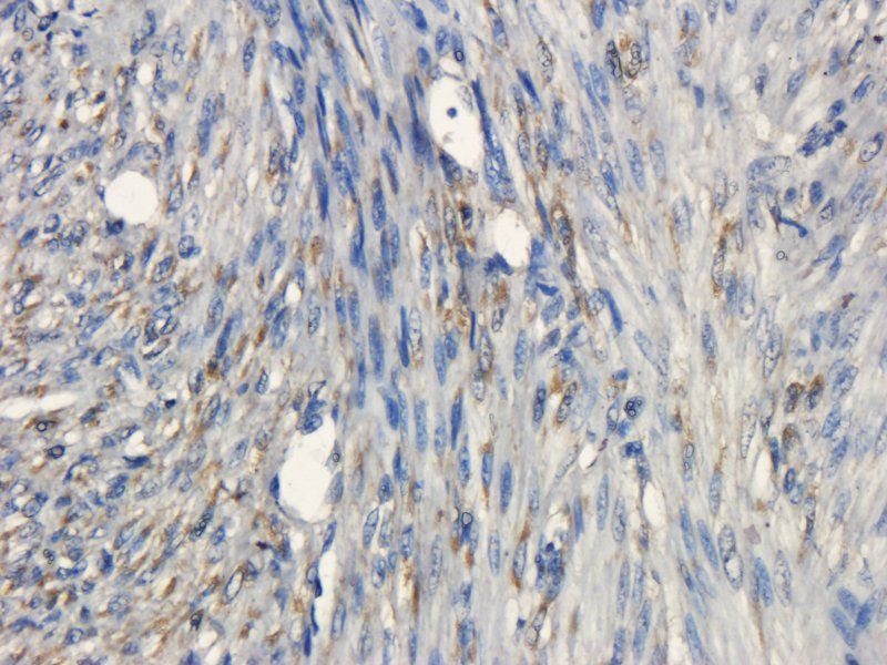
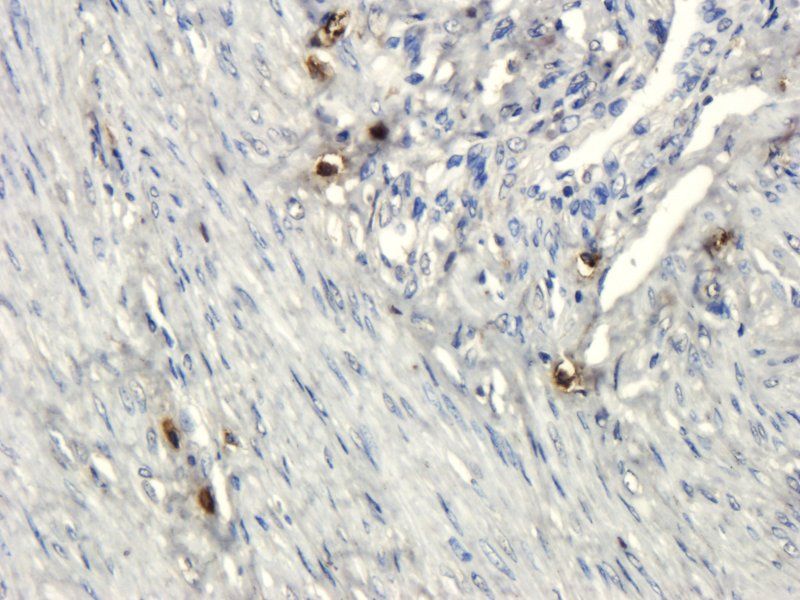
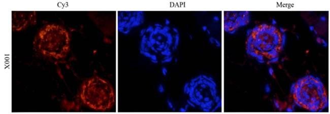
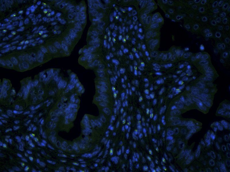
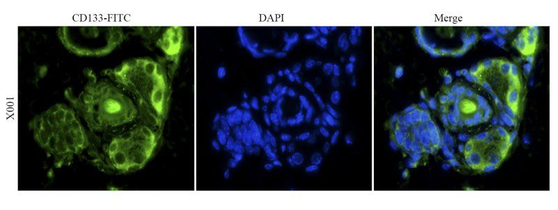
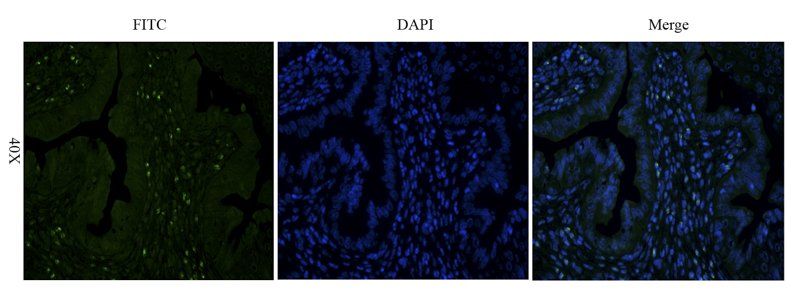
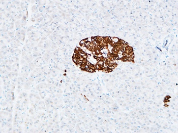
IHC analysis of PROM1 using anti-PROM1 antibody. PROM1 was detected in paraffin-embedded section of human mammary cancer tissue. Heat mediated antigen retrieval was performed in EDTA buffer (pH8.0, epitope retrieval solution). The tissue section was blocked with 10% goat serum. The tissue section was then incubated with 1 µg/ml rabbit anti-PROM1 Antibody overnight at 4°C. Biotinylated goat anti-rabbit IgG was used as secondary antibody and incubated for 30 minutes at 37°C. The tissue section was developed using Strepavidin-Biotin-Complex (SABC) with DAB as the chromogen.
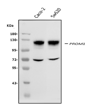
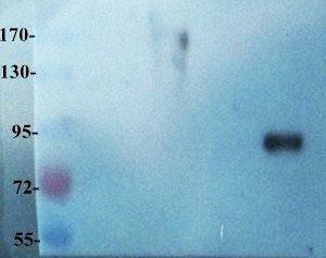
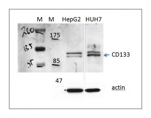
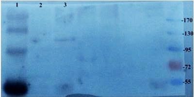
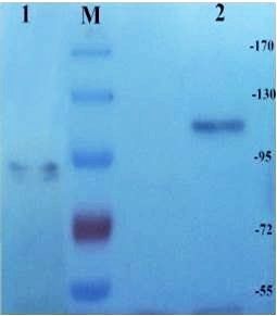
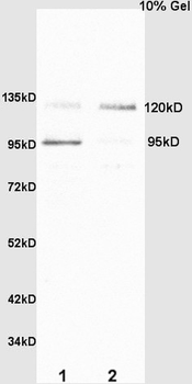
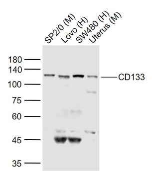
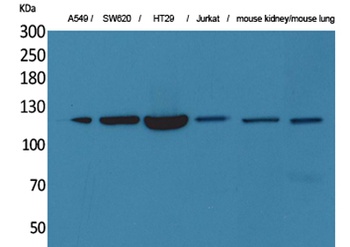
Western blot analysis of PROM1 using anti-PROM1 antibody. Electrophoresis was performed on a 5-20% SDS-PAGE gel at 70V (Stacking gel) / 90V (Resolving gel) for 2-3 hours. The sample well of each lane was loaded with 50 ug of sample under reducing conditions. Lane 1: human CACO-2 whole cell lysates, Lane 2: human SW620 whole cell lysates. After Electrophoresis, proteins were transferred to a Nitrocellulose membrane at 150mA for 50-90 minutes. Blocked the membrane with 5% Non-fat Milk/ TBS for 1.5 hour at RT. The membrane was incubated with rabbit anti-PROM1 antigen affinity purified polyclonal antibody at 0.5 µg/mL overnight at 4°C, then washed with TBS-0.1% Tween 3 times with 5 minutes each and probed with a goat anti-rabbit IgG-HRP secondary antibody at a dilution of 1:10000 for 1.5 hour at RT. The signal is developed using an Enhanced Chemiluminescent detection (ECL) kit with Tanon 5200 system. A specific band was detected for PROM1 at approximately 120 KD. The expected band size for PROM1 is at 120 KD.
Quick Database Links
Gene Symbol
Prominin-1
UniProt
UniProt Details
− No UniProt data available
Documents Download
Datasheet
Product Information
Request a Document
Protocol Information
WB
Western Blot (IB, immunoblot)
IHC
Immunohistochemistry
FC
Flow Cytometry
PROM1 Rabbit Polyclonal Antibody (orb215910)

- 0.0
Based on 0 reviews
Participating in our Biorbyt product reviews program enables you to support fellow scientists by sharing your firsthand experience with our products.
Login to Submit a ReviewAvailable Sizes
Select a size below
Free Secondary Antibody (20 ul)0/0
Please add an antibody product to your cart first.