
You have no items in your shopping cart.
Description
Research Area
Disease Biomarkers, Immunology & Inflammation; Cell Biology, Stem Cell & Developmental Biology
Images & Validation
−Item 1 of 5
| Tested Applications | IHC, WB |
|---|---|
| Dilution Range | WB: 1:500-1:1000, IHC: 1:100-1:250 |
| Reactivity | Human |
Key Properties
−| Antibody Type | Primary Antibody |
|---|---|
| Host | Rabbit |
| Clonality | Polyclonal |
| Isotype | IgG |
| Immunogen | Synthetic peptide corresponding to amino acids from an internal sequence of the human Prominin-1 (CD133) protein. |
| Target | CD133 |
| Purification | Antigen Affinity Purified |
| Conjugation | Unconjugated |
Storage & Handling
−| Storage | Maintain refrigerated at 2-8°C for up to 2 weeks. For long term storage store at -20°C in small aliquots to prevent freeze-thaw cycles. |
|---|---|
| Form/Appearance | Liquid |
| Buffer/Preservatives | PBS + 0.02% NaN3 |
| Expiration Date | 12 months from date of receipt. |
| Disclaimer | For research use only |
Alternative Names
−PROM1, Antigen AC133, Prominin-like protein 1, PROML1, MSTP061
Similar Products
−- Item 1 of 30
CD133 Rabbit Polyclonal Antibody [orb99113]
ELISA, ICC, IF, IHC-P, WB
Human, Mouse, Rat
Rabbit
Polyclonal
Unconjugated
100 μg - Item 1 of 9
- Item 1 of 5
CD133 Rabbit Polyclonal Antibody [orb526485]
FC, WB
Rat
Human, Mouse
Rabbit
Polyclonal
Unconjugated
50 μl, 100 μl, 200 μl - Item 1 of 6
CD133 Rabbit Polyclonal Antibody [orb1294396]
IF, IHC, WB
Human, Mouse, Rat
Rabbit
Polyclonal
Unconjugated
100 μl, 25 μl - Item 1 of 5


Quality Guarantee
Explore bioreagents carefree to elevate your research. All our products are rigorously tested for performance. If a product does not perform as described on its datasheet, our scientific support team will provide expert troubleshooting, a prompt replacement, or a refund. For full details, please see our Terms & Conditions and Buying Guide. Contact us at [email protected].

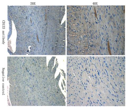
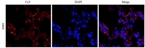
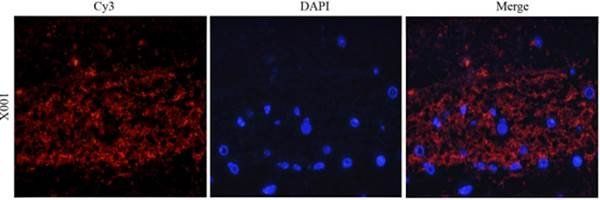
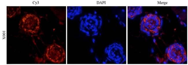
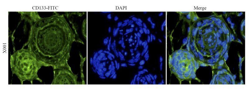
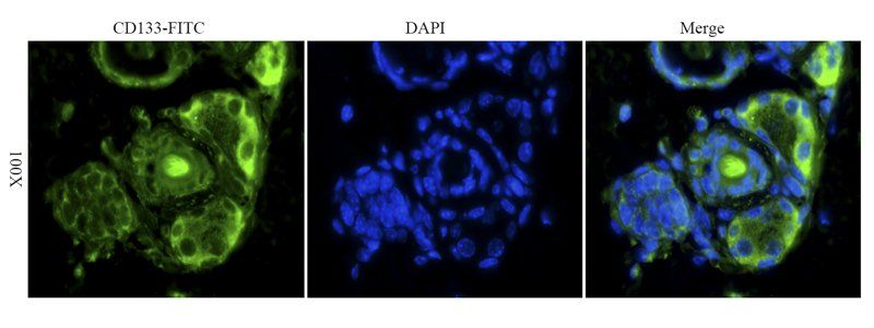
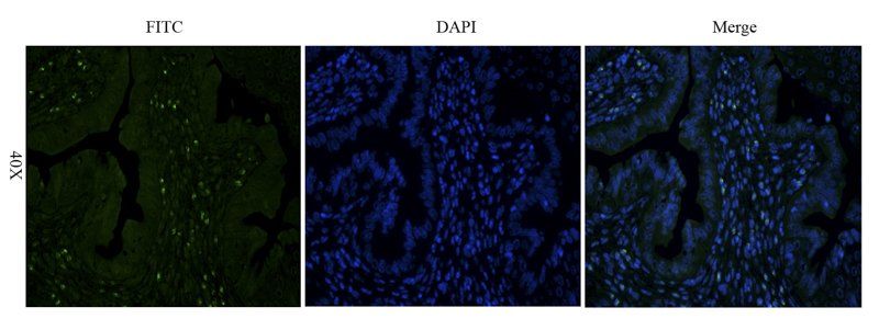
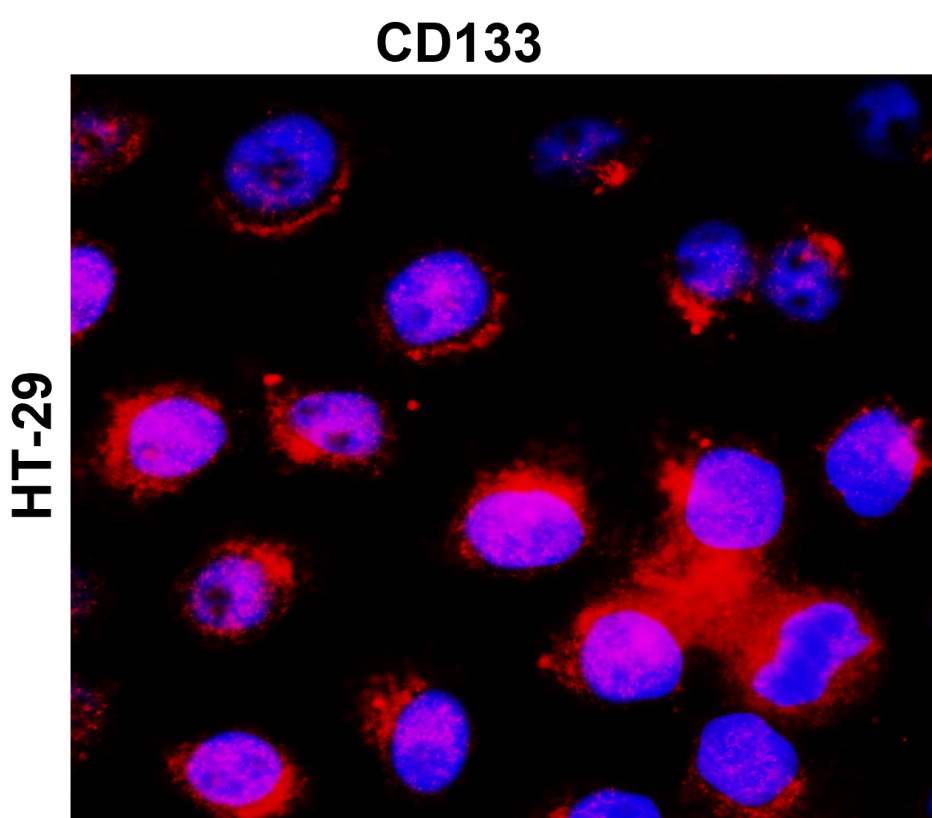
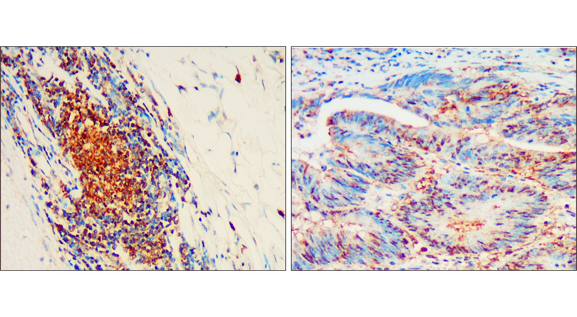
Chromatin Immunoprecipitation showing staining of CD133 in Bone Marrow Mononuclear Cells by immunofluorescence.

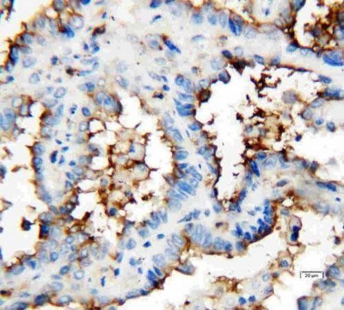
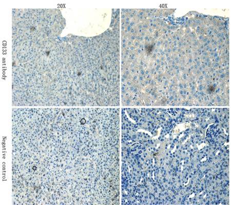
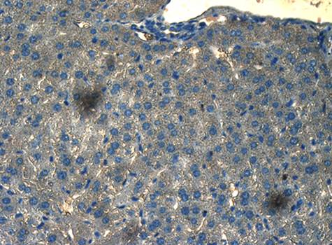
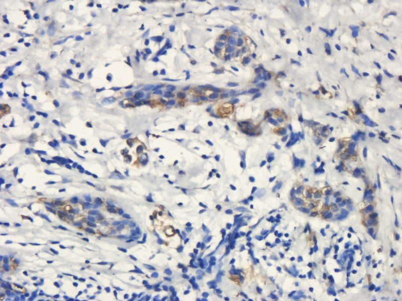
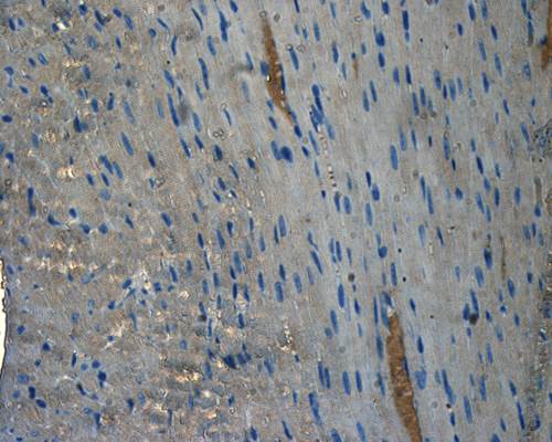
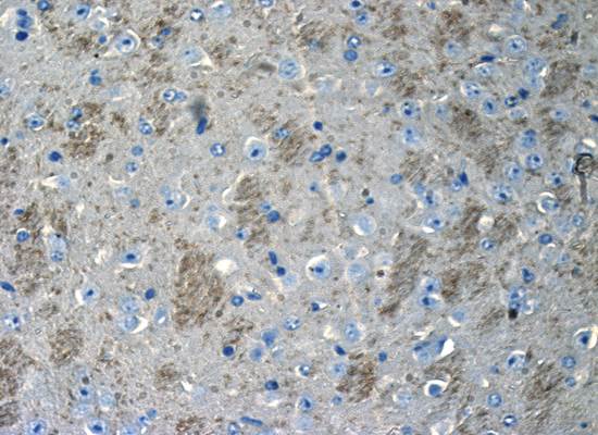
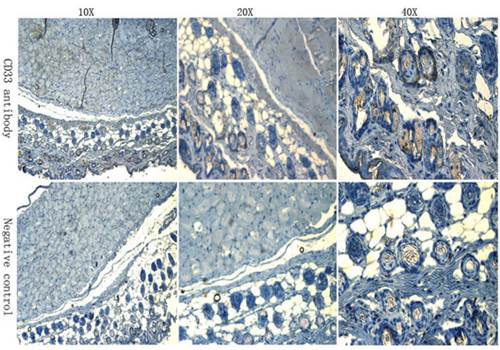
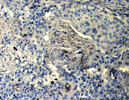
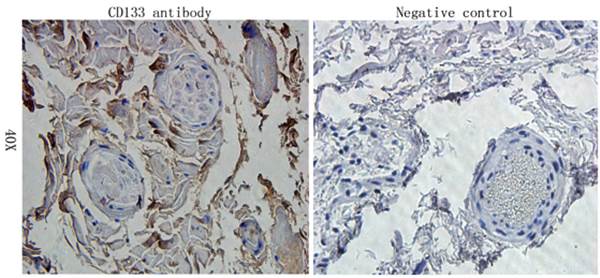
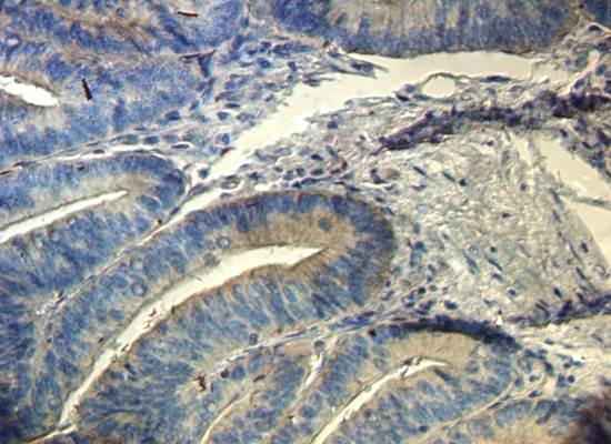
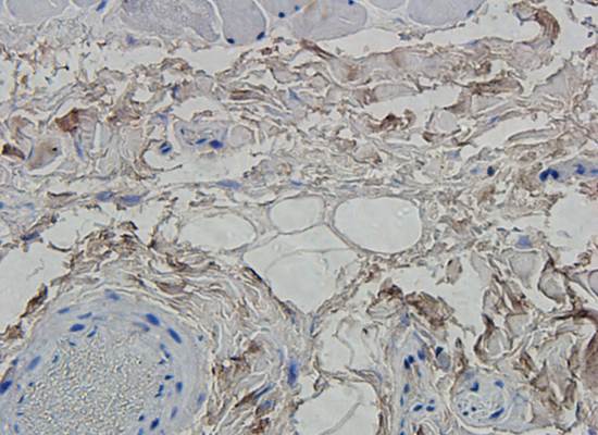
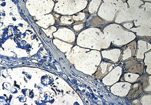
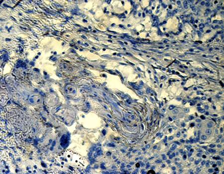
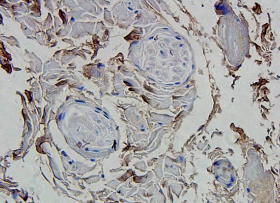
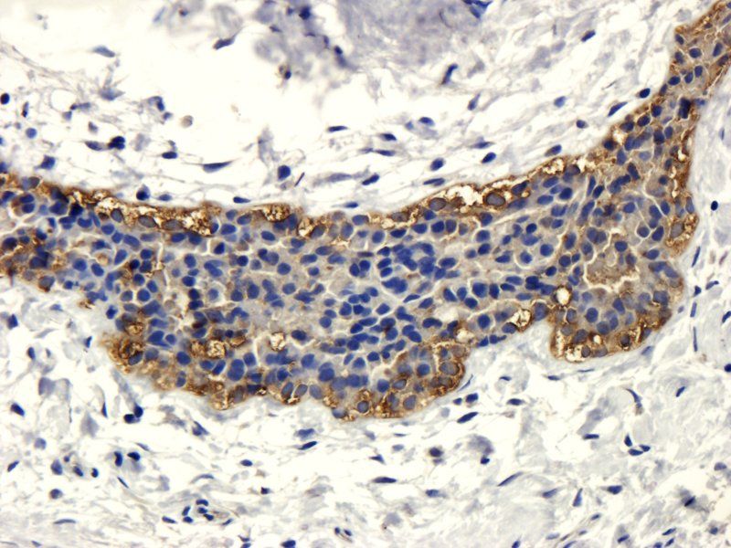
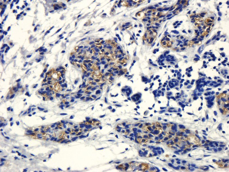
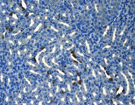
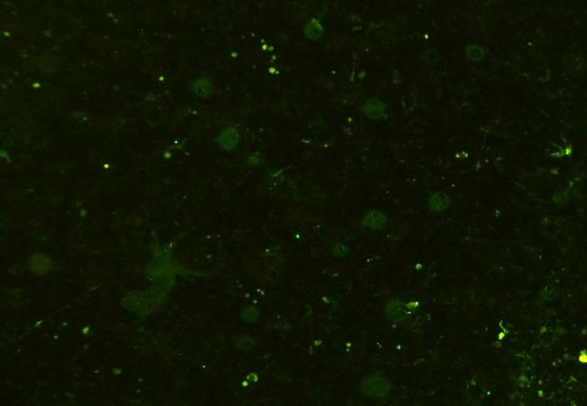
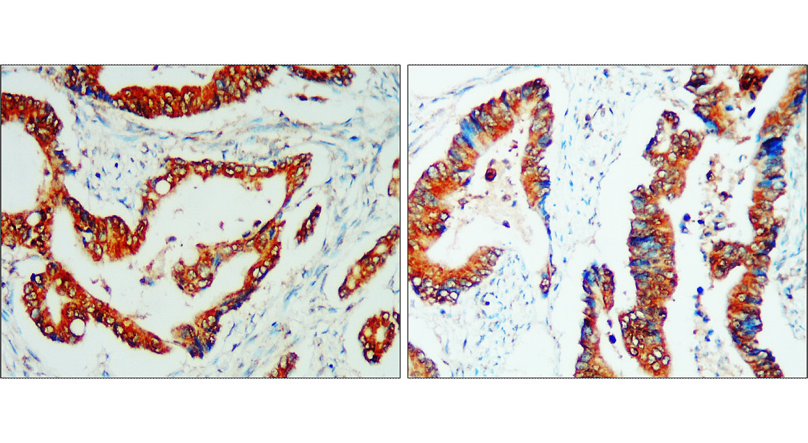
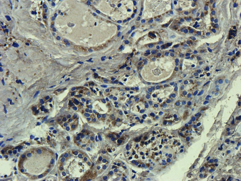
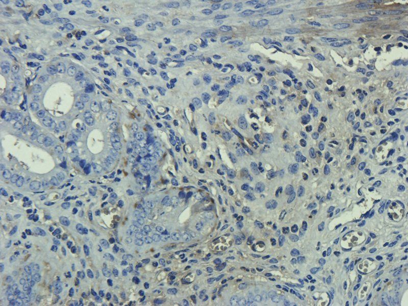
Immunohistochemistry of human hepatocarcinoma (HC) tissue showing specific staining of CD133.

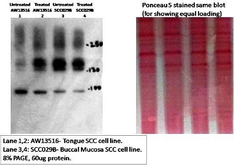
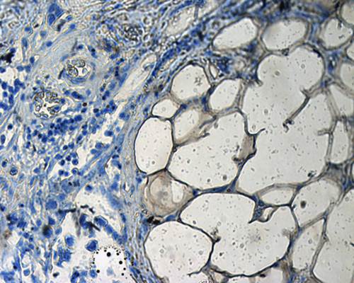
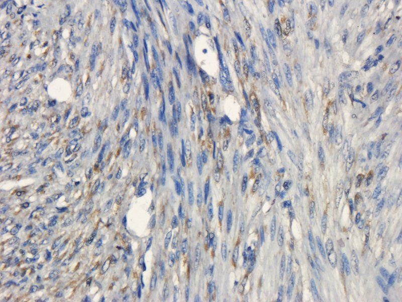
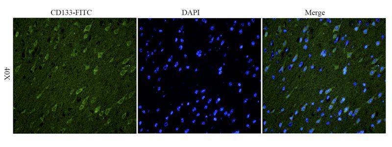
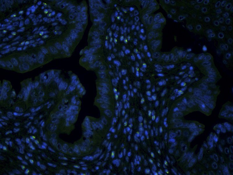
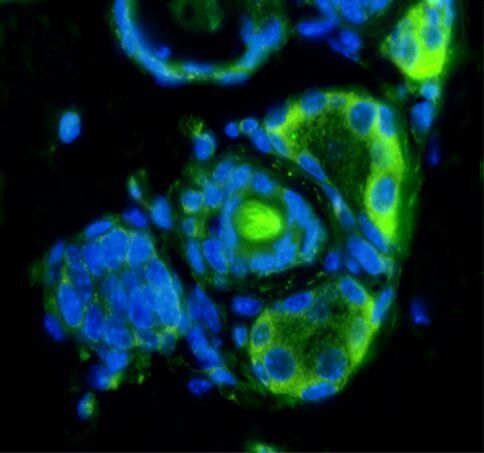
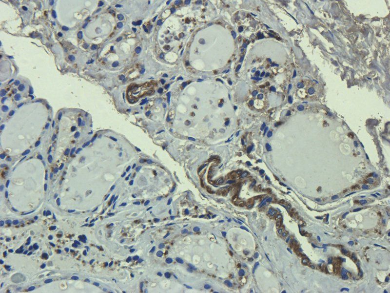
Immunostaining of normal ovarian and tumor tissue sections showing specific labeling of CD133. Staining of CD133 in OSE layer (A, B) as well as cortex (C) reveals specific CD133+ cells with relatively higher cell numbers in BL and HG. Area within dotted lines in BN OSE (A) are magnified in (B) while elliptical/spindle shaped CD133+ cells in cortex from various fields were represented in the composite image in (C) of BN and HG. Large CD133+ cells in cortex were also observed.

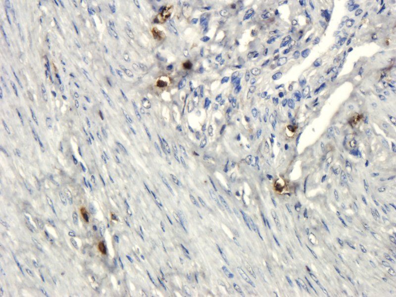
Immunostaining of normal ovarian and tumor tissue sections showing specific labeling of CD133. Staining of CD133 in OSE layer (A, B) as well as cortex (C) reveals specific CD133+ cells with relatively higher cell numbers in BL and HG. Area within dotted lines in BN OSE (A) are magnified in (B) while elliptical/spindle shaped CD133+ cells in cortex from various fields were represented in the composite image in (C) of BN and HG. Large CD133+ cells in cortex were also observed.

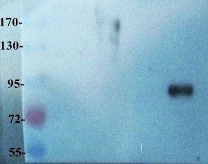
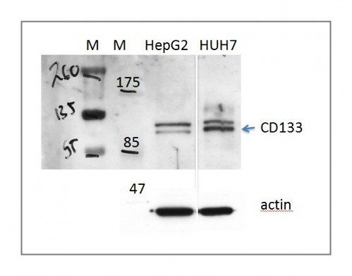
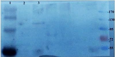
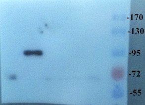
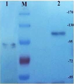
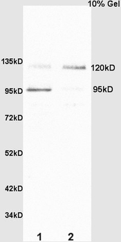
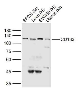
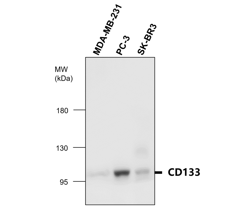
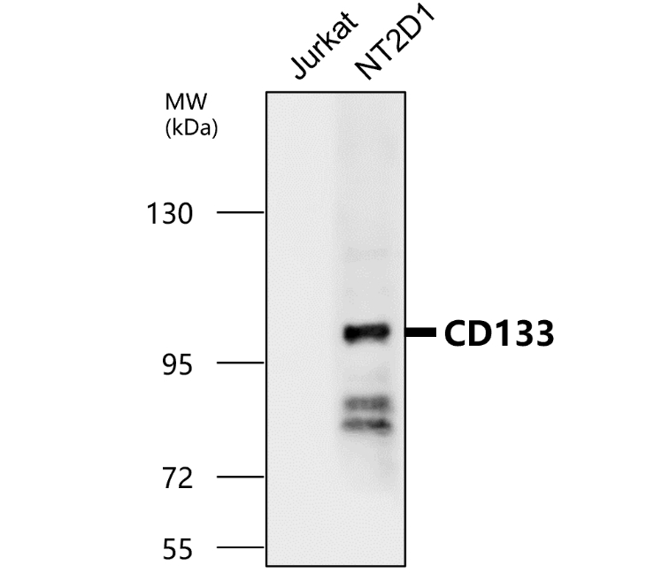
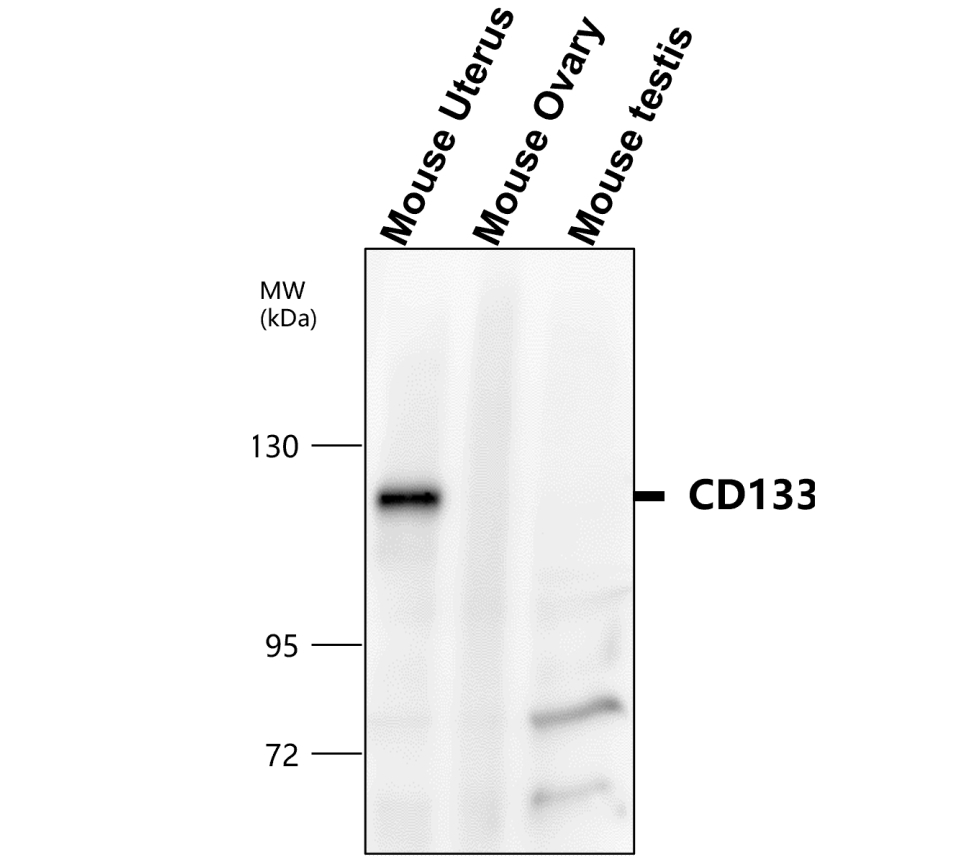
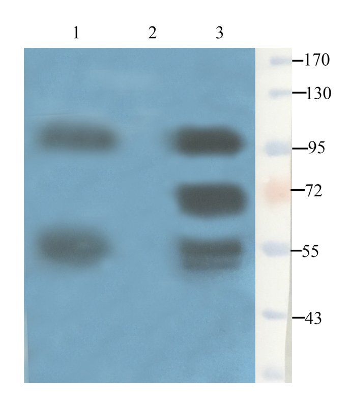
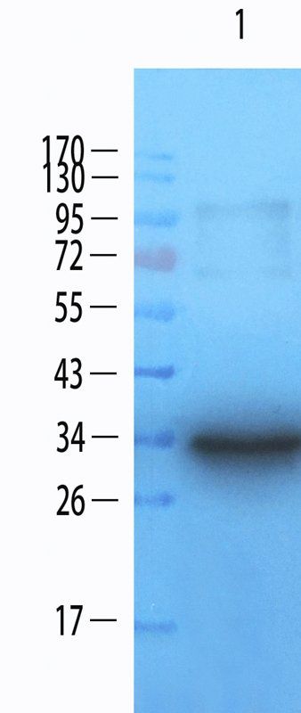
Western Blot of Y79 cells showing specific immunolabeling of human CD133. Band is at ~120 kDa due to protein glycosylation.
Quick Database Links
Gene Symbol
CD133
UniProt
UniProt Details
− No UniProt data available
Documents Download
Datasheet
Product Information
Request a Document
Protocol Information
WB
Western Blot (IB, immunoblot)
IHC
Immunohistochemistry
CD133 Antibody (orb1821778)

- 0.0
Based on 0 reviews
Participating in our Biorbyt product reviews program enables you to support fellow scientists by sharing your firsthand experience with our products.
Login to Submit a ReviewAvailable Sizes
Select a size below
Choose Conjugation or Carrier Free Version
Free Secondary Antibody (20 ul)0/0
Please add an antibody product to your cart first.
 Usually dispatched within 5-10 working days