You have no items in your shopping cart.
Featured
Description
Research Area
Signal Transduction
Images & Validation
−Item 1 of 4
| Tested Applications | ELISA, IF, IHC, WB |
|---|---|
| Dilution Range | IF: 1:50-200 Western Blot: 1/500 - 1/2000. Immunohistochemistry: 1/100 - 1/300. ELISA: 1/20000. Not yet tested in other applications. |
| Reactivity | Human, Mouse, Rat |
Key Properties
−| Clonality | Polyclonal |
|---|---|
| Isotype | IgG |
| Immunogen | Synthesized phospho-peptide around the phosphorylation site of human Akt (phospho Thr308) |
| Molecular Weight | 55 |
| Purification | The antibody was affinity-purified from rabbit antiserum by affinity-chromatography using epitope-specific immunogen. |
| Conjugation | Unconjugated |
Storage & Handling
−| Storage | Maintain refrigerated at 2-8°C for up to 2 weeks. For long term storage store at -20°C in small aliquots to prevent freeze-thaw cycles. |
|---|---|
| Buffer/Preservatives | PBS with 0.02% sodium azide and 50% glycerol pH 7.4. |
| Concentration | 1 mg/ml |
| Expiration Date | 12 months from date of receipt. |
| Disclaimer | For research use only |
Alternative Names
−Anti-AKT1 antibody, anti-PKB antibody, anti-RAC antibody, anti-RAC-alpha serine/threonine-protein kinase antibody, anti-Protein kinase B antibody, anti-PKB antibody, anti-Protein kinase B alpha antibody, anti-PKB alpha antibody, anti-Proto-oncogene c-Akt antibody, anti-RAC-PK-alpha antibody, anti-AKT2 antibody, anti-RAC-beta serine/threonine-protein kinase antibody, anti- antibody
Similar Products
−- Item 1 of 2

phospho-AKT1 + AKT2 + AKT3 (Thr308+Thr309+Thr305) Rabbit pAb, Pacific Blue conjugated [orb2824805]
FC, ICC, IF
Bovine, Canine, Gallus, Porcine, Rabbit, Sheep
Human, Mouse, Rat
Rabbit
Polyclonal
Pacific Blue
100 μlphospho-AKT1 + AKT2 + AKT3 (Thr308+Thr309+Thr305) Rabbit pAb, BF700 conjugated [orb2824806]
FC, ICC, IF
Bovine, Canine, Gallus, Porcine, Rabbit, Sheep
Human, Mouse, Rat
Rabbit
Polyclonal
BF700
100 μlphospho-AKT1 + AKT2 + AKT3 (Thr308+Thr309+Thr305) Rabbit pAb, IRDye 800CW conjugated [orb2824807]
WB
Bovine, Canine, Gallus, Porcine, Rabbit, Sheep
Human, Mouse, Rat
Rabbit
Polyclonal
IRDye800
100 μlphospho-AKT1 (Thr308) Rabbit pAb, Pacific Blue conjugated [orb2825098]
FC, IF
Bovine, Canine, Gallus, Porcine, Rabbit, Sheep
Human, Mouse, Rat
Rabbit
Polyclonal
Pacific Blue
100 μl
Quality Guarantee
Explore bioreagents carefree to elevate your research. All our products are rigorously tested for performance. If a product does not perform as described on its datasheet, our scientific support team will provide expert troubleshooting, a prompt replacement, or a refund. For full details, please see our Terms & Conditions and Buying Guide. Contact us at [email protected].
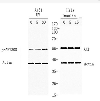
Western blot analysis of lysates from A431 and Hela cells treated with UV or insulin 0.01U/ml, using AKT p-308 Antibody. Primary Antibody was diluted at 1:1000 4°C over night, secondary antibody was diluted at 1:10000, 37° 1hour.
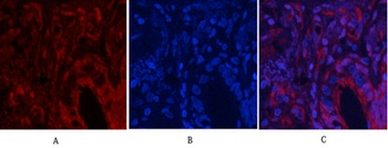
Immunofluorescence analysis of human-lung tissue. 1, Akt (phospho Thr308) Polyclonal Antibody (red) was diluted at 1:200 (4°C, overnight). 2, Cy3 labled Secondary antibody was diluted at 1:300 (room temperature, 50min).3, Picture B: DAPI (blue) 10min. Picture A:Target. Picture B: DAPI. Picture C: merge of A + B.
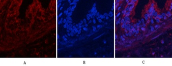
Immunofluorescence analysis of human-lung tissue. 1, Akt (phospho Thr308) Polyclonal Antibody (red) was diluted at 1:200 (4°C, overnight). 2, Cy3 labled Secondary antibody was diluted at 1:300 (room temperature, 50min).3, Picture B: DAPI (blue) 10min. Picture A:Target. Picture B: DAPI. Picture C: merge of A + B.
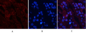
Immunofluorescence analysis of human-stomach tissue. 1, Akt (phospho Thr308) Polyclonal Antibody (red) was diluted at 1:200 (4°C, overnight). 2, Cy3 labled Secondary antibody was diluted at 1:300 (room temperature, 50min).3, Picture B: DAPI (blue) 10min. Picture A:Target. Picture B: DAPI. Picture C: merge of A + B.
Documents Download
Datasheet
Product Information
Request a Document
Protocol Information
WB
Western Blot (IB, immunoblot)
IHC
Immunohistochemistry
IF
Immunofluorescence
ELISA
Enzyme-linked Immunosorbent Assay (EIA)
Akt (phospho Thr308) rabbit pAb Antibody (orb764338)

- 0.0
Based on 0 reviews
Participating in our Biorbyt product reviews program enables you to support fellow scientists by sharing your firsthand experience with our products.
Login to Submit a ReviewAvailable Sizes
Select a size below
Choose Conjugation or Carrier Free Version
Free Secondary Antibody (20 ul)0/0
Please add an antibody product to your cart first.