You have no items in your shopping cart.
GRP94 Antibody
SKU: orb1822453
Description
Research Area
Cancer Biology, Cell Biology, Disease Biomarkers, Metabolism Research, Protein Biochemistry, Signal Transduction
Images & Validation
−Item 1 of 3
| Tested Applications | ICC, IF, IHC, IP, WB |
|---|---|
| Dilution range | WB (1:1000), ICC/IF (1:120), IP (1:80); optimal dilutions for assays should be determined by the user. |
| Reactivity | Bovine, Human, Mouse, Rat |
| Application Notes |
Key Properties
−| Host | Rabbit |
|---|---|
| Clonality | Polyclonal |
| Immunogen | Synthetic peptide corresponding to the sequence near the C-terminus of mouse GRP94 |
| Target | GRP94 |
| Molecular Weight | 94kDa |
| Purification | Peptide Affinity Purified |
| Conjugation | Unconjugated |
Storage & Handling
−| Storage | Maintain refrigerated at 2-8°C for up to 2 weeks. For long term storage store at -20°C in small aliquots to prevent freeze-thaw cycles. |
|---|---|
| Buffer/Preservatives | PBS pH 7.4, 50% glycerol, 0.09% sodium azide *Storage buffer changes when conjugated |
| Concentration | 1 mg/ml |
| Disclaimer | For research use only |
Alternative Names
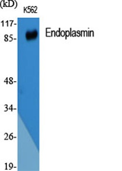
−HSP90B1, GP96, TRA1, ECGP, 94 kDa glucose regulated protein, 94 kDa glucose-regulated protein, Endoplasmin, Endothelial cell (HBMEC) glycoprotein, ENPL_HUMAN, Glucose regulated protein 94kDa, gp96, gp96 homolog, GRP 94, GRP-94, Heat shock protein 90 kDa beta member 1, heat shock protein 90kDa beta (Grp94), member 1, Heat shock protein, 90 kDa, beta, 1, Stress inducible tumor rejection antigen GP96, tumor rejection antigen (gp96) 1, Tumor rejection antigen 1, Tumor rejection antigen gp96, Tumor rejection antigen-1 (gp96)
Similar Products
−- Item 1 of 11
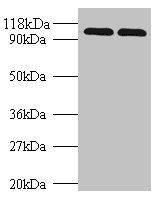
GRP94 Rabbit Polyclonal Antibody [orb10752]
FC, IF, IHC-Fr, IHC-P, WB
Bovine, Canine, Equine, Gallus, Porcine, Rabbit
Human, Mouse, Rat
Rabbit
Polyclonal
Unconjugated
50 μl, 100 μl, 200 μl - Item 1 of 7
GRP94/HSP90B1 Antibody [orb315145]
FC, ICC, IF, IHC, WB
Human, Mouse, Rat
Rabbit
Polyclonal
Unconjugated
100 μg - Item 1 of 4
HSP90B1 Antibody (N-term) [orb1930773]
FC, IF, IHC-P, WB
Bovine, Gallus, Monkey, Porcine, Rat
Hamster, Human, Mouse
Rabbit
Polyclonal
Unconjugated
100 μl, 50 μl - Item 1 of 5
- Item 1 of 4
Endoplasmin rabbit pAb Antibody [orb765138]
ELISA, IF, IHC, WB
Human, Mouse
Polyclonal
Unconjugated
100 μl, 50 μl

Quality Guarantee
Explore bioreagents carefree to elevate your research. All our products are rigorously tested for performance. If a product does not perform as described on its datasheet, our scientific support team will provide expert troubleshooting, a prompt replacement, or a refund. For full details, please see our Terms & Conditions and Buying Guide. Contact us at [email protected].
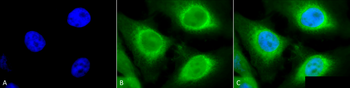
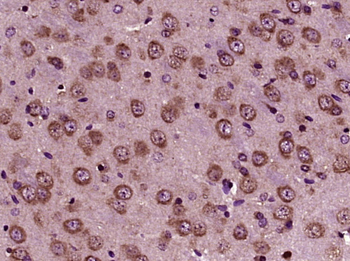
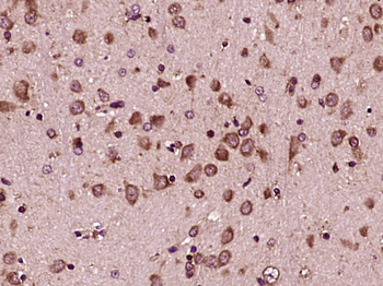
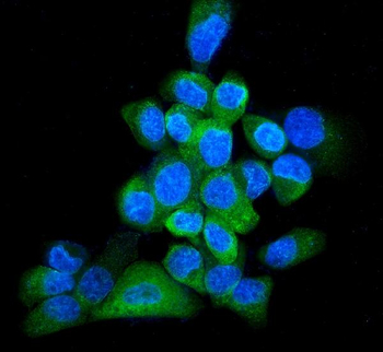
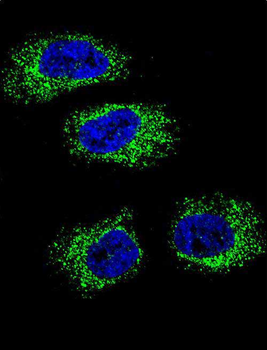
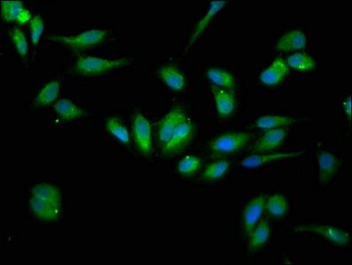
Immunocytochemistry/Immunofluorescence analysis using Rabbit Anti-GRP94 Polyclonal Antibody. Tissue: Heat Shocked Cervical cancer cell line (HeLa). Species: Human. Fixation: 2% Formaldehyde for 20 min at RT. Primary Antibody: Rabbit Anti-GRP94 Polyclonal Antibody at 1:120 for 12 hours at 4°C. Secondary Antibody: FITC Goat Anti-Rabbit (green) at 1:200 for 2 hours at RT. Counterstain: DAPI (blue) nuclear stain at 1:40000 for 2 hours at RT. Localization: Endoplasmic reticulum lumen. Melanosome. Magnification: 100x. (A) DAPI (blue) nuclear stain. (B) Anti-GRP94 Antibody. (C) Composite. Heat Shocked at 42°C for 1h.
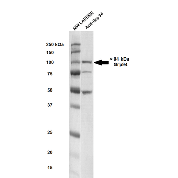
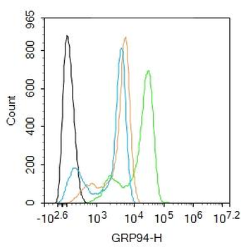
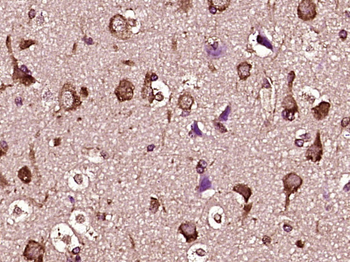
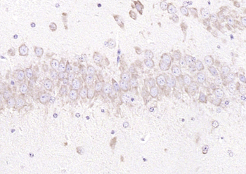
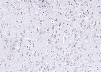
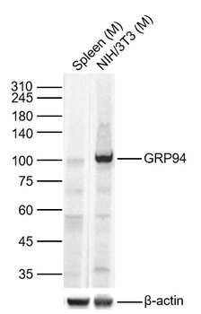
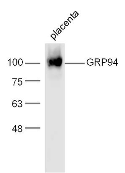
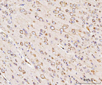
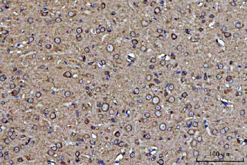
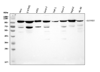
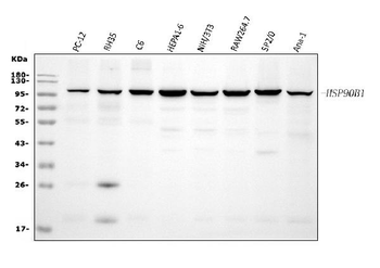
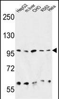
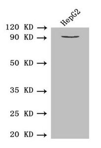
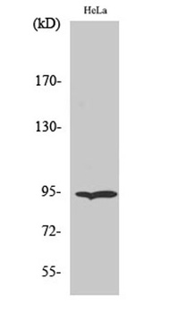
Western blot analysis of Rat brain cell lysates showing detection of ~ 94-100 kDa GRP94 protein using Rabbit Anti-GRP94 Polyclonal Antibody. Lane 1: MW ladder. Lane 2: Anti-GRP94 (1:250). Load: 20 μg. Block: 5% milk + TBST for 1 hour at RT. Primary Antibody: Rabbit Anti-GRP94 Polyclonal Antibody at 1:250 for 1 hour at RT. Secondary Antibody: Goat Anti-Rabbit HRP antibody at 1:50-1:100 for 1 hour at RT. Color Development: TMB solution for 5 min at RT. Predicted/Observed Size: ~ 94-100 kDa. Other Band (s): ~50, ~75 kDa.
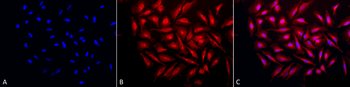
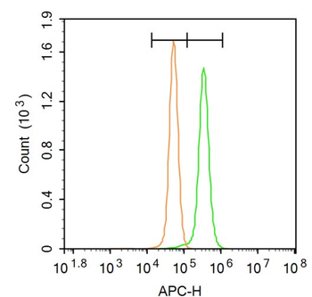
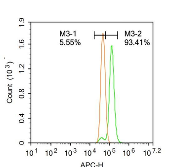
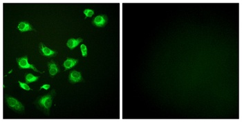
Immunocytochemistry/Immunofluorescence analysis using Rabbit Anti-GRP94 Polyclonal Antibody. Tissue: Heat Shocked Cervical cancer cell line (HeLa). Species: Human. Fixation: 2% Formaldehyde for 20 min at RT. Primary Antibody: Rabbit Anti-GRP94 Polyclonal Antibody at 1:120 for 12 hours at 4°C. Secondary Antibody: APC Goat Anti-Rabbit (red) at 1:200 for 2 hours at RT. Counterstain: DAPI (blue) nuclear stain at 1:40000 for 2 hours at RT. Localization: Endoplasmic reticulum lumen. Melanosome. Magnification: 20x. (A) DAPI (blue) nuclear stain. (B) Anti-GRP94 Antibody. (C) Composite. Heat Shocked at 42°C for 1h.
Quick Database Links
UniProt Details
− No UniProt data available
NCBI Gene Details
− No NCBI Gene data available
NCBI Reference Sequences
−Associated Accession Numbers
Curated reference sequences for the gene transcript and protein product| Protein | NP_035761.1 |
|---|
Documents Download
Datasheet
Product Information
Request a Document
Protocol Information
WB
Western Blot (IB, immunoblot)
IHC
Immunohistochemistry
IF
Immunofluorescence
ICC
Immunocytochemistry
IP
Immunoprecipitation
GRP94 Antibody (orb1822453)

- 0.0
Based on 0 reviews
Participating in our Biorbyt product reviews program enables you to support fellow scientists by sharing your firsthand experience with our products.
Login to Submit a ReviewAvailable Sizes
Select a size below