You have no items in your shopping cart.
Description
Research Area
Cancer Biology
Images & Validation
−Item 1 of 8
| Tested Applications | ELISA, NeA, WB |
|---|---|
| Reactivity | Human |
| Application Notes |
Key Properties
−| Antibody Type | Primary Antibody |
|---|---|
| Host | Rabbit |
| Clonality | Polyclonal |
| Immunogen | Produced from sera of rabbits pre-immunized with highly pure (>98%) recombinant hTIMP-1 (human Tissue Inhibitor of Metalloproteinases-1). Human TIMP-1 specific antibody was purified by affinity chromatography employing immobilized hTIMP-1 matrix. |
| Target | TIMP1 |
| Conjugation | Unconjugated |
Storage & Handling
−| Storage | Maintain refrigerated at 2-8°C for up to 2 weeks. For long term storage store at -20°C in small aliquots to prevent freeze-thaw cycles. |
|---|---|
| Form/Appearance | Lyophilized |
| Concentration | batch dependent |
| Expiration Date | 12 months from date of receipt. |
| Disclaimer | For research use only |
Alternative Names
−EPA, EPO, HCI, CLGI, TIMP, Metalloproteinase inhibitor 1, Erythroid-potentiating activity, EPA
Similar Products
−- Item 1 of 18
TIMP1 Rabbit Polyclonal Antibody [orb195994]
ELISA, ICC, IF, IHC-P, WB
Bovine, Canine, Guinea pig, Human, Mouse, Porcine, Rat, Sheep
Rabbit
Polyclonal
Unconjugated
100 μg - Item 1 of 10
TIMP-1 Mouse Monoclonal Antibody [orb500828]
FC, ICC
Mouse, Rat
Human
Mouse
Monoclonal
Unconjugated
50 μl, 100 μl, 200 μl, 200 μg - Item 1 of 7
TIMP1 Rabbit Polyclonal Antibody [orb11483]
ELISA, ICC, IF, IHC-P, WB
Rabbit
Polyclonal
Unconjugated
100 μg - Item 1 of 5
TIMP-1 Rabbit Polyclonal Antibody [orb100174]
WB
Bovine, Canine, Mouse, Porcine, Rabbit, Sheep
Human
Rabbit
Polyclonal
Unconjugated
100 μl, 200 μl, 50 μl - Item 1 of 5
TIMP-1 Rabbit Polyclonal Antibody [orb313247]
WB
Bovine, Canine, Porcine, Rat, Sheep
Human
Rabbit
Polyclonal
Unconjugated
50 μl, 100 μl, 200 μl
Quality Guarantee
Explore bioreagents carefree to elevate your research. All our products are rigorously tested for performance. If a product does not perform as described on its datasheet, our scientific support team will provide expert troubleshooting, a prompt replacement, or a refund. For full details, please see our Terms & Conditions and Buying Guide. Contact us at [email protected].
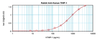
To detect hTIMP-1 by sandwich ELISA (using 100 ul/well antibody solution) a concentration of 0.5 - 2.0 ug/ml of this antibody is required. This antigen affinity purified antibody, in conjunction with Biotinylated Anti-Human TIMP-1 (orb1272680) as a detection antibody, allows the detection of at least 0.2 - 0.4 ng/well of recombinant hTIMP-1.
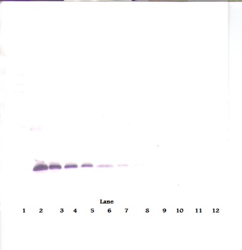
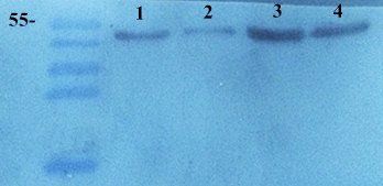
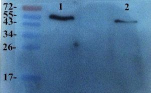
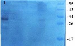
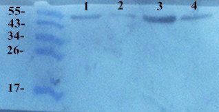
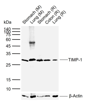
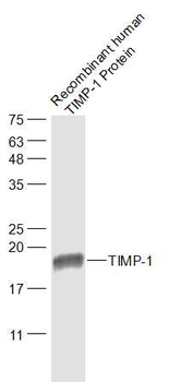
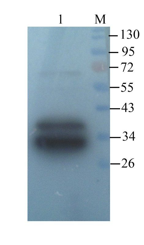
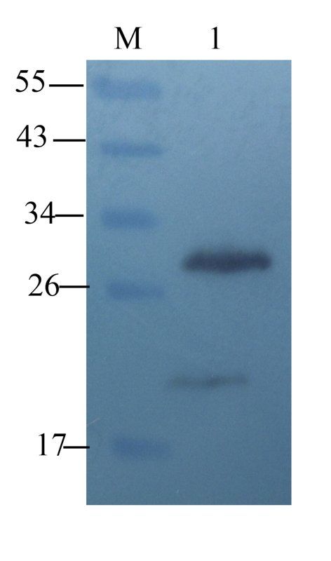
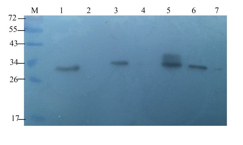
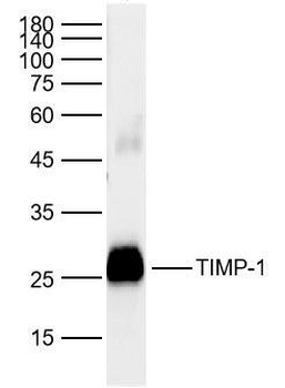
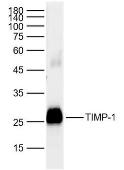
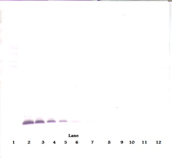
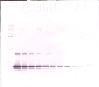
To detect hTIMP-1 by Western Blot analysis this antibody can be used at a concentration of 0.1 - 0.2 ug/ml. Used in conjunction with compatible secondary reagents the detection limit for recombinant hTIMP-1 is 1.5 - 3.0 ng/lane, under either reducing or non-reducing conditions.
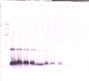
To detect hTIMP-1 by Western Blot analysis this antibody can be used at a concentration of 0.1 - 0.2 ug/ml. Used in conjunction with compatible secondary reagents the detection limit for recombinant hTIMP-1 is 1.5 - 3.0 ng/lane, under either reducing or non-reducing conditions.
To detect hTIMP-1 by Western Blot analysis this antibody can be used at a concentration of 0.1 - 0.2 ug/ml. Used in conjunction with compatible secondary reagents the detection limit for recombinant hTIMP-1 is 1.5 - 3.0 ng/lane, under either reducing or non-reducing conditions.
To detect hTIMP-1 by Western Blot analysis this antibody can be used at a concentration of 0.1 - 0.2 ug/ml. Used in conjunction with compatible secondary reagents the detection limit for recombinant hTIMP-1 is 1.5 - 3.0 ng/lane, under either reducing or non-reducing conditions.
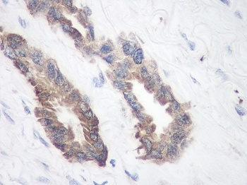
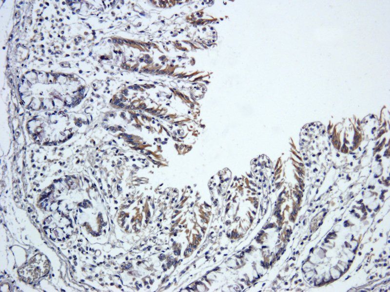
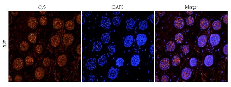
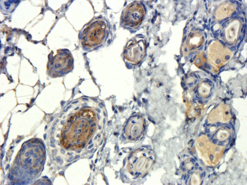
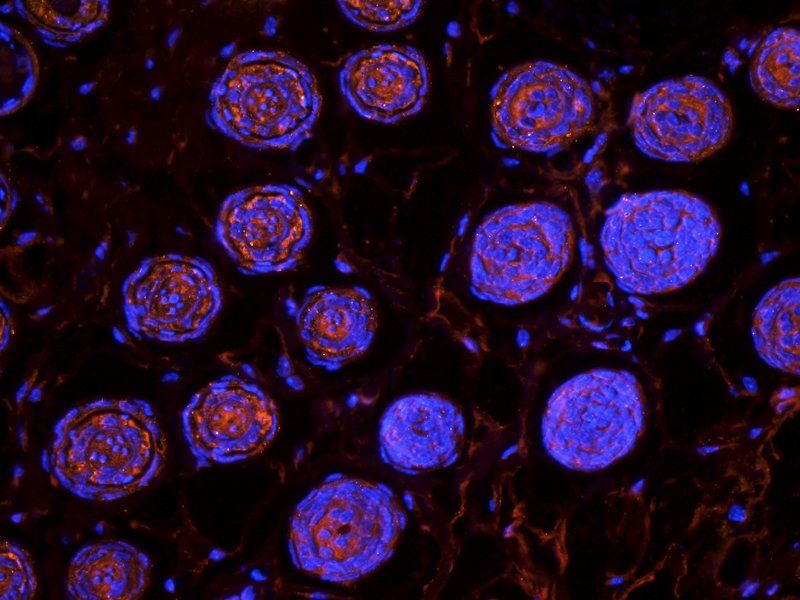
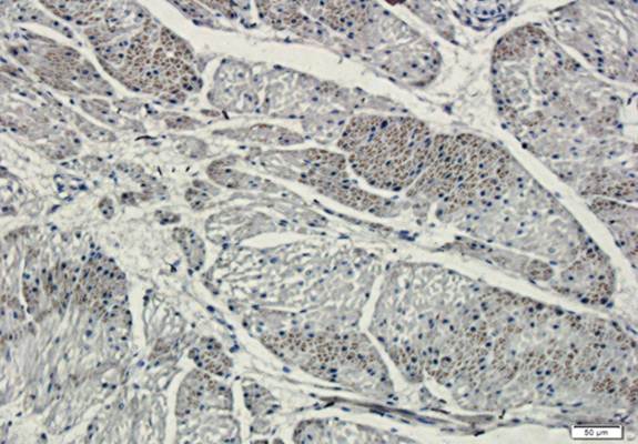
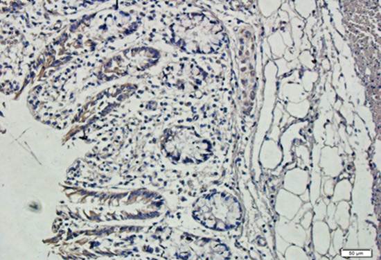
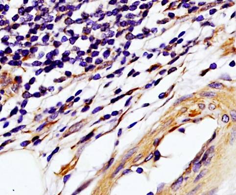
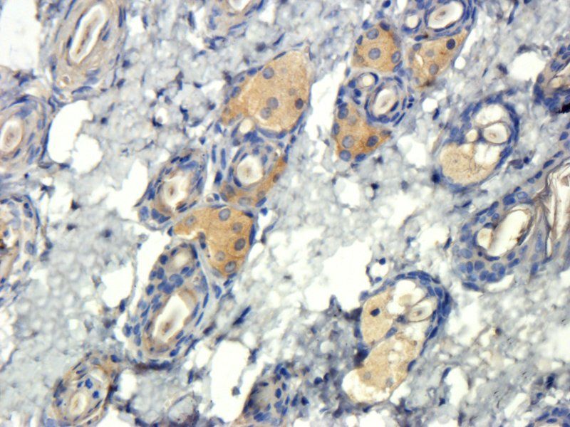
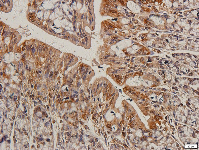
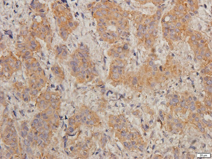
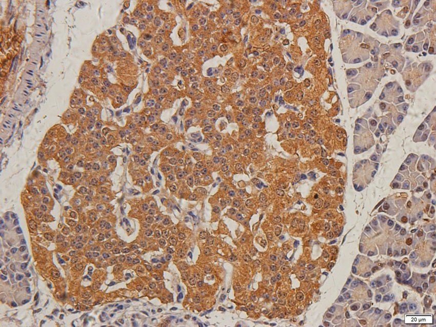
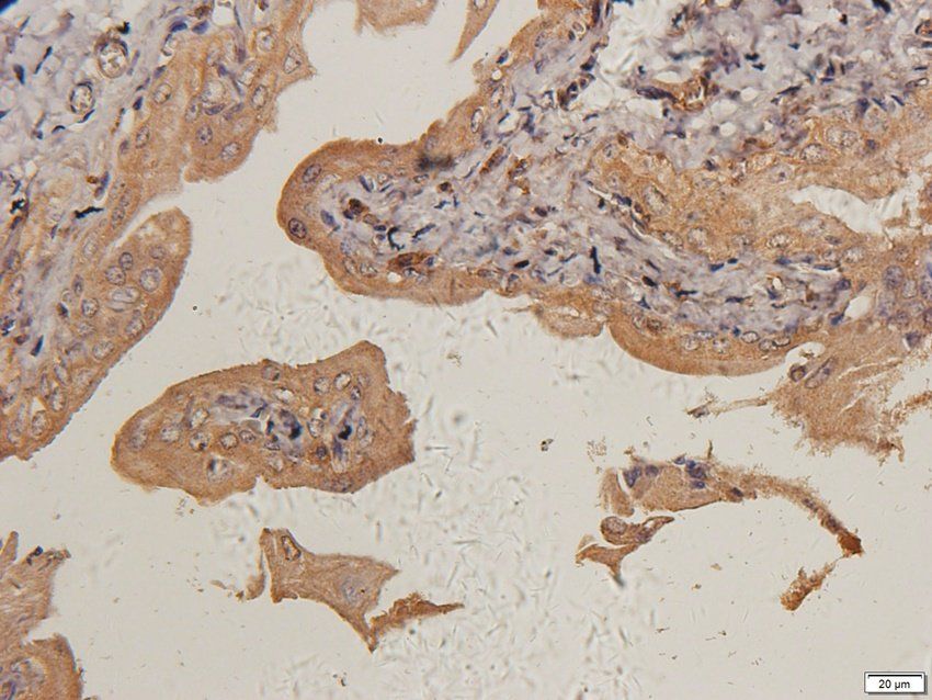
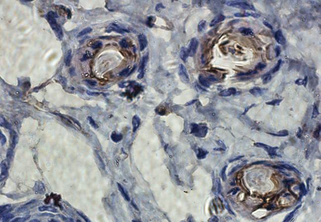
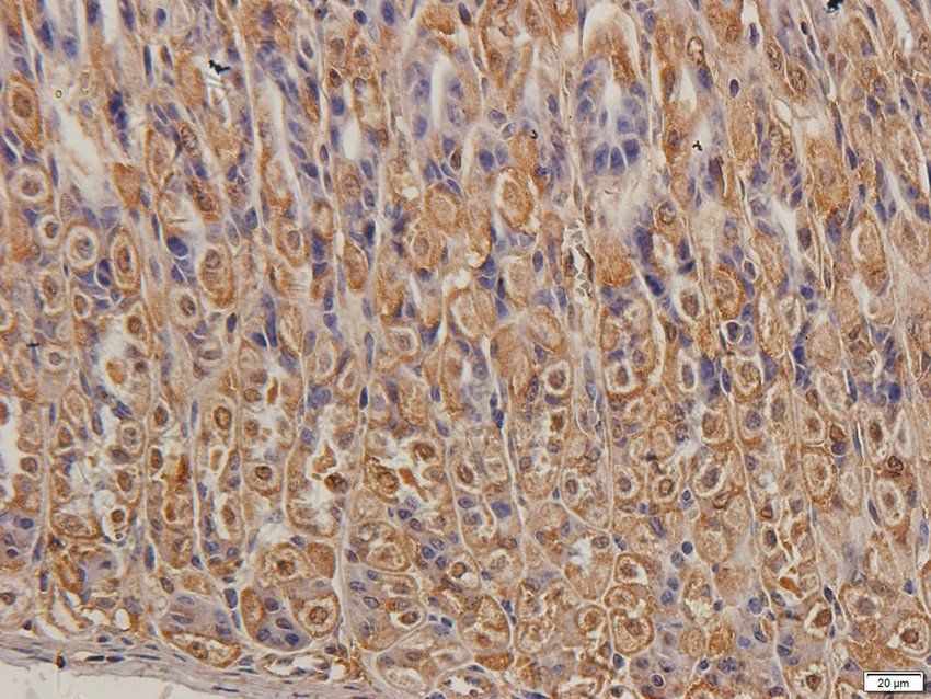
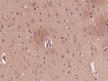
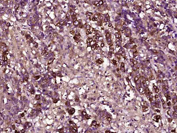
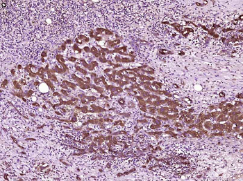
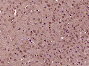
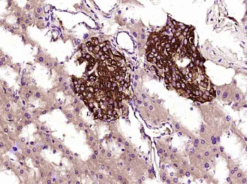
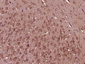
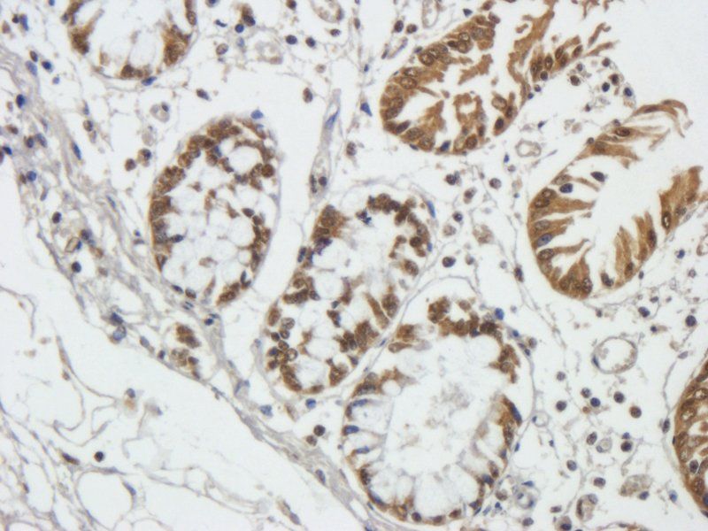
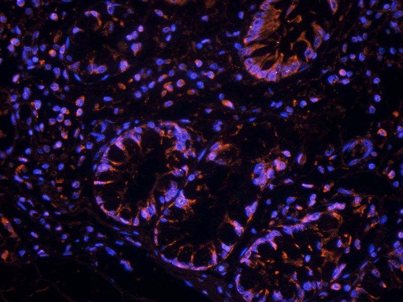
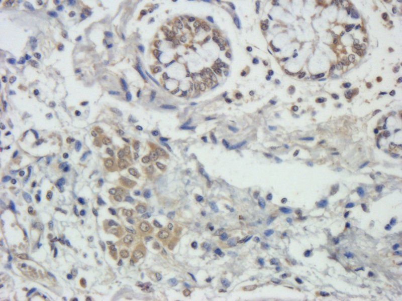
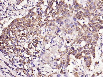
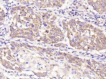
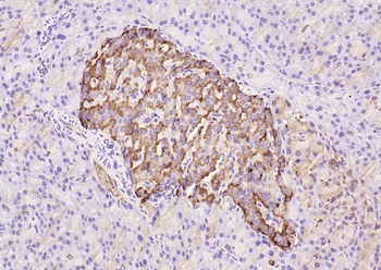
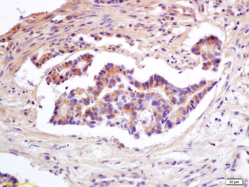
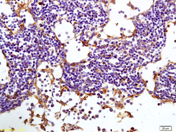
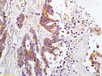
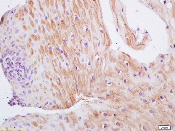
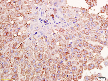
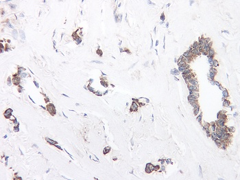
This antibody stained formalin-fixed, paraffin-embedded sections of human breast invasive ductal carcinoma. The recommended concentration is 0.1 ug/ml with an overnight incubation at 4 °C. An HRP-labeled polymer detection system was used with a DAB chromogen. Optimal results were achieved with a proteinase K antigen retrieval. Optimal concentrations and conditions may vary.
This antibody stained formalin-fixed, paraffin-embedded sections of human breast invasive ductal carcinoma. The recommended concentration is 0.1 ug/ml with an overnight incubation at 4 °C. An HRP-labeled polymer detection system was used with a DAB chromogen. Optimal results were achieved with a proteinase K antigen retrieval. Optimal concentrations and conditions may vary.
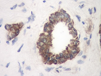
This antibody stained formalin-fixed, paraffin-embedded sections of human breast invasive ductal carcinoma. The recommended concentration is 0.1 ug/ml with an overnight incubation at 4 °C. An HRP-labeled polymer detection system was used with a DAB chromogen. Optimal results were achieved with a proteinase K antigen retrieval. Optimal concentrations and conditions may vary.
Documents Download
Datasheet
Product Information
Request a Document
Protocol Information
WB
Western Blot (IB, immunoblot)
ELISA
Enzyme-linked Immunosorbent Assay (EIA)
TIMP1 Antibody (orb1272681)

- 0.0
Based on 0 reviews
Participating in our Biorbyt product reviews program enables you to support fellow scientists by sharing your firsthand experience with our products.
Login to Submit a ReviewAvailable Sizes
Select a size below
Free Secondary Antibody (20 ul)0/0
Please add an antibody product to your cart first.