You have no items in your shopping cart.
Description
Research Area
Signal Transduction
Images & Validation
−Item 1 of 5
| Tested Applications | IHC-P, WB |
|---|---|
| Dilution Range | WB - 1:2000, IHC-P - 1:25 |
| Reactivity | Human, Mouse |
| Predicted Reactivity | Other |
Key Properties
−| Host | Rabbit |
|---|---|
| Clonality | Polyclonal |
| Isotype | Rabbit IgG |
| Immunogen | This TBK antibody is generated from rabbits immunized with a KLH conjugated synthetic peptide between 150-181 amino acids from human TBK. Antigen Region: 150-181 aa. |
| Target | TBK1 {ECO:0000303|PubMed:10581243, ECO:0000312|HGNC:HGNC:11584} |
| Molecular Weight | 83642 Da |
| Conjugation | Unconjugated |
Storage & Handling
−| Storage | Maintain refrigerated at 2-8°C for up to 2 weeks. For long term storage store at -20°C in small aliquots to prevent freeze-thaw cycles |
|---|---|
| Form/Appearance | Purified polyclonal antibody supplied in PBS with 0.09% (W/V) sodium azide. This antibody is purified through a protein A column, followed by peptide affinity purification. |
| Expiration Date | 12 months from date of receipt. |
| Disclaimer | For research use only |
Alternative Names
−Serine/threonine-protein kinase TBK1, NF-kappa-B-activating kinase, T2K, TANK-binding kinase 1, TBK1, NAK
Similar Products
−- Item 1 of 1
Phospho-TBK1 (Ser172) Rabbit Polyclonal Antibody [orb7056]
IF, IHC-Fr, IHC-P, WB
Mouse
Human, Mouse, Rat
Rabbit
Polyclonal
Unconjugated
50 μl, 100 μl - Item 1 of 3
TBK1 (Phospho-S172) Rabbit Polyclonal Antibody [orb515281]
IHC, WB
Human, Mouse, Rat
Rabbit
Polyclonal
Unconjugated
30 μl, 100 μl, 200 μl, 50 μl - Item 1 of 2
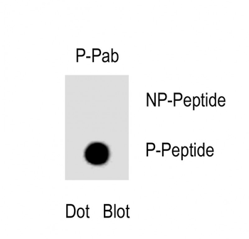
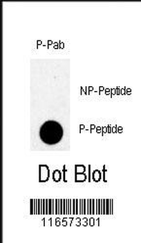
Phospho-TBK(S172) Antibody [orb1931085]
DOT, WB
Mouse, Other
Human
Rabbit
Polyclonal
Unconjugated
50 μl, 100 μl - Item 1 of 2
Phospho-TBK1 (Ser172) Recombinant Rabbit Monoclonal Antibody [orb1499322]
WB
Bovine, Canine, Equine, Gallus, Mouse, Rabbit, Rat
Human
Rabbit
Recombinant
Unconjugated
50 μl, 100 μl, 25 μl - Item 1 of 1
Phospho-NAK/TBK1/NAK Rabbit Monoclonal Antibody [orb866372]
IP, WB
Human, Mouse, Rat
Rabbit
Monoclonal
Unconjugated
100 μl
Quality Guarantee
Explore bioreagents carefree to elevate your research. All our products are rigorously tested for performance. If a product does not perform as described on its datasheet, our scientific support team will provide expert troubleshooting, a prompt replacement, or a refund. For full details, please see our Terms & Conditions and Buying Guide. Contact us at [email protected].
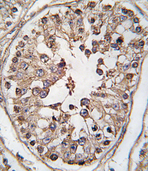
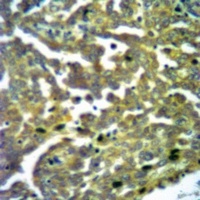
Formalin-fixed and paraffin-embedded human testis tissue reacted with TBK-pS172, which was peroxidase-conjugated to the secondary antibody, followed by DAB staining. This data demonstrates the use of this antibody for immunohistochemistry; clinical relevance has not been evaluated.
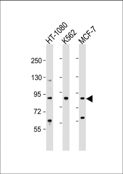
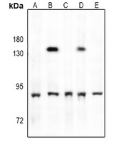
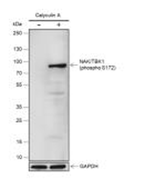
All lanes: Anti-TBK Antibody (S172) at 1:2000 dilution. Lane 1: HT-1080 whole cell lysate. Lane 2: K562 whole cell lysate. Lane 3: MCF-7 whole cell lysate. Lysates/proteins at 20 µg per lane. Secondary Goat Anti-Rabbit IgG, (H+L), Peroxidase conjugated at 1/10000 dilution. Predicted band size: 84 kDa. Blocking/Dilution buffer: 5% NFDM/TBST.
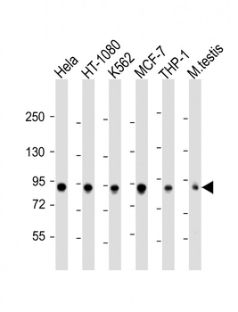
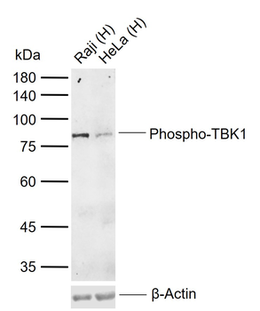
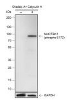
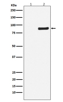
All lanes: Anti-TBK1 Antibody (S172) at 1:2000 dilution. Lane 1: Hela whole cell lysate. Lane 2: HT-1080 whole cell lysate. Lane 3: K562 whole cell lysate. Lane 4: MCF-7 whole cell lysate. Lane 5: THP-1 whole cell lysate. Lane 6: mouse testis lysate. Lysates/proteins at 20 µg per lane. Secondary Goat Anti-Rabbit IgG, (H+L), Peroxidase conjugated at 1/10000 dilution. Predicted band size: 84kDa. Blocking/Dilution buffer: 5% NFDM/TBST.
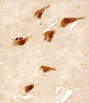
Staining TBK in human brain tissue sections by Immunohistochemistry (IHC-P - paraformaldehyde-fixed, paraffin-embedded sections). Tissue was fixed with formaldehyde and blocked with 3% BSA for 0.5 hour at room temperature; antigen retrieval was by heat mediation with a citrate buffer (pH6). Samples were incubated with primary antibody (1/25) for 1 hours at 37°C. A undiluted biotinylated goat polyvalent antibody was used as the secondary antibody.
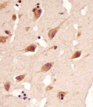
Staining TBK1 in human brain tissue sections by Immunohistochemistry (IHC-P - paraformaldehyde-fixed, paraffin-embedded sections). Tissue was fixed with formaldehyde and blocked with 3% BSA for 0.5 hour at room temperature; antigen retrieval was by heat mediation with a citrate buffer (pH6). Samples were incubated with primary antibody (1/25) for 1 hours at 37°C. A undiluted biotinylated goat polyvalent antibody was used as the secondary antibody.
Quick Database Links
Gene Symbol
TBK1 {ECO:0000303|PubMed:10581243, ECO:0000312|HGNC:HGNC:11584}
UniProt
RefSeq (Protein):NP_037386.1
UniProt Details
− No UniProt data available
NCBI Reference Sequences
−Associated Accession Numbers
Curated reference sequences for the gene transcript and protein product| Protein | NP_037386.1 |
|---|
Documents Download
Datasheet
Product Information
Request a Document
Protocol Information
WB
Western Blot (IB, immunoblot)
IHC-P
Immunohistochemistry Paraffin
TBK1 Antibody (S172) (orb1928791)

- 0.0
Based on 0 reviews
Participating in our Biorbyt product reviews program enables you to support fellow scientists by sharing your firsthand experience with our products.
Login to Submit a ReviewAvailable Sizes
Select a size below
Choose Conjugation or Carrier Free Version
Free Secondary Antibody (20 ul)0/0
Please add an antibody product to your cart first.