You have no items in your shopping cart.
SNX1 Antibody
SKU: orb622711
Description
Images & Validation
−Item 1 of 8
| Tested Applications | FC, ICC, IF, IHC, WB |
|---|---|
| Dilution Range | WB: 1:1,000-1:2,000 FC: 1:50-1:100 ICC/IF: 1:50-1:200 IHC: 1:50-1:200 |
| Reactivity | Human, Mouse, Rat |
Key Properties
−| Antibody Type | Primary Antibody |
|---|---|
| Host | Rabbit |
| Clonality | Recombinant |
| Clone No. | JG40-06 |
| Immunogen | Recombinant protein within human SNX1 aa 50-250. |
| Molecular Weight | Calculated MW 63 kDa |
| Purity | ProA affinity purified. |
| Conjugation | Unconjugated |
Storage & Handling
−| Storage | Store at -20˚C. |
|---|---|
| Form/Appearance | Liquid |
| Buffer/Preservatives | 1*TBS (pH7.4), 1% rAlbumin, 40% Glycerol, 0.05% Sodium Azide |
| Expiration Date | 12 months from date of receipt. |
| Disclaimer | For research use only |
Alternative Names
−HsT17379 antibody/ MGC8664 antibody/ SNX 1 antibody/ SNX 1a antibody/ Snx1 antibody/ SNX1_HUMAN antibody/ SNX1A antibody/ Sorting nexin 1 antibody/ Sorting nexin 1A antibody/ Sorting nexin-1 antibody/ Vps5 antibody
Similar Products
−- Item 1 of 6
SNX1 Rabbit Polyclonal Antibody [orb1474817]
ELISA, FC, ICC, IF, IHC, WB
Human
Rabbit
Polyclonal
Unconjugated
100 μg - Item 1 of 4
SNX1 Antibody [orb20425]
ELISA, ICC, IHC, WB
Bovine, Canine, Mouse, Rat
Human
Goat
Polyclonal
Unconjugated
100 μg - Item 1 of 2
SNX1 rabbit pAb Antibody [orb766350]
ELISA, IHC, WB
Human, Mouse, Rat
Polyclonal
Unconjugated
100 μl, 50 μl - Item 1 of 3
SNX1 Rabbit Polyclonal Antibody [orb331258]
WB
Bovine, Canine, Equine, Guinea pig, Mouse, Rabbit, Rat
Human
Rabbit
Polyclonal
Unconjugated
100 μl - Item 1 of 2
SNX1 Rabbit Polyclonal Antibody [orb330156]
WB
Bovine, Canine, Equine, Guinea pig, Mouse, Porcine, Rabbit, Rat, Yeast, Zebrafish
Human
Rabbit
Polyclonal
Unconjugated
100 μl


Quality Guarantee
Explore bioreagents carefree to elevate your research. All our products are rigorously tested for performance. If a product does not perform as described on its datasheet, our scientific support team will provide expert troubleshooting, a prompt replacement, or a refund. For full details, please see our Terms & Conditions and Buying Guide. Contact us at [email protected].
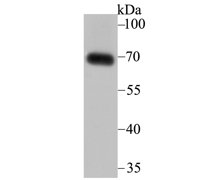
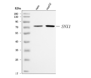
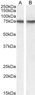
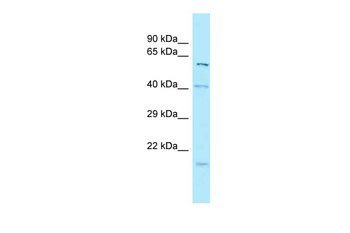
Western blot analysis of SNX1 on human skin tissue lysate using anti-SNX1 antibody at 1/2000 dilution
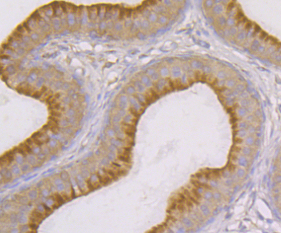
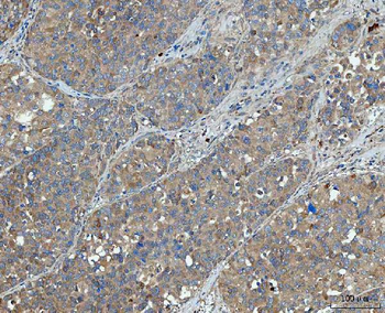
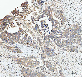
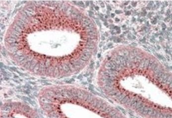
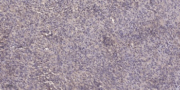
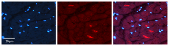
Immunohistochemical analysis of paraffin-embedded rat epididymis tissue using anti-SNX1 antibody. Counter stained with hematoxylin
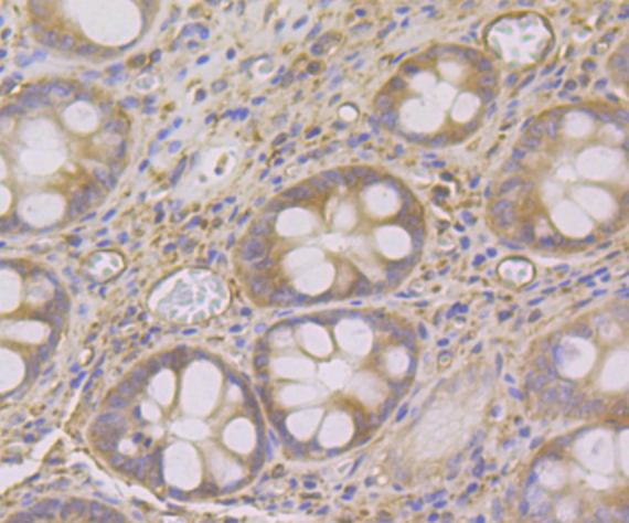
Immunohistochemical analysis of paraffin-embedded human colon tissue using anti-SNX1 antibody. Counter stained with hematoxylin

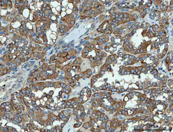
Immunohistochemical analysis of paraffin-embedded human breast cancer tissue using anti-SNX1 antibody. Counter stained with hematoxylin
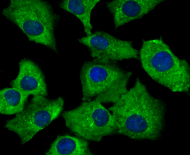
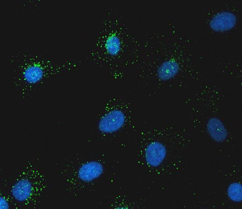
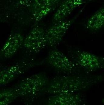
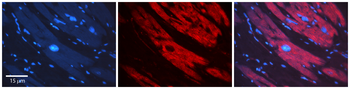
ICC staining SNX1 in A549 cells (green). The nuclear counter stain is DAPI (blue). Cells were fixed in paraformaldehyde, permeabilised with 0.25% Triton X100/PBS

ICC staining SNX1 in SH-SY-5Y cells (green). The nuclear counter stain is DAPI (blue). Cells were fixed in paraformaldehyde, permeabilised with 0.25% Triton X100/PBS

ICC staining SNX1 in SiHa cells (green). The nuclear counter stain is DAPI (blue). Cells were fixed in paraformaldehyde, permeabilised with 0.25% Triton X100/PBS
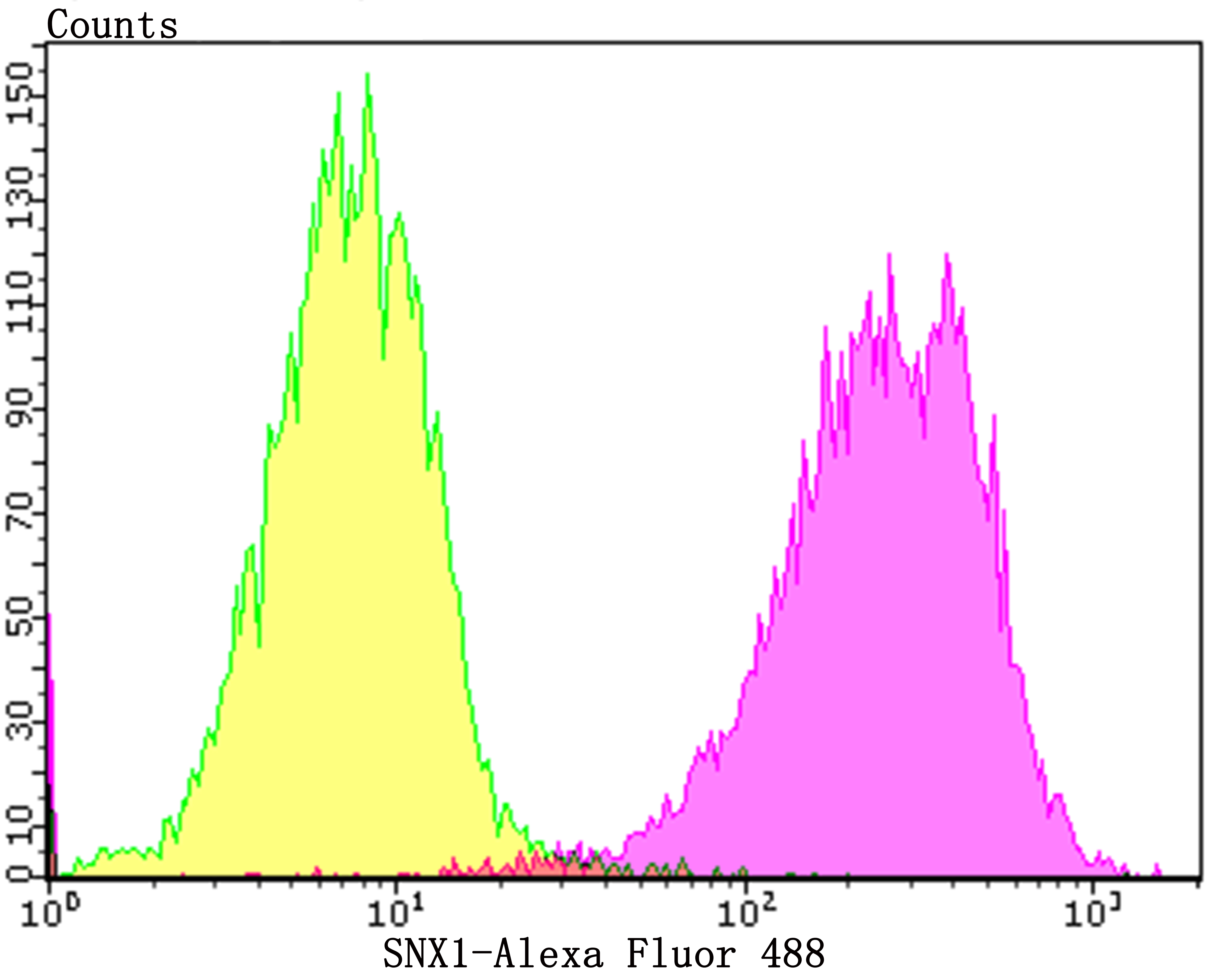
Flow cytometric analysis of SiHa cells with SNX1 antibody at 1/100 dilution (purple) compared with an unlabelled control (cells without incubation with primary antibody; yellow). Alexa Fluor 488-conjugated goat anti-rabbit IgG was used as the secondary antibody
Quick Database Links
UniProt
UniProt Details
− No UniProt data available
Documents Download
Datasheet
Product Information
Request a Document
Protocol Information
WB
Western Blot (IB, immunoblot)
IHC
Immunohistochemistry
FC
Flow Cytometry
IF
Immunofluorescence
ICC
Immunocytochemistry
SNX1 Antibody (orb622711)

- 0.0
Based on 0 reviews
Participating in our Biorbyt product reviews program enables you to support fellow scientists by sharing your firsthand experience with our products.
Login to Submit a ReviewAvailable Sizes
Select a size below