You have no items in your shopping cart.
Featured
Description
Research Area
Infectious Disease & Virology, Signal Transduction
Images & Validation
−Item 1 of 4
| Tested Applications | IF, IHC-P, WB |
|---|---|
| Dilution Range | IF - 1:10-50, WB - 1:2000, IHC-P - 1:50-100 |
| Reactivity | Human, Rat |
| Predicted Reactivity | Mouse, Other |
Key Properties
−| Antibody Type | Primary Antibody |
|---|---|
| Host | Rabbit |
| Clonality | Polyclonal |
| Isotype | Rabbit IgG |
| Immunogen | Synthetic Peptide |
| Target | SMURF2 |
| Molecular Weight | 86196 |
| Conjugation | Unconjugated |
Storage & Handling
−| Storage | Maintain refrigerated at 2-8°C for up to 2 weeks. For long term storage store at -20°C in small aliquots to prevent freeze-thaw cycles |
|---|---|
| Form/Appearance | Purified polyclonal antibody supplied in PBS with 0.09% (W/V) sodium azide. This antibody is prepared by Saturated Ammonium Sulfate (SAS) precipitation followed by dialysis against PBS. |
| Expiration Date | 12 months from date of receipt. |
| Disclaimer | For research use only |
Alternative Names
−Anti-E3 ubiquitin-protein ligase SMURF2 antibody
Quality Guarantee
Explore bioreagents carefree to elevate your research. All our products are rigorously tested for performance. If a product does not perform as described on its datasheet, our scientific support team will provide expert troubleshooting, a prompt replacement, or a refund. For full details, please see our Terms & Conditions and Buying Guide. Contact us at [email protected].
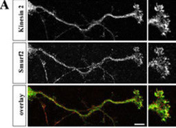
Hippocampal neurons were fixed at stage 3, stained with anti-Smurf2 (red) and anti-Kinesin-2 (green) antibodies, and analyzed by confocal microscopy. The panels show single confocal planes.
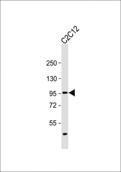
Anti-SMURF2 Antibody (C-term) at 1:2000 dilution + C2C12 whole cell lysate. Lysates/proteins at 20 µg per lane. Secondary Goat Anti-Rabbit IgG, (H+L), Peroxidase conjugated at 1/10000 dilution. Predicted band size: 86 kDa. Blocking/Dilution buffer: 5% NFDM/TBST.
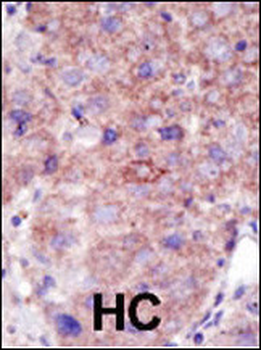
Formalin-fixed and paraffin-embedded human cancer tissue reacted with the primary antibody, which was peroxidase-conjugated to the secondary antibody, followed by DAB staining. This data demonstrates the use of this antibody for immunohistochemistry; clinical relevance has not been evaluated. BC = breast carcinoma; HC = hepatocarcinoma.
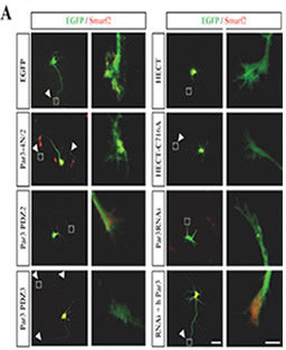
Hippocampal neurons were transfected 2 h after plating with expression vectors for EGFP, EGFP-tagged Par3-4N/2, Par3-PDZ2, Par3-PDZ3, Smurf2-HECT (HECT), Smurf2-HECT-C716A (HECT CA), and shRNA directed against mPar3 (Par3 RNAi), or vectors for the anti-Par3 shRNA and human Myc-Par3 (RNAi + h Par3) (green). Transfected cells were analyzed at 3 d.i.v. by staining with an anti-Smurf2 antibody (red). Axons are marked by arrowheads. The marked growth cones are shown at a higher magnification.
Quick Database Links
UniProt Details
− No UniProt data available
NCBI Reference Sequences
−Associated Accession Numbers
Curated reference sequences for the gene transcript and protein product| Protein | NP_073576.1 |
|---|
Documents Download
Datasheet
Product Information
Request a Document
Protocol Information
WB
Western Blot (IB, immunoblot)
IHC-P
Immunohistochemistry Paraffin
IF
Immunofluorescence
SMURF2 Antibody (C-term) (orb33681)

- 0.0
Based on 0 reviews
Participating in our Biorbyt product reviews program enables you to support fellow scientists by sharing your firsthand experience with our products.
Login to Submit a ReviewAvailable Sizes
Select a size below
Choose Conjugation or Carrier Free Version
Free Secondary Antibody (20 ul)0/0
Please add an antibody product to your cart first.