You have no items in your shopping cart.
Description
Research Area
Signal Transduction
Images & Validation
−Item 1 of 7
| Tested Applications | FC, IHC-P, WB |
|---|---|
| Dilution Range | WB - 1:2000, IHC-P-Leica - 1:500, FC - 1:10-50, IHC - 1:1000 |
| Reactivity | Human, Mouse, Rat |
| Predicted Reactivity | Rabbit |
Key Properties
−| Host | Rabbit |
|---|---|
| Clonality | Polyclonal |
| Isotype | Rabbit IgG |
| Immunogen | This S100B antibody is generated from rabbits immunized with S100B recombinant protein. |
| Target | S100B {ECO:0000303|PubMed:6487634, ECO:0000312|HGNC:HGNC:10500} |
| Molecular Weight | 10713 Da |
| Conjugation | Unconjugated |
Storage & Handling
−| Storage | Maintain refrigerated at 2-8°C for up to 2 weeks. For long term storage store at -20°C in small aliquots to prevent freeze-thaw cycles |
|---|---|
| Form/Appearance | Purified polyclonal antibody supplied in PBS with 0.09% (W/V) sodium azide. This antibody is purified through a protein A column, followed by peptide affinity purification. |
| Expiration Date | 12 months from date of receipt. |
| Disclaimer | For research use only |
Alternative Names
−Protein S100-B, S-100 protein beta chain, S-100 protein subunit beta, S100 calcium-binding protein B, S100B
Similar Products
−- Item 1 of 7
S100B Rabbit Polyclonal Antibody [orb11345]
IF, IHC-Fr, IHC-P, WB
Bovine, Gallus, Rabbit
Human
Rabbit
Polyclonal
Unconjugated
50 μl, 100 μl, 200 μl - Item 1 of 7
S100B Mouse Monoclonal Antibody [orb500644]
FC, IF, IHC-Fr, IHC-P, WB
Mouse, Rat
Human, Mouse, Rat
Mouse
Monoclonal
Unconjugated
50 μl, 100 μl, 200 μl, 200 μg - Item 1 of 7
S100 beta Recombinant Rabbit Monoclonal Antibody [orb612224]
FC, ICC, IF, IHC-Fr, IHC-P, WB
Mouse, Rat, Zebrafish
Human, Mouse, Rat
Rabbit
Recombinant
Unconjugated
50 μl, 100 μl, 25 μl - Item 1 of 1
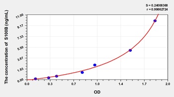
Human S100 Calcium Binding Protein B (S100B) ELISA Kit [orb776476]
Human
0.16-10 ng/mL
0.058 ng/mL
96 T, 48 T - Item 1 of 4
S100B Antibody [orb388636]
IF, IHC, WB
Bovine, Human, Mouse, Rat
Mouse
Monoclonal
Unconjugated
20 μg, 100 μg, 100 μg (without BSA and Azide)

Quality Guarantee
Explore bioreagents carefree to elevate your research. All our products are rigorously tested for performance. If a product does not perform as described on its datasheet, our scientific support team will provide expert troubleshooting, a prompt replacement, or a refund. For full details, please see our Terms & Conditions and Buying Guide. Contact us at [email protected].
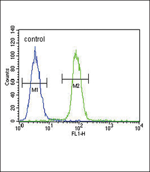
S100B Antibody flow cytometric analysis of A375 cells (right histogram) compared to a negative control cell (left histogram). FITC-conjugated goat-anti-rabbit secondary antibodies were used for the analysis.
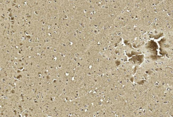
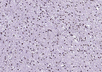
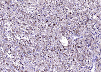
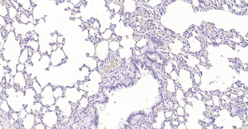
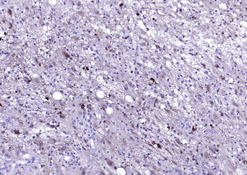
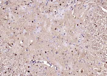
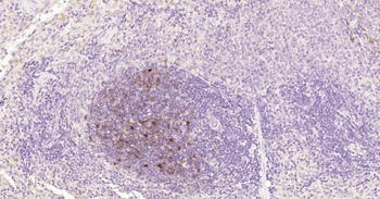
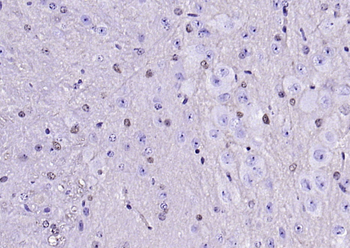
Immunohistochemical analysis of paraffin-embedded Human brain section using Pink1. Diluted at 1:1000 dilution. A undiluted biotinylated goat polyvalent antibody was used as the secondary, followed by DAB staining.
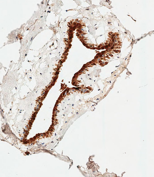
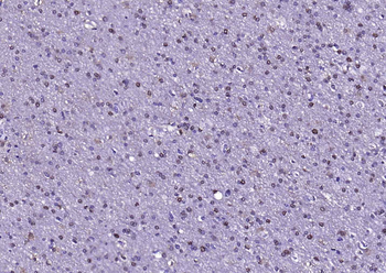
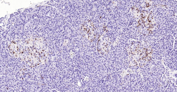
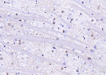
Immunohistochemical analysis of paraffin-embedded Human breast tissue was performed on the Leica BOND RXm. Tissue was fixed with formaldehyde at room temperature, antigen retrieval was by heat mediation with a EDTA buffer (pH9.0). Samples were incubated with primary antibody (1:500) for 1 hours at room temperature. A undiluted biotinylated CRF Anti-Polyvalent HRP Polymer antibody was used as the secondary antibody.
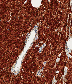
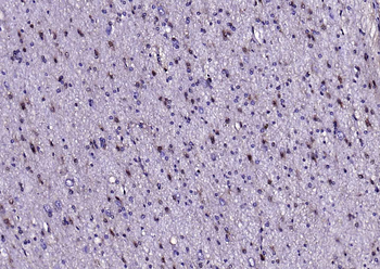
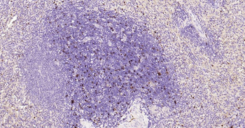
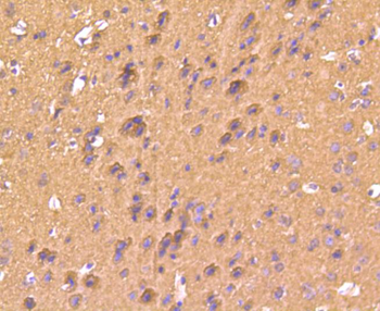
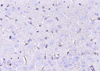
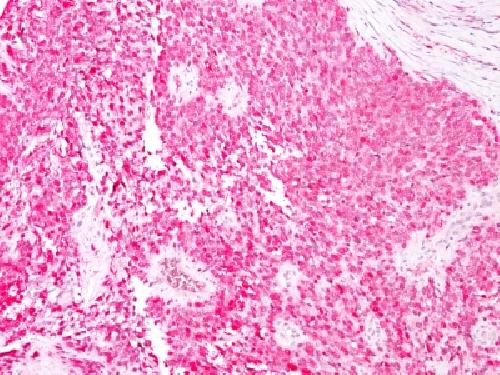
Immunohistochemical analysis of paraffin-embedded Human melanoma tissue was performed on the Leica BOND RXm. Tissue was fixed with formaldehyde at room temperature, antigen retrieval was by heat mediation with a EDTA buffer (pH9.0). Samples were incubated with primary antibody (1:500) for 1 hours at room temperature. A undiluted biotinylated CRF Anti-Polyvalent HRP Polymer antibody was used as the secondary antibody.
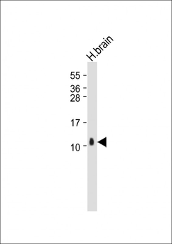
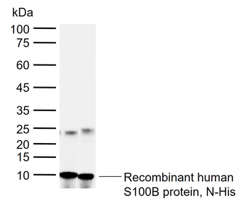
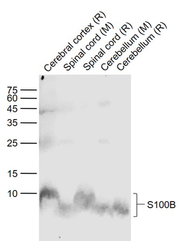
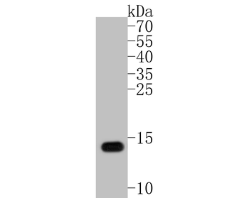
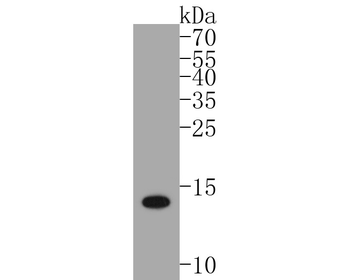
Anti-S100B Antibody at 1:2000 dilution + Human brain whole tissue lysate. Lysates/proteins at 20 µg per lane. Secondary Goat Anti-Rabbit IgG, (H+L), Peroxidase conjugated at 1/10000 dilution. Predicted band size: 11 kDa. Blocking/Dilution buffer: 5% NFDM/TBST.
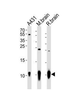
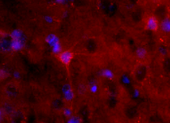
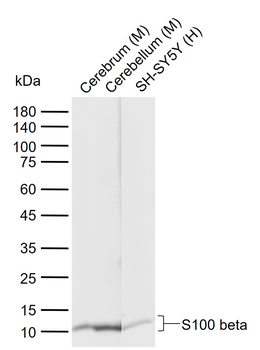
Western blot analysis of lysates from A431 cell line, mouse brain, rat brain tissue (from left to right), using S100B Antibody. Diluted at 1:1000 at each lane. A goat anti-rabbit IgG H&L (HRP) at 1:10000 dilution was used as the secondary antibody. Lysates at 20 ug per lane.
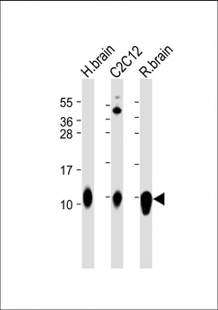
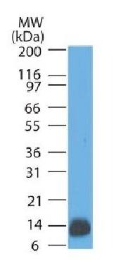
All lanes: Anti-S100B Antibody at 1:2000 dilution. Lane 1: Human brain lysate. Lane 2: C2C12 whole cell lysate. Lane 3: Rat brain lysate. Lysates/proteins at 20 µg per lane. Secondary Goat Anti-Rabbit IgG, (H+L), Peroxidase conjugated at 1/10000 dilution. Predicted band size: 11 kDa. Blocking/Dilution buffer: 5% NFDM/TBST.
Quick Database Links
Gene Symbol
S100B {ECO:0000303|PubMed:6487634, ECO:0000312|HGNC:HGNC:10500}
UniProt
RefSeq (Protein):NP_006263.1
UniProt Details
− No UniProt data available
NCBI Reference Sequences
−Associated Accession Numbers
Curated reference sequences for the gene transcript and protein product| Protein | NP_006263.1 |
|---|
Documents Download
Datasheet
Product Information
Request a Document
Protocol Information
WB
Western Blot (IB, immunoblot)
IHC-P
Immunohistochemistry Paraffin
FC
Flow Cytometry
S100B Antibody (orb1930681)

- 0.0
Based on 0 reviews
Participating in our Biorbyt product reviews program enables you to support fellow scientists by sharing your firsthand experience with our products.
Login to Submit a ReviewAvailable Sizes
Select a size below
Free Secondary Antibody (20 ul)0/0
Please add an antibody product to your cart first.