You have no items in your shopping cart.
Rabbit IgG (H&L) Antibody Rhodamine Conjugated Pre-Adsorbed
SKU: orb347644
Description
Images & Validation
−Item 1 of 3
| Tested Applications | FC, FLISA, IF |
|---|---|
| Dilution Range | FLISA: 1:10,000 - 1:50,000, FC: 1:500 - 1:2,500, IF: 1:1,000 - 1:5,000 |
| Reactivity | Rabbit |
| Application Notes |
Key Properties
−| Antibody Type | Secondary Antibody |
|---|---|
| Host | Goat |
| Clonality | Polyclonal |
| Isotype | IgG |
| Immunogen | Rabbit IgG whole molecule |
| Purity | This product was prepared from monospecific antiserum by immunoaffinity chromatography using Rabbit IgG coupled to agarose beads followed by solid phase adsorption(s) to remove any unwanted reactivities. Assay by immunoelectrophoresis resulted in a single precipitin arc against anti-Goat Serum, Rabbit IgG and Rabbit Serum. No reaction was observed against Bovine, Chicken, Goat, Guinea Pig, Hamster, Horse, Human, Mouse, Rat and Sheep Serum Proteins. |
| Conjugation | TRITC |
Storage & Handling
−| Storage | Store vial at 4° C prior to restoration. For extended storage aliquot contents and freeze at -20° C or below. Avoid cycles of freezing and thawing. Centrifuge product if not completely clear after standing at room temperature. This product is stable for several weeks at 4° C as an undiluted liquid. Dilute only prior to immediate use. |
|---|---|
| Form/Appearance | Lyophilized |
| Buffer/Preservatives | Preservative: 0.01% (w/v) Sodium Azide. Stabilizer: 10 mg/mL Bovine Serum Albumin (rAlbumin) - Immunoglobulin and Protease free; Buffer: 0.02 M Potassium Phosphate, 0.15 M Sodium Chloride, pH 7.2 |
| Concentration | 1.0 mg/mL |
| Expiration Date | 12 months from date of receipt. |
| Disclaimer | For research use only |
Alternative Names
−Goat anti-Rabbit IgG Antibody Rhodamine Conjugation, Goat anti-Rabbit IgG Rhodamine Conjugated Antibody
Similar Products
−- Item 1 of 6
Rabbit IgG (H&L) Antibody Rhodamine Conjugated Pre-Adsorbed [orb347751]
FC, FLISA, IF
Rabbit
Donkey
Polyclonal
TRITC
1 mg - Item 1 of 2
Rabbit IgG (H&L) Antibody Pre-Adsorbed [orb347715]
DOT, FC, FLISA, IF
Rabbit
Guinea pig
Polyclonal
TRITC
1 mg - Item 1 of 2
Rabbit IgG (H&L) Antibody Pre-Adsorbed [orb347722]
DOT, FC, FLISA, IF
Rabbit
Mouse
Polyclonal
TRITC
1 mg - Item 1 of 2
Rabbit IgG (H&L) Antibody Rhodamine Conjugated Pre-Adsorbed [orb347651]
DOT, FC, FLISA, IF
Rabbit
Goat
Polyclonal
TRITC
2 mg - Item 1 of 1
Rabbit IgG (H&L) Antibody Rhodamine Conjugated Pre-Adsorbed [orb347729]
FC, FLISA, IF
Rabbit
Rat
Polyclonal
TRITC
1 mg
Quality Guarantee
Explore bioreagents carefree to elevate your research. All our products are rigorously tested for performance. If a product does not perform as described on its datasheet, our scientific support team will provide expert troubleshooting, a prompt replacement, or a refund. For full details, please see our Terms & Conditions and Buying Guide. Contact us at [email protected].
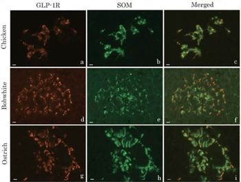
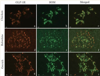
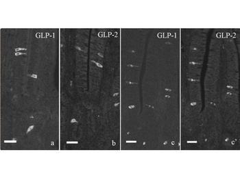
Double immunofluorescence images of glucagon-like peptide-1 receptor (GLP-1R, a, d, g) and somatostatin (SOM, b, e, h) in the pancreatic islets of chickens (a–c), northern bobwhites (d–f), and ostriches (g–i). Figures c, f, and i show merged images of a and b, d and e, and g and h, respectively. Almost every SOM-immunoreactive cell in the pancreatic islets of three avian species also demonstrated GLP-1R immunoreactivity. Bars indicate 10 µm.
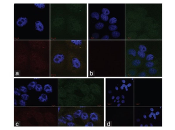
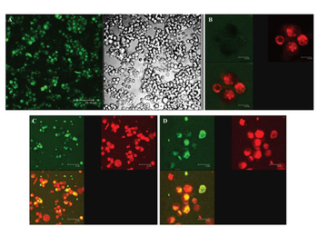
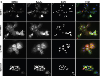
Fluorescent micrographs of MCF-7 cells unexposed to either solvent (a) exposed to 0.5% Methanol (b) and 0.5% DMSO (c). D is negative control. BCL-2 was stained with monoclonal anti-BCL-2 and Goat Anti-Mouse IgG FITC conjugated (Upper Right Quadrant- showing the nuclear and cytoplasmic expression of the oncogene BCL-2). BAX was stained with polyclonal rabbit anti-BAX and Goat anti-Rabbit IgG Rhodamine conjugated (Lower Left quadrant- showing the nuclear and cytoplasmic expression of BAX). Nucleus was counterstained with DAPI (Upper left quadrant). The lower right quadrant shows the combined image. 2D shows fluorescent micrographs of MCF-7 cells unexposed to either solvent. Cells were incubated with the secondary antibodies but not the primary antibodies to reveal non-specific staining. The nucleus was counterstained with DAPI (Upper left quadrant). The lower right quadrant represents the combined image.
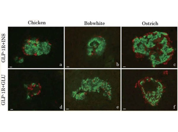
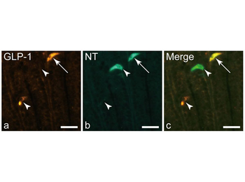
Merged images of double immunofluorescence pictures of glucagon-like peptide-1 receptor (red) and insulin (green) (GLP-1R + INS, a–c), and glucagon-like peptide-1 receptor (red) and glucagon (green) (GLP-1R + GLU, d–f) in the pancreatic islets of chickens (a and d), northern bobwhites (b and e), and ostriches (c and f). Islet cells showing either insulin or glucagon immunoreactivity were immunonegative to glucagon-like peptide-1 receptor. Bars indicate 10 µm.
Documents Download
Datasheet
Product Information
Request a Document
Protocol Information
FC
Flow Cytometry
IF
Immunofluorescence
Rabbit IgG (H&L) Antibody Rhodamine Conjugated Pre-Adsorbed (orb347644)

- 0.0
Based on 0 reviews
Participating in our Biorbyt product reviews program enables you to support fellow scientists by sharing your firsthand experience with our products.
Login to Submit a ReviewAvailable Sizes
Select a size below
Free Secondary Antibody (20 ul)0/0
Please add an antibody product to your cart first.