You have no items in your shopping cart.
Rabbit IgG (H&L) Antibody Biotin Conjugated Pre-Adsorbed
SKU: orb347727
Description
Images & Validation
−Item 1 of 2
| Tested Applications | ELISA, IHC, WB |
|---|---|
| Dilution Range | ELISA: 1:20,000 - 1:100,000, IHC: 1:1,000 - 1:5,000, WB: 1:2,000 - 1:10,000 |
| Reactivity | Rabbit |
| Application Notes |
Key Properties
−| Antibody Type | Secondary Antibody |
|---|---|
| Host | Mouse |
| Clonality | Polyclonal |
| Isotype | IgG |
| Immunogen | Rabbit IgG whole molecule |
| Purity | This product was prepared from monospecific antiserum by immunoaffinity chromatography using Rabbit IgG coupled to agarose beads followed by solid phase adsorption(s) to remove any unwanted reactivities. Assay by immunoelectrophoresis resulted in a single precipitin arc against anti-biotin, anti-Mouse Serum, Rabbit IgG and Rabbit Serum. No reaction was observed against Human, Goat and Mouse Serum Proteins. |
| Conjugation | Biotin |
Storage & Handling
−| Storage | Store vial at 4° C prior to restoration. For extended storage aliquot contents and freeze at -20° C or below. Avoid cycles of freezing and thawing. Centrifuge product if not completely clear after standing at room temperature. This product is stable for several weeks at 4° C as an undiluted liquid. Dilute only prior to immediate use. |
|---|---|
| Form/Appearance | Lyophilized |
| Buffer/Preservatives | Preservative: 0.01% (w/v) Sodium Azide. Stabilizer: 10 mg/mL Bovine Serum Albumin (rAlbumin) - Immunoglobulin and Protease free; Buffer: 0.02 M Potassium Phosphate, 0.15 M Sodium Chloride, pH 7.2 |
| Concentration | 1.0 mg/mL |
| Expiration Date | 12 months from date of receipt. |
| Disclaimer | For research use only |
Alternative Names
−Mouse Anti-Rabbit IgG Biotin Conjugated Antibody, Mouse Anti Rabbit IgG Antibody Biotin Conjugation
Similar Products
−- Item 1 of 2
Sheep IgG (H&L) Secondary Antibody Biotin Conjugated Pre-Adsorbed [orb347939]
DOT, ELISA, IHC, WB
Sheep
Rabbit
Polyclonal
Biotin
1 mg - Item 1 of 2
Sheep IgG (H&L) Secondary Antibody Biotin Conjugated Pre-Adsorbed [orb347940]
DOT, ELISA, IHC, WB
Sheep
Rabbit
Polyclonal
Biotin
500 μg - Item 1 of 2
Rabbit IgG (H&L) Secondary Antibody Biotin Conjugated Pre-Adsorbed [orb347656]
ELISA, IHC, WB
Rabbit
Goat
Polyclonal
Biotin
1 mg - Item 1 of 2
Human IgG (H&L) Antibody Biotin Conjugated Pre-Adsorbed [orb347311]
ELISA, IHC, WB
Human
Rabbit
Polyclonal
Biotin
2 mg - Item 1 of 1
Rat IgG (H&L) Antibody Biotin Conjugated Pre-Adsorbed [orb347872]
ELISA, IHC, WB
Rat
Rabbit
Polyclonal
Biotin
1 mg


Quality Guarantee
Explore bioreagents carefree to elevate your research. All our products are rigorously tested for performance. If a product does not perform as described on its datasheet, our scientific support team will provide expert troubleshooting, a prompt replacement, or a refund. For full details, please see our Terms & Conditions and Buying Guide. Contact us at [email protected].

GRK5 regulates dendritic development. (A and B) Hippocampal neuron cultures transfected at DIV5 were observed at DIV8. Total dendritic branch tip numbers (TDBTN) and total dendrite length of transfected neurons were measured. For each group, 40–60 (A) or 30–40 (B) neurons from three independent cultures were analyzed. One-way ANOVA followed by Tukey–Kramer posthoc test. (C and D) Hippocampal neurons were transfected at DIV9 and observed at DIV17. Boxed regions are enlarged below each image. For each group, 30–40 dendrites of 8–10 neurons from three independent cultures were analyzed. Protrusion and spine number were measured. (C) GFP was cotransfected with GRK5 variants to visualize dendritic spines (one-way ANOVA followed by Tukey–Kramer posthoc test). (D) Neuron cultures transfected with control or GRK5 RNAi constructs (Student's t test). Bars, 10 µm. Error bars indicate SEM. *, P < 0.03; **, P < 0.01; ***, P < 0.001. Ctrl, control.

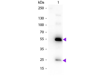
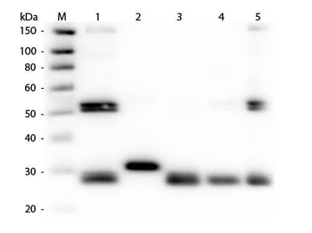
Mouse Anti-Rabbit IgG biotin conjugated antibody. Peri-Urethral Prostate Tissues Exhibit Fibroblastic and Myofibroblastic Cell Populations. Peri-urethral prostate tissues from patients 1007 and 0516 were explanted and primary fibroblasts were isolated and grown to monolayer cultures. Photomicrographs demonstrate fibroblastic morphology for 0516 primary cells but mixed fibroblastic and myofibroblastic morphologies for patient 1007. Cells from both cultures were then stained for collagen 1 (COL1) (PE-cy5-conjugated Ab, red), α-smooth muscle actin (αSMA) (fluorescein-conjugated Ab, green), or the nuclei counterstained with DAPI (blue). Merged images show that primary cells from patient 1007 exhibited high levels of co-localized COL1 and αSMA protein expression (yellow) consistent with a myofibroblastic phenotype. Control mouse IgG2a and rabbit IgG biotin conjugate were used at 1∶2000 dilution. All images were captured at 400X in visible light on brightfield settings.
Documents Download
Datasheet
Product Information
Request a Document
Protocol Information
WB
Western Blot (IB, immunoblot)
IHC
Immunohistochemistry
ELISA
Enzyme-linked Immunosorbent Assay (EIA)
Rabbit IgG (H&L) Antibody Biotin Conjugated Pre-Adsorbed (orb347727)

- 0.0
Based on 0 reviews
Participating in our Biorbyt product reviews program enables you to support fellow scientists by sharing your firsthand experience with our products.
Login to Submit a ReviewAvailable Sizes
Select a size below
Free Secondary Antibody (20 ul)0/0
Please add an antibody product to your cart first.