You have no items in your shopping cart.
Phospho-TAK1 (Thr184 + Thr187) Rabbit Polyclonal Antibody
SKU: orb7050
Featured
Description
Research Area
Signal Transduction
Images & Validation
−Item 1 of 2
| Tested Applications | IF, IHC-Fr, IHC-P |
|---|---|
| Dilution Range | IHC-P=1:50-1000, IHC-F=1:50-1000, IF=1:100-500 |
| Reactivity | Rat |
| Predicted Reactivity | Bovine, Equine, Gallus, Human, Mouse, Porcine, Rabbit |
Related Conjugates & Formulations
−Key Properties
−| Antibody Type | Primary Antibody |
|---|---|
| Host | Rabbit |
| Clonality | Polyclonal |
| Isotype | IgG |
| Immunogen | KLH conjugated Synthesised phosphopeptide derived from human TAK1 around the phosphorylation site of Thr184/187 IQ(p-T)HM(p-T)NN |
| Target | MAP3K7 |
| Molecular Weight | 67 kDa |
| Purification | Affinity purified by Protein A |
| Conjugation | Unconjugated |
Storage & Handling
−| Storage | Maintain refrigerated at 2-8°C for up to 2 weeks. For long term storage store at -20°C in small aliquots to prevent freeze-thaw cycles. |
|---|---|
| Form/Appearance | Liquid |
| Buffer/Preservatives | 0.01M TBS (pH7.4) with 1% rAlbumin, 0.02% Proclin300 and 50% Glycerol. |
| Concentration | 1mg/ml |
| Expiration Date | 12 months from date of receipt. |
| Disclaimer | For research use only |
Alternative Names
−CSCF; FMD2; MEKK7; TAK1; TGF1a; B430101B05; M3K7_HUMAN; MAP3K7; Transforming growth factor-beta-activated kinase 1 (TGF-beta-activated kinase 1); 2.7.11.25; M3K7_MOUSE; M3K7_RAT; mitogen-activated protein kinase kinase kinase 7; TGF-beta activated kinase 1; MAP3K7 | TAK1 (phospho-T184/T187); p-TAK1; phospho-TAK1; p-MAP3K7; MAP3K7 | TAK1 (phospho-Thr184/Thr187)
Similar Products
−
Phospho-TAK1 (Thr184 + Thr187) Rabbit Polyclonal Antibody (FITC) [orb9898]
IF
Bovine, Equine, Gallus, Human, Mouse, Porcine, Rabbit
Rat
Rabbit
Polyclonal
FITC
100 μlPhospho-TAK1 (Thr184 + Thr187) Rabbit Polyclonal Antibody (Cy3) [orb982678]
IF
Bovine, Equine, Gallus, Human, Mouse, Porcine, Rabbit
Rat
Rabbit
Polyclonal
Cy3
100 μlPhospho-TAK1 (Thr184 + Thr187) Rabbit Polyclonal Antibody (Cy5) [orb899908]
IF
Bovine, Equine, Gallus, Human, Mouse, Porcine, Rabbit
Rat
Rabbit
Polyclonal
Cy5
100 μlPhospho-TAK1 (Thr184 + Thr187) Rabbit Polyclonal Antibody (AP) [orb900239]
IHC-Fr, IHC-P
Bovine, Equine, Gallus, Human, Mouse, Porcine, Rabbit
Rat
Rabbit
Polyclonal
AP
100 μlPhospho-TAK1 (Thr184 + Thr187) Rabbit Polyclonal Antibody (PE) [orb505738]
IF
Bovine, Equine, Gallus, Human, Mouse, Porcine, Rabbit
Rat
Rabbit
Polyclonal
PE
100 μl
Quality Guarantee
Explore bioreagents carefree to elevate your research. All our products are rigorously tested for performance. If a product does not perform as described on its datasheet, our scientific support team will provide expert troubleshooting, a prompt replacement, or a refund. For full details, please see our Terms & Conditions and Buying Guide. Contact us at [email protected].
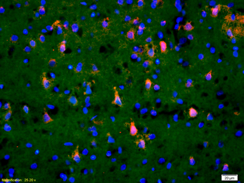
Tissue/Cell: rat brain tissue, 4% Paraformaldehyde-fixed and paraffin-embedded, Antigen retrieval: citrate buffer (0.01M, pH6.0), Boiling bathing for 15 min, Blocking buffer (normal goat serum) at 37°C for 20 min, Incubation: Anti-Phospho-TAK1 (Thr184/187) Polyclonal Antibody, Unconjugated (orb7050) 1:200, overnight at 4°C, The secondary antibody was Goat Anti-Rabbit IgG, Cy3 conjugated (orb868589) used at 1:200 dilution for 40 minutes at 37°C. DAPI (5 ug/ml, blue) was used to stain the cell nuclei.
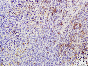
Tissue/Cell: rat spleen tissue, 4% Paraformaldehyde-fixed and paraffin-embedded, Antigen retrieval: citrate buffer (0.01M, pH6.0), Boiling bathing for 15 min, Block endogenous peroxidase by 3% Hydrogen peroxide for 30 min, Blocking buffer (normal goat serum) at 37°C for 20 min, Incubation: Anti-Phospho-TAK1 (Thr184/187) Polyclonal Antibody, Unconjugated (orb7050) 1:200, overnight at 4°C, followed by conjugation to the secondary antibody and DAB staining.
Quick Database Links
Gene Symbol
MAP3K7
UniProt
UniProt Details
− No UniProt data available
Documents Download
Datasheet
Product Information
Request a Document
Protocol Information
IHC-P
Immunohistochemistry Paraffin
IHC-Fr
Immunohistochemistry Frozen
IF
Immunofluorescence
Zheng, Xichuan et al. TAK1 accelerates transplant arteriosclerosis in rat aortic allografts by inducing autophagy in vascular smooth muscle cells Atherosclerosis, 343, 19/10/2023 (2022)
Phospho-TAK1 (Thr184 + Thr187) Rabbit Polyclonal Antibody (orb7050)

- 0.0
Based on 0 reviews
Participating in our Biorbyt product reviews program enables you to support fellow scientists by sharing your firsthand experience with our products.
Login to Submit a ReviewAvailable Sizes
Select a size below
Free Secondary Antibody (20 ul)0/0
Please add an antibody product to your cart first.