You have no items in your shopping cart.
Mouse IgG (H&L) Antibody Rhodamine Conjugated Pre-Adsorbed
SKU: orb347355
Description
Images & Validation
−Item 1 of 4
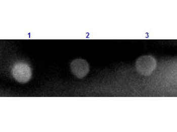
| Tested Applications | DOT, FC, FLISA, IF |
|---|---|
| Dilution Range | FLISA: 1:10,000 - 1:50,000, FC: 1:500 - 1:2,500, IF: 1:1,000 - 1:5,000 |
| Reactivity | Mouse |
| Application Notes |
Key Properties
−| Antibody Type | Secondary Antibody |
|---|---|
| Host | Goat |
| Clonality | Polyclonal |
| Isotype | IgG |
| Immunogen | Mouse IgG whole molecule |
| Purity | This product was prepared from monospecific antiserum by immunoaffinity chromatography using Mouse IgG coupled to agarose beads followed by solid phase adsorption(s) to remove any unwanted reactivities. Assay by immunoelectrophoresis resulted in a single precipitin arc against anti-Goat Serum, Mouse IgG and Mouse Serum. No reaction was observed against Bovine, Chicken, Goat, Guinea Pig, Hamster, Horse, Human, Rabbit, Rat and Sheep Serum Proteins. |
| Conjugation | TRITC |
Storage & Handling
−| Storage | Store vial at 4° C prior to restoration. For extended storage aliquot contents and freeze at -20° C or below. Avoid cycles of freezing and thawing. Centrifuge product if not completely clear after standing at room temperature. This product is stable for several weeks at 4° C as an undiluted liquid. Dilute only prior to immediate use. |
|---|---|
| Form/Appearance | Lyophilized |
| Buffer/Preservatives | Preservative: 0.01% (w/v) Sodium Azide. Stabilizer: 10 mg/mL Bovine Serum Albumin (rAlbumin) - Immunoglobulin and Protease free; Buffer: 0.02 M Potassium Phosphate, 0.15 M Sodium Chloride, pH 7.2 |
| Concentration | 1.0 mg/mL |
| Expiration Date | 12 months from date of receipt. |
| Disclaimer | For research use only |
Alternative Names
−goat anti-Mouse IgG rhodamine conjugated Antibody, goat anti-Mouse IgG Antibody TRITC conjugation
Similar Products
−- Item 1 of 2
Rabbit IgG (H&L) Antibody Pre-Adsorbed [orb347722]
DOT, FC, FLISA, IF
Rabbit
Mouse
Polyclonal
TRITC
1 mg - Item 1 of 1
Mouse IgG (H&L) Antibody Rhodamine Conjugated Pre-Adsorbed [orb347504]
DOT, FC, FLISA, IF
Mouse
Rabbit
Polyclonal
TRITC
2 mg - Item 1 of 1
Mouse IgG (H&L) Antibody Rhodamine Conjugated Pre-Adsorbed [orb347371]
FC, FLISA, IF, WB
Mouse
Goat
Polyclonal
TRITC
2 mg 
F(ab')2 Mouse IgG (H&L) Antibody Rhodamine Conjugated Pre-Adsorbed [orb348274]
FC, FLISA, IF
Mouse
Rabbit
Polyclonal
TRITC
500 μlF(ab')2 Mouse IgG (H&L) Antibody Rhodamine Conjugated Pre-Adsorbed [orb348282]
FC, FLISA, IF
Mouse
Donkey
Polyclonal
TRITC
500 μl
Quality Guarantee
Explore bioreagents carefree to elevate your research. All our products are rigorously tested for performance. If a product does not perform as described on its datasheet, our scientific support team will provide expert troubleshooting, a prompt replacement, or a refund. For full details, please see our Terms & Conditions and Buying Guide. Contact us at [email protected].
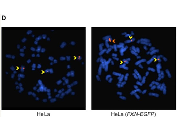
Characterization of HeLa (FXN-EGFP) stable cell lines. The probe was prepared using purified DNA from RP11-265B8, which was labeled by nick translation with digoxigenin. The labeled DNA was ethanol precipitated together with human COT1 DNA and resuspended in 50% formamide, 10% dextran sulfate, and 2×SSC to a concentration of 40 ng/ µl. The probe was denatured by heating at 75°C, followed by preannealing at 37°C and Hybridization was at 37°C. The probe was detected with mouse anti-digoxigenin antibody followed by rhodamine conjugated anti-mouse antibody. (D) Determination of transgenic fragment integration site by FISH. Rhodamine-labeled RP11-265B8 was hybridized onto metaphase chromosomes (DAPI stained) of HeLa (left) and HeLa (FXN-EGFP) (right) cells. Three hybridization signals (yellow arrows) corresponded to the endogenous FXN gene. The presence of one additional brighter signal (orange arrow) establishes the presence of a single integration site containing multiple copies of the FXN-EGFP transgene.
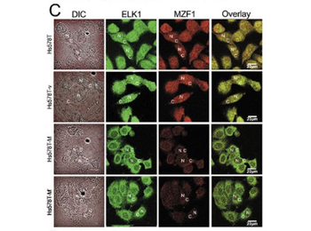
Disrupting the interaction between MZF1 and ELK1 by MZF160-72 interrupts EMT in Hs578T cells. (C) Immunofluorescence staining showed the distribution of the ELK1 and MZF1 proteins. The cells were fixed and stained with antibodies against ELK1 and MZF1 [1:400] followed by the appropriate FITC-or rhodamine-conjugated secondary antibodies. Confocal slices of 0.5 and 0.6 µm were obtained, and images were taken through the center of the nucleus. "N" indicates the nucleus, and "C" indicates the cytosol.
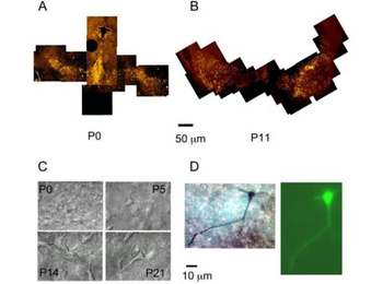
Distribution, morphology, and double staining of SN DA neurons. (A) TH staining of rat midbrain neurons in transversal sections of 200 µm thickness. Sections were cut, fixed, and labeled with a fluorescent anti-TH antibody as described in the methods. TH-antibody staining of a P0 rat. Migrating cells from the fourth ventricle can still be observed. Bar is 50 µm. (B) TH-antibody staining of a P11 rat. The left-hand side SN at P11 is more rostral and shows a widespread localization of DA neurons. The right-hand side SN at P11 is more caudal and shows already the typical SN compacta band in the dorsal aspect, and a smaller number of displaced dopamine neurons outside the compacta area. Dorsal is up and ventral down. (C) Infrared IR-DIC optics images of SN neurons. Rat slices prepared according to the methods section. Infrared pictures of neurons in the SN as observed during the electrophysiological recording. (D) Double stained SN DA neuron at P3. The image on the left shows the TH staining. The image on the right shows the LFY filled neuron.
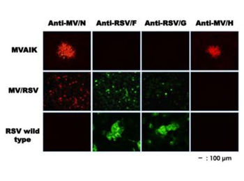
Immunostaining of Vero cells infected with MVAIK, MV/RSV, and RSV. Monoclonal antibodies against RSV F or G protein and secondary antibody against mouse IgG conjugated with FITC were used for the detection of RSV F or G protein. Monoclonal antibody against MV HA protein and secondary antibody against mouse IgG conjugated with rhodamine were used for the detection of MV HA. The expression of measles N protein was stained with monoclonal antibody against measles N protein and secondary antibody conjugated with Alexa Fluor 568.
Documents Download
Datasheet
Product Information
Request a Document
Protocol Information
Mouse IgG (H&L) Antibody Rhodamine Conjugated Pre-Adsorbed (orb347355)

- 0.0
Based on 0 reviews
Participating in our Biorbyt product reviews program enables you to support fellow scientists by sharing your firsthand experience with our products.
Login to Submit a ReviewAvailable Sizes
Select a size below
Free Secondary Antibody (20 ul)0/0
Please add an antibody product to your cart first.