You have no items in your shopping cart.
Description
Research Area
Signal Transduction
Images & Validation
−Item 1 of 6
| Tested Applications | ELISA, FC, IF, WB |
|---|---|
| Dilution Range | ELISA: 1:4000, WB: 0.5-3 μg/ml, IHC-P: 10ug/ml |
| Reactivity | Human, Mouse |
| Predicted Reactivity | Bovine |
| Application Notes |
Key Properties
−| Clonality | Polyclonal |
|---|---|
| Target | RBP1 |
| Protein Sequence | VEGVVCKQVFKKVQ |
| Molecular Weight | 22.3 |
| Purification | Purified from goat serum by ammonium sulphate precipitation followed by antigen affinity chromatography using the immunizing peptide. |
| Conjugation | Unconjugated |
Storage & Handling
−| Storage | Maintain refrigerated at 2-8°C for up to 2 weeks. For long term storage store at -20°C in small aliquots to prevent freeze-thaw cycles. |
|---|---|
| Buffer/Preservatives | Supplied at 0.5 mg/ml in Tris saline, 0.02% sodium azide, pH 7.3 with 0.5% bovine serum albumin. Aliquot and store at -20°C. Minimize freezing and thawing. |
| Expiration Date | 12 months from date of receipt. |
| Disclaimer | For research use only |
Alternative Names
−anti RBP1 antibody, anti CRBP antibody, anti RBPC antibody, anti CRBP1 antibody, anti CRABP-I antibody, anti retinol binding protein 1, cellular antibody, anti retinol-binding protein 1, cellular antibody, anti CRBPI antibody
Similar Products
−- Item 1 of 8
RBP1 Rabbit Polyclonal Antibody [orb1676418]
ELISA, FC, ICC, IF, IHC, WB
Human, Mouse, Rat
Rabbit
Polyclonal
Unconjugated
100 μg - Item 1 of 1
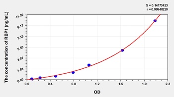
Human Retinol Binding Protein 1, Cellular (RBP1) ELISA Kit [orb777073]
Human
0.16-10 ng/mL
0.059 ng/mL
48 T, 96 T - Item 1 of 5
- Item 1 of 5
- Item 1 of 5


Quality Guarantee
Explore bioreagents carefree to elevate your research. All our products are rigorously tested for performance. If a product does not perform as described on its datasheet, our scientific support team will provide expert troubleshooting, a prompt replacement, or a refund. For full details, please see our Terms & Conditions and Buying Guide. Contact us at [email protected].
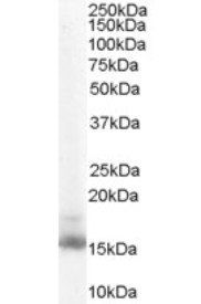
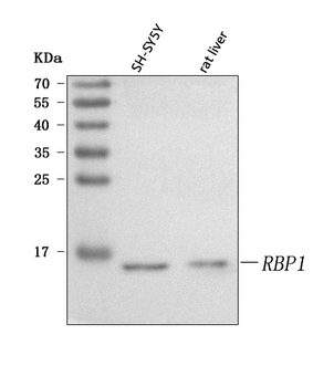
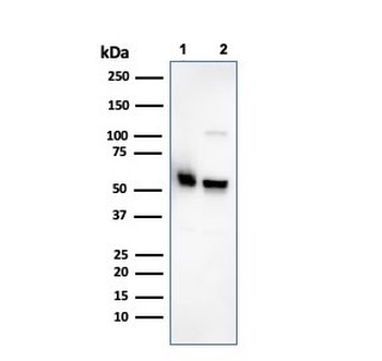
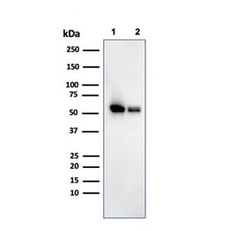
Western blot analysis of NIH3T3 lysate using RBP1 antibody
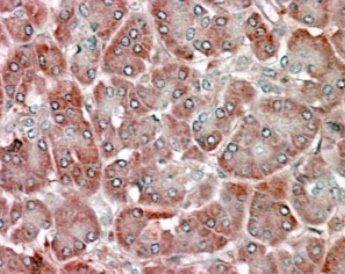
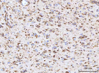
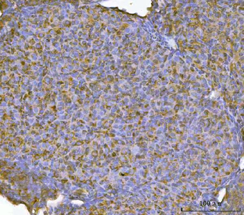
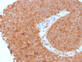
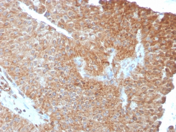
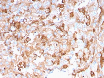
Immunohistochemical staining of Human Pancreas using RBP1 antibody
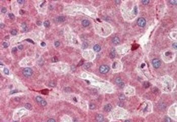
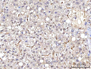
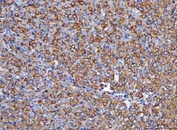
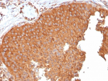
Immunohistochemical staining of Human Liver using RBP1 antibody
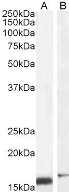
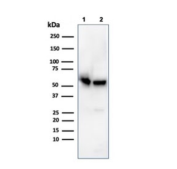
WB analysis of NIH3T3 (A) and U251 (B) cell lysate using RBP1 antibody
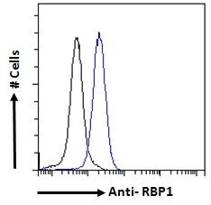
Flow cytometric analysis of HeLa cells using RBP1 antibody
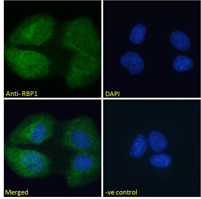
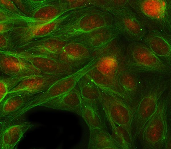
Immunofluorescence analysis of U2OS cells using RBP1 antibody
Documents Download
Datasheet
Product Information
Request a Document
Protocol Information
WB
Western Blot (IB, immunoblot)
FC
Flow Cytometry
IF
Immunofluorescence
ELISA
Enzyme-linked Immunosorbent Assay (EIA)
RBP1 Antibody (orb18987)

- 0.0
Based on 0 reviews
Participating in our Biorbyt product reviews program enables you to support fellow scientists by sharing your firsthand experience with our products.
Login to Submit a ReviewAvailable Sizes
Select a size below
Choose Conjugation or Carrier Free Version
Free Secondary Antibody (20 ul)0/0
Please add an antibody product to your cart first.