You have no items in your shopping cart.
Description
Research Area
Neuroscience
Images & Validation
−Item 1 of 9
| Tested Applications | ELISA, FC, IF, IHC |
|---|---|
| Dilution Range | ELISA: 1:16000, IHC-P: 2.5μg/ml, IF/ICC: 10μg/ml, FACS: 10ug/ml |
| Reactivity | Human |
| Predicted Reactivity | Bovine, Canine, Mouse, Rat |
| Application Notes |
Key Properties
−| Host | Goat |
|---|---|
| Clonality | Polyclonal |
| Target | NOS1 |
| Protein Sequence | ESKKDTDEVFSS |
| Molecular Weight | 161; 164.6; 125 |
| Purification | Purified from goat serum by ammonium sulphate precipitation followed by antigen affinity chromatography using the immunizing peptide. |
| Conjugation | Unconjugated |
Storage & Handling
−| Storage | Maintain refrigerated at 2-8°C for up to 2 weeks. For long term storage store at -20°C in small aliquots to prevent freeze-thaw cycles. |
|---|---|
| Buffer/Preservatives | Supplied at 0.5 mg/ml in Tris saline, 0.02% sodium azide, pH 7.3 with 0.5% bovine serum albumin. Aliquot and store at -20°C. Minimize freezing and thawing. |
| Expiration Date | 12 months from date of receipt. |
| Disclaimer | For research use only |
Alternative Names
−anti NOS1 antibody, anti nitric oxide synthase 1 (neuronal) antibody, anti NOS antibody, anti neuronal nitric oxide synthase antibody, anti PnNOS antibody, anti penile neuronal nitric oxide synthase antibody, anti penile neuronal NOS antibody, anti IHPS1 antibody, anti nNOS antibody, anti nitric oxide synthase 1, neuronal antibody
Similar Products
−- Item 1 of 8
NNOS Recombinant Rabbit Monoclonal Antibody [orb1499394]
WB
Human, Mouse
Rat
Rabbit
Recombinant
Unconjugated
50 μl, 100 μl - Item 1 of 4
NNOS Rabbit Polyclonal Antibody [orb158006]
IF, IHC-Fr, IHC-P, WB
Bovine, Canine, Gallus, Human, Porcine, Rat, Sheep
Mouse
Rabbit
Polyclonal
Unconjugated
50 μl, 100 μl, 200 μl - Item 1 of 5
- Item 1 of 4
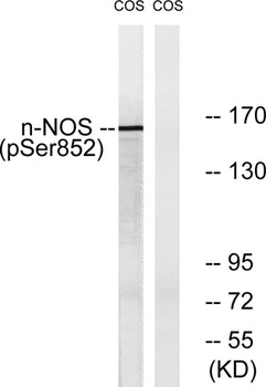
NOS1 (phospho Ser852) rabbit pAb Antibody [orb769272]
ELISA, IF, IHC, WB
Human, Monkey, Mouse, Rat
Polyclonal
Unconjugated
100 μl, 50 μl - Item 1 of 1
Human Nitric Oxide Synthase 1, Neuronal (NOS1) ELISA Kit [orb775483]
Human
0.16-10 ng/mL
0.062 ng/mL
48 T, 96 T


Quality Guarantee
Explore bioreagents carefree to elevate your research. All our products are rigorously tested for performance. If a product does not perform as described on its datasheet, our scientific support team will provide expert troubleshooting, a prompt replacement, or a refund. For full details, please see our Terms & Conditions and Buying Guide. Contact us at [email protected].
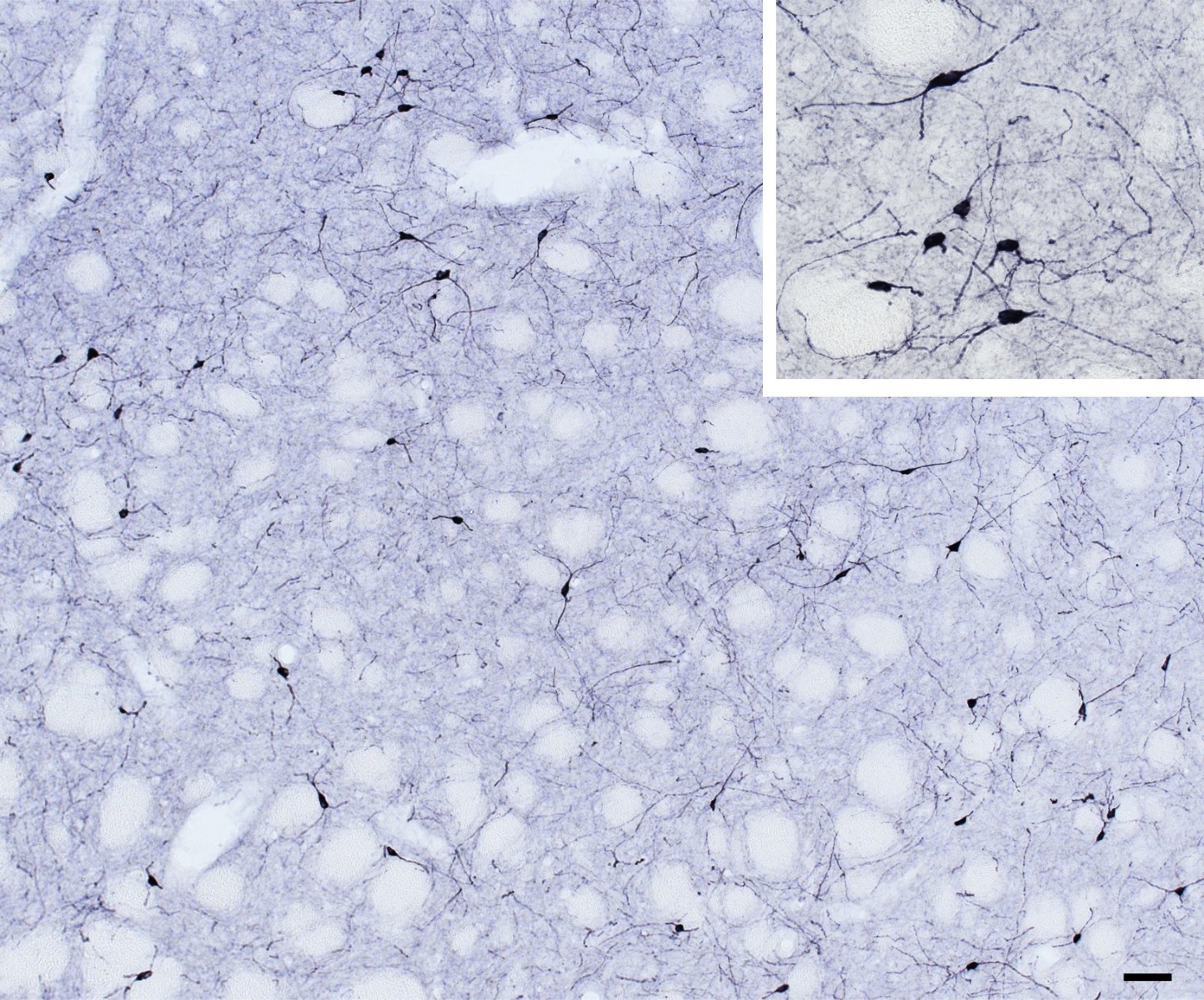
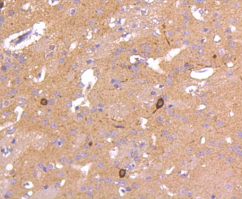
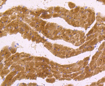
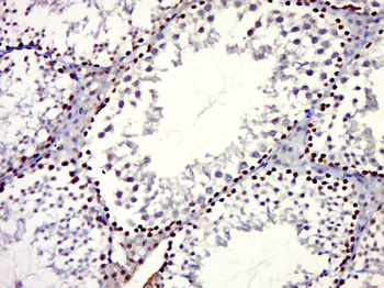
Immunohistochemical staining of mouse caudate-putamen using NOS1 antibody
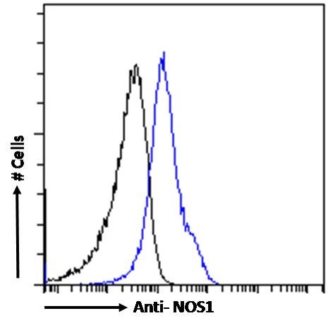
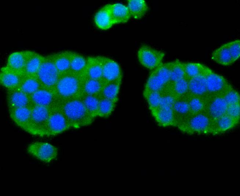
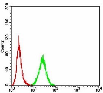
Flow cytometric analysis of Kelly cells using NOS1 antibody
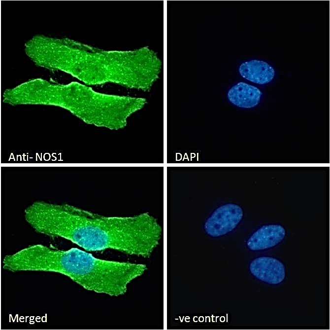
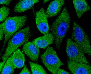
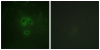
Immunofluorescence analysis of HeLa cells using NOS1 antibody
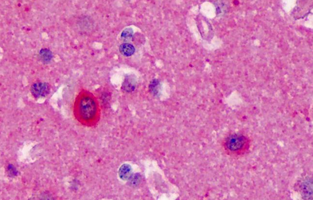
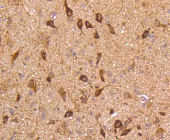
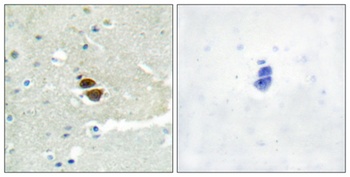
Immunohistochemical staining of Human Cortex using NOS1 antibody
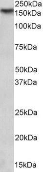
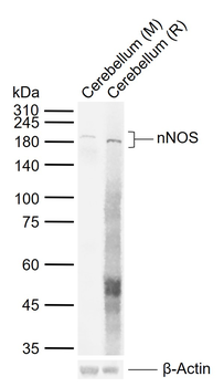
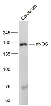
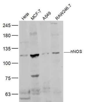
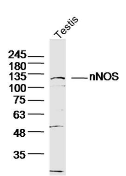
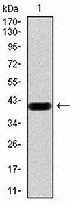
1 µg/mL staining of Mouse Brain lysate (35 µg protein in RIPA buffer). Detected by chemiluminescence.
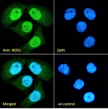
Immunofluorescence analysis of paraformaldehyde fixed HeLa cells, permeabilized with 0.15% Triton. Primary incubation 1 hr (10 µg/mL) followed by Alexa Fluor 488 secondary antibody (2 µg/mL), showing nuclear staining. The nuclear stain is DAPI (blue). Negative control: Unimmunized goat IgG (10 µg/mL) followed by Alexa Fluor 488 secondary antibody (2 µg/mL).
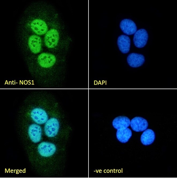
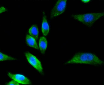
Immunofluorescence analysis of paraformaldehyde fixed U2OS cells, permeabilized with 0.15% Triton. Primary incubation 1 hr (10 µg/mL) followed by Alexa Fluor 488 secondary antibody (2 µg/mL), showing nuclear staining. The nuclear stain is DAPI (blue). Negative control: Unimmunized goat IgG (10 µg/mL) followed by Alexa Fluor 488 secondary antibody (2 µg/mL).
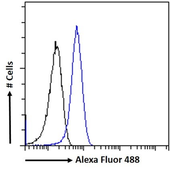
Flow cytometric analysis of paraformaldehyde fixed HeLa cells (blue line), permeabilized with 0.5% Triton. Primary incubation 1 hr (10 µg/mL) followed by Alexa Fluor 488 secondary antibody (1 µg/mL). IgG control: Unimmunized goat IgG (black line) followed by Alexa Fluor 488 secondary antibody.
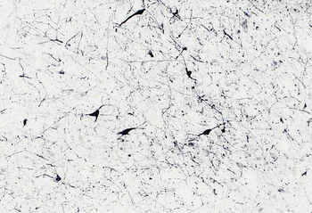
Immunostaining of 30 µm thick cryosections of PFA-perfused Human Hypothalamus, antigen retrieval with citrate buffer Ph 6 at 80C for 30 min, HRP-staining with Ni-DAB after Biotin-SP-antigoat amplification.
Documents Download
Datasheet
Product Information
Request a Document
Protocol Information
IHC
Immunohistochemistry
FC
Flow Cytometry
IF
Immunofluorescence
ELISA
Enzyme-linked Immunosorbent Assay (EIA)
NOS1 Antibody (orb18337)

- 0.0
Based on 0 reviews
Participating in our Biorbyt product reviews program enables you to support fellow scientists by sharing your firsthand experience with our products.
Login to Submit a ReviewAvailable Sizes
Select a size below
Choose Conjugation or Carrier Free Version
Free Secondary Antibody (20 ul)0/0
Please add an antibody product to your cart first.