You have no items in your shopping cart.
Description
Images & Validation
−Item 1 of 7
| Tested Applications | ELISA, IF, IHC, WB |
|---|---|
| Dilution Range | ELISA: 1:24,800-1:44,800, IHC: 1:100, IF: 5-15µg/mL |
| Reactivity | Human |
| Application Notes |
Key Properties
−| Antibody Type | Primary Antibody |
|---|---|
| Host | Rabbit |
| Clonality | Polyclonal |
| Immunogen | Glucagon antibody was prepared from whole rabbit serum produced by repeated immunizations with a synthetic peptide corresponding to an internal portion of human Glucagon. |
| Target | GCG |
| Purity | This affinity purified antibody is directed against human Glucagon. This product was affinity purified from monospecific antiserum by immunoaffinity purification. A BLAST analysis was used to suggest cross-reactivity with the antigen based on 100% homology with the immunizing sequence to human, chimpanzee, and bonobo. |
| Conjugation | Unconjugated |
Storage & Handling
−| Storage | Store vial at -20° C prior to opening. Aliquot contents and freeze at -20° C or below for extended storage. Avoid cycles of freezing and thawing. Centrifuge product if not completely clear after standing at room temperature. This product is stable for several weeks at 4° C as an undiluted liquid. Dilute only prior to immediate use. |
|---|---|
| Form/Appearance | Liquid (sterile filtered) |
| Buffer/Preservatives | Preservative: 0.01% (w/v) Sodium Azide. Stabilizer: None; Buffer: 0.02 M Potassium Phosphate, 0.15 M Sodium Chloride, pH 7.2 |
| Concentration | 1.18 mg/mL |
| Expiration Date | 12 months from date of receipt. |
| Dry Ice Shipping | Please note: This product requires shipment on dry ice. A dry ice surcharge will apply. |
| Disclaimer | For research use only |
Alternative Names
−Rabbit Anti-Glucagon, Pro-glucagon, Glicentin, Oxyntomodulin, OXM, OXY, Glucagon
Similar Products
−- Item 1 of 12
GLP-1 Mouse Monoclonal Antibody [orb10721]
IF, IHC-Fr, IHC-P
Mouse, Rat
Human, Mouse, Rat
Mouse
Monoclonal
Unconjugated
200 μl, 200 μg, 50 μl, 100 μl - Item 1 of 7
GLP2 Rabbit Polyclonal Antibody [orb10724]
ICC, IF, IHC-P, WB
Guinea pig, Human, Mouse, Porcine, Rat
Rabbit
Polyclonal
Unconjugated
100 μg - Item 1 of 6
Glucagon Antibody (C-term) [orb1929798]
FC, IF, IHC-P, WB
Human
Rabbit
Polyclonal
Unconjugated
50 μl, 100 μl - Item 1 of 4
GCG rabbit pAb Antibody [orb765282]
ELISA, IF, IHC, WB
Human, Monkey, Mouse, Rat
Polyclonal
Unconjugated
100 μl, 50 μl - Item 1 of 3
GLP-1 (7-36) Rabbit Polyclonal Antibody [orb10719]
IF, IHC-Fr, IHC-P
Bovine, Porcine, Sheep
Human, Mouse, Rat
Rabbit
Polyclonal
Unconjugated
50 μl, 100 μl, 200 μl


Quality Guarantee
Explore bioreagents carefree to elevate your research. All our products are rigorously tested for performance. If a product does not perform as described on its datasheet, our scientific support team will provide expert troubleshooting, a prompt replacement, or a refund. For full details, please see our Terms & Conditions and Buying Guide. Contact us at [email protected].
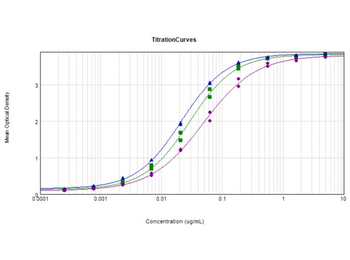
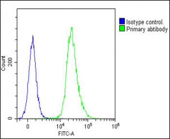
ELISA Results of Rabbit Anti-Glucagon Antibody. Each well was coated with 1 µg of conjugate. The starting concentration of antibody in the dilution series was 5 µg/ml. The titer is 1:34800 Glucagon - Free peptide [Green Line], 1:47200 Glucagon Standard - BSA conjugated [Blue Line], and 1:20500 Glucagon - BSA conjugated [Purple Line]. Each point on the Y-axis represents a 3-fold dilution. 3% Fish Gel (p/n orb348587), HRP conjugated Goat anti-Rabbit IgG (H&L) (p/n orb347673), and TMB substrate were used for detection.
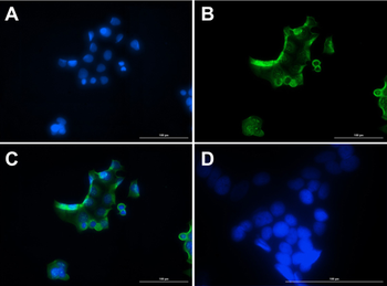
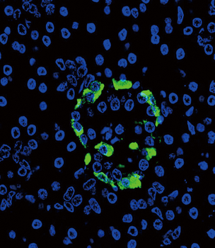
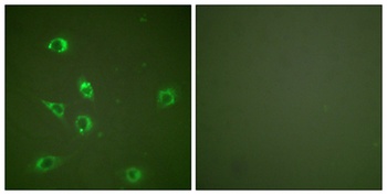
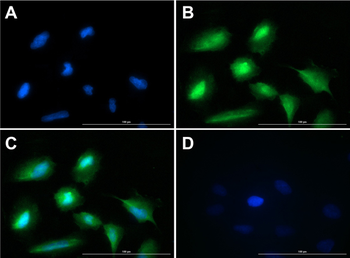
Immunofluorescence of Rabbit Anti-Glucagon Antibody. Cell Line: MCF7 cells. Fixative: 100% Methanol. Permeabilization: 0.3% Triton X-100. Primary Antibody: Anti-Glucagon at 15 µg/ml overnight at 2-8°C. Secondary Antibody: Goat Anti-Rabbit IgG DyLight™488 at 5 µl/mL for 1hr at RT. Nuclear Counterstain: DAPI. Staining: (A). DAPI. (B). Anti-Glucagon + DyLight™488 secondary. (C). Merge A + B. (D). secondary only. Localization expected: Cytoplasm.
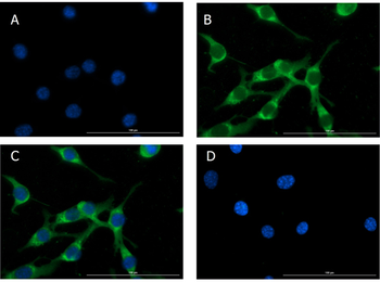
Immunofluorescence of Rabbit Anti-Glucagon Antibody. Cell Line: NIH/3T3 cells. Fixative: 100% Methanol. Permeabilization: Triton X-100. Primary Antibody: Anti-Glucagon at 15 µg/ml overnight at 2-8°C. Secondary Antibody: Goat Anti-Rabbit IgG DyLight™488 at 5 µl/mL for 1hr at RT. Nuclear Counterstain: DAPI. Staining: (A). DAPI. (B). Anti-Glucagon + DyLight™488 secondary. (C). Merge A + B. (D). secondary only. Localization expected: Cytoplasm.
Immunofluorescence of Rabbit Anti-Glucagon Antibody. Cell Line: NIH/3T3 cells. Fixative: 4% PFA. Permeabilization: Triton X-100. Primary Antibody: Anti-Glucagon at 15 µg/ml overnight at 2-8°C. Secondary Antibody: Goat Anti-Rabbit IgG DyLight™488 at 5 µl/mL for 1hr at RT. Nuclear Counterstain: DAPI. Staining: (A). DAPI. (B). Anti-Glucagon + DyLight™488 secondary. (C). Merge A + B. (D). secondary only. Localization expected: Cytoplasm.
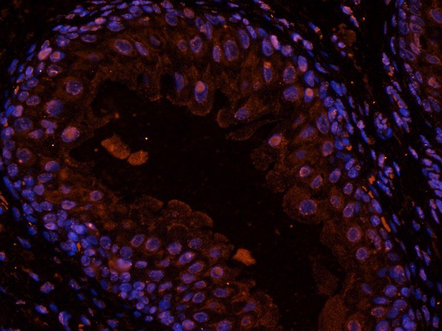
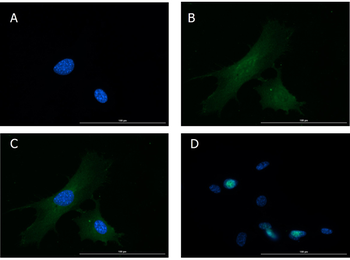
Immunofluorescence of Rabbit Anti-Glucagon Antibody. Cell Line: U20S cells. Fixative: 4% PFA. Permeabilization: 0.3% Triton X-100. Primary Antibody: Anti-Glucagon at 15 µg/ml overnight at 2-8°C. Secondary Antibody: Goat Anti-Rabbit IgG DyLight™488 at 5 µl/mL for 1hr at RT. Nuclear Counterstain: DAPI. Staining: (A). DAPI. (B). Anti-Glucagon + DyLight™488 secondary. (C). Merge A + B. (D). secondary only. Localization expected: Cytoplasm.
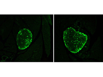
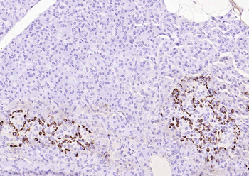
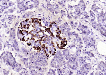
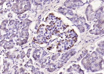
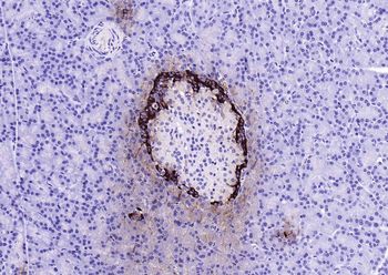
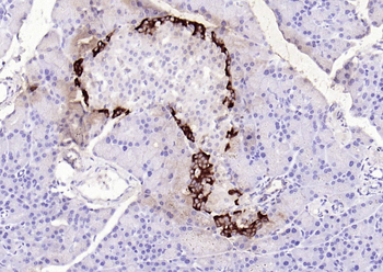
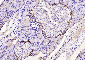
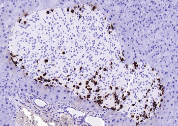
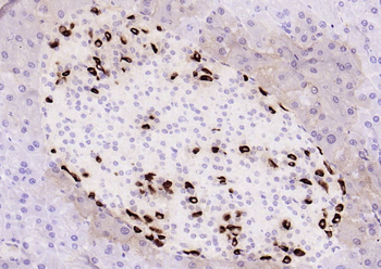
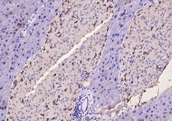
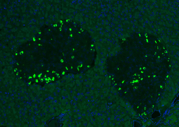
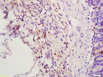
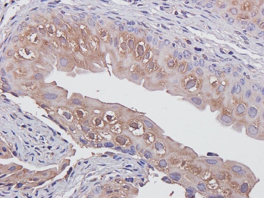
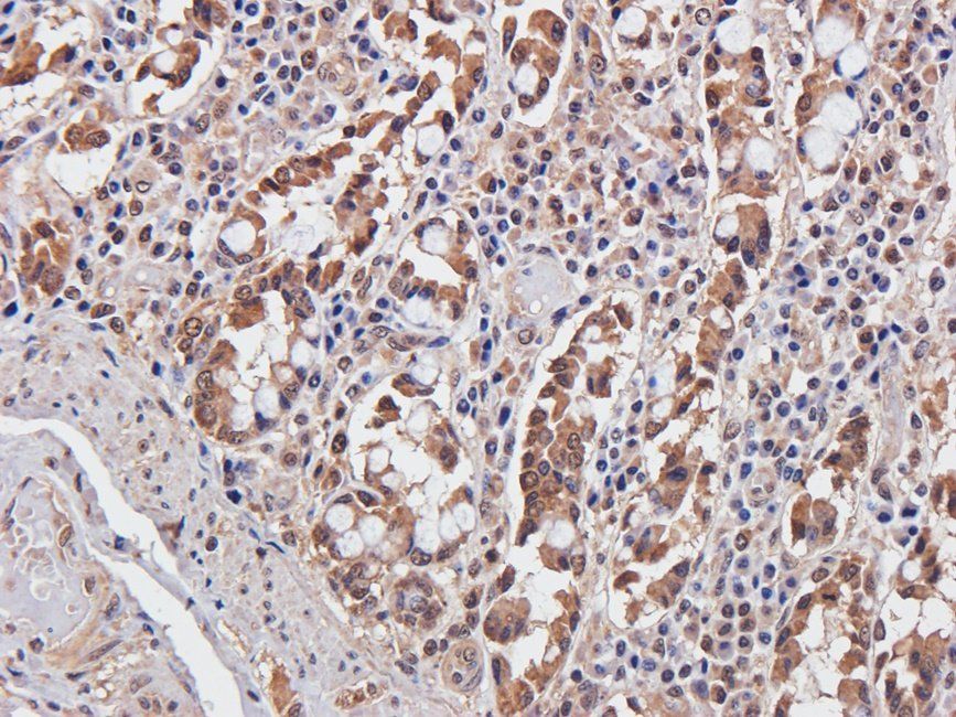
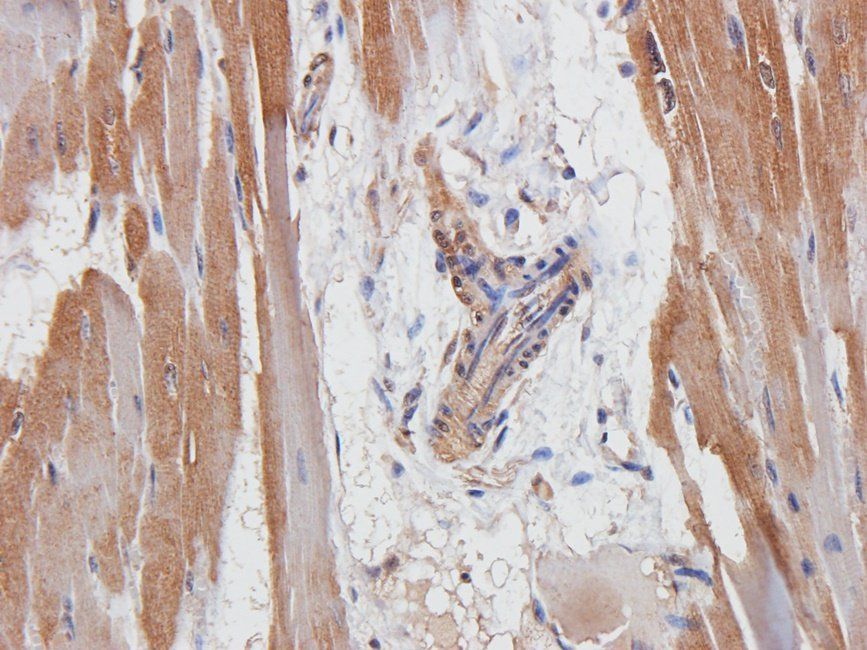
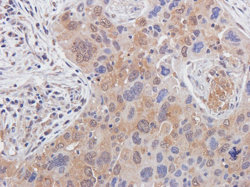
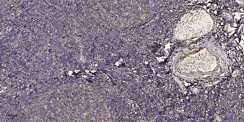
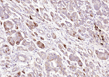
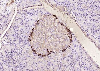
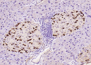
Immunohistochemistry results using Rabbit Anti-Glucagon Antibody. Tissue: alpha cells in CD1 mouse pancreatic islets. Fixation: 4% paraformaldehyde. Antigen Retrieval: 10 mM Sodium Citrate buffer for 10 mins at 95-100°C. Blocking: PBS, 1% ovalbumin, 0.3% Triton X-100. Primary Antibody: Anti-Glucagon at 1:100 overnight at RT. Secondary Antibody: Anti-Rabbit Alexa Fluor 488 at 1:500 for 1hr at RT. Original magnification 20x.
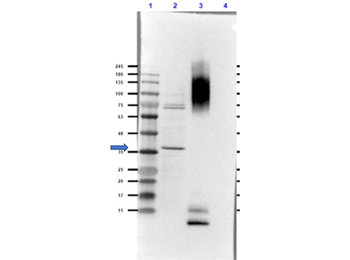
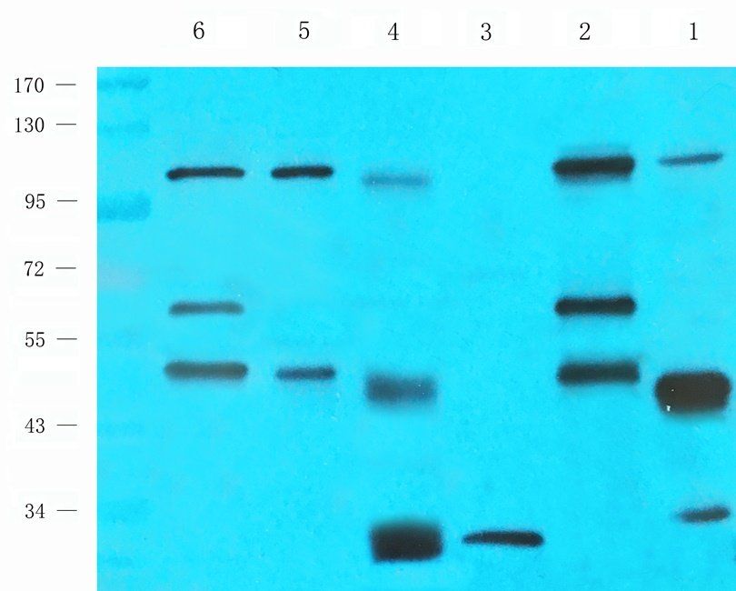
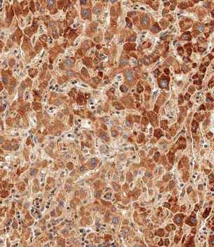
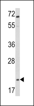
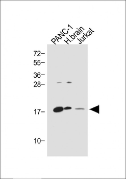
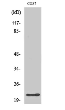
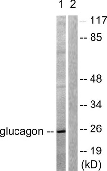
Western Blot of Rabbit Anti-Glucagon Antibody. Lane 1: Opal Prestained Molecular Weight. Lane 2: COS-7 Lysate - reduced (20 µg). Lane 3: BSA Conjugated Glucagon peptide - reduced (0.02 µg). Lane 4: Insulin - reduced (0.05 µg). Primary Antibody: Anti-Glucagon [Rabbit] Antibody at 1.0 µg/ml overnight at 2-8°C. Secondary Antibody: Goat Anti-Rabbit IgG (MX10) Peroxidase conjugated at 1:70000 for 30 mins at RT. Block: Blocking Buffer for Fluorescent Western Blotting (p/n orb348637) for 1hr at RT. Expected MW: ~21kDa. Observed MW: endogenous detection in COS-7 Lysate at ~35kDa. Glucagon peptide is detected at the MW of BSA. No cross-reactivity with insulin is observed. Exposure: 25 sec.
Quick Database Links
UniProt Details
− No UniProt data available
NCBI Reference Sequences
−Associated Accession Numbers
Curated reference sequences for the gene transcript and protein product| Protein | NP_002045.1 |
|---|
Documents Download
Datasheet
Product Information
Request a Document
Protocol Information
WB
Western Blot (IB, immunoblot)
IHC
Immunohistochemistry
IF
Immunofluorescence
ELISA
Enzyme-linked Immunosorbent Assay (EIA)
GCG Antibody (orb1784557)

- 0.0
Based on 0 reviews
Participating in our Biorbyt product reviews program enables you to support fellow scientists by sharing your firsthand experience with our products.
Login to Submit a ReviewAvailable Sizes
Select a size below
Choose Conjugation or Carrier Free Version
Free Secondary Antibody (20 ul)0/0
Please add an antibody product to your cart first.