You have no items in your shopping cart.
Featured
Description
Research Area
Cell Biology
Images & Validation
−Item 1 of 7
| Tested Applications | ELISA, ICC, IF, IHC-P, WB |
|---|---|
| Reactivity | Human, Mouse, Rat |
| Application Notes |
Key Properties
−| Antibody Type | Primary Antibody |
|---|---|
| Host | Rabbit |
| Clonality | Polyclonal |
| Isotype | IgG |
| Immunogen | Caspase-8 antibody was raised against a 16 amino acid synthetic peptide from near the carboxy terminus of human Caspase-8 isoform A.The immunogen is located within the last 50 amino acids of Caspase-8. |
| Target | CASP8 |
| Molecular Weight | 55 kDa |
| Purification | Caspase-8 Antibody is affinity chromatography purified via peptide column. |
| Conjugation | Unconjugated |
Storage & Handling
−| Storage | Maintain refrigerated at 2-8°C for up to 2 weeks. For long term storage store at -20°C in small aliquots to prevent freeze-thaw cycles. |
|---|---|
| Form/Appearance | Liquid |
| Buffer/Preservatives | Caspase-8 Antibody is supplied in PBS containing 0.02% sodium azide. |
| Concentration | 1 mg/mL |
| Expiration Date | 12 months from date of receipt. |
| Disclaimer | For research use only |
Alternative Names
−Caspase-8 Antibody: CAP4, MACH, MCH5, FLICE, ALPS2B, Casp-8, Caspase-8, Apoptotic cysteine protease, CASP-8
Similar Products
−- Item 1 of 8
Caspase 8 Mouse Monoclonal Antibody [orb500937]
WB
Rat
Human, Mouse
Mouse
Monoclonal
Unconjugated
50 μl, 100 μl, 200 μl, 200 μg - Item 1 of 8
Caspase 8/CASP8 Rabbit Polyclonal Antibody [orb1728223]
FC, ICC, IHC, IHC-Fr, WB
Human, Mouse, Rat
Rabbit
Polyclonal
Unconjugated
100 μg - Item 1 of 4
Caspase 8 Rabbit Polyclonal Antibody [orb10241]
FC, IF, IHC-Fr, IHC-P, WB
Bovine, Canine, Equine, Mouse, Porcine
Human, Rat
Rabbit
Polyclonal
Unconjugated
100 μl, 200 μl, 50 μl - Item 1 of 7
Cleaved-Caspase-8 (D384) Polyclonal Antibody [orb1416163]
IF, IHC-P, WB
Human
Rabbit
Polyclonal
Unconjugated
100 μl - Item 1 of 7
Caspase-8 Polyclonal Antibody [orb1414446]
IF, IHC-P, WB
Human, Mouse, Rat
Rabbit
Polyclonal
Unconjugated
100 μl

Quality Guarantee
Explore bioreagents carefree to elevate your research. All our products are rigorously tested for performance. If a product does not perform as described on its datasheet, our scientific support team will provide expert troubleshooting, a prompt replacement, or a refund. For full details, please see our Terms & Conditions and Buying Guide. Contact us at [email protected].
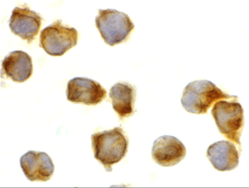
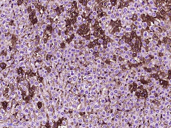
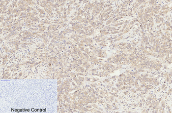
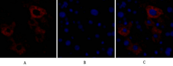
Immunocytochemistry of caspase-8 in Jurkat cells with caspase-8 antibody at 2 µg/mL.
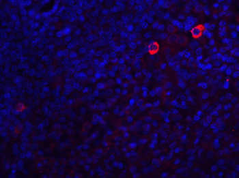
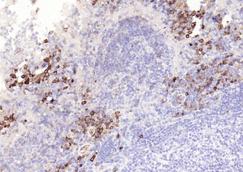
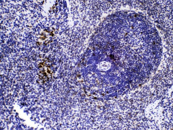
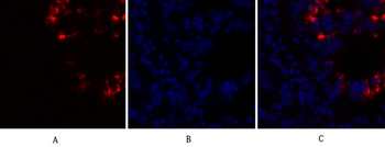
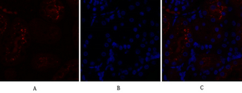
Immunofluorescence of Caspase-8 in human spleen tissue with Caspase-8 antibody at 20 µg/mL. Red: Caspase-8 Antibody (orb1239264). Blue: DAPI staining.
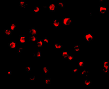
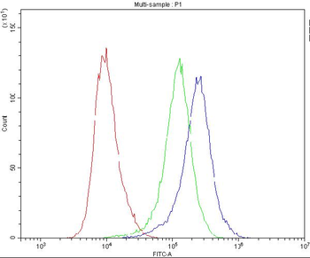
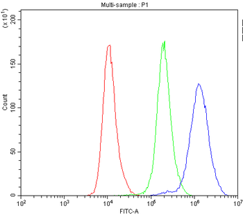
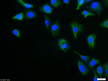
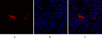
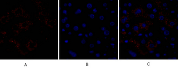
Immunofluorescence of Caspase-8 in Jurkat cells with Caspase-8 antibody at 20 µg/mL.
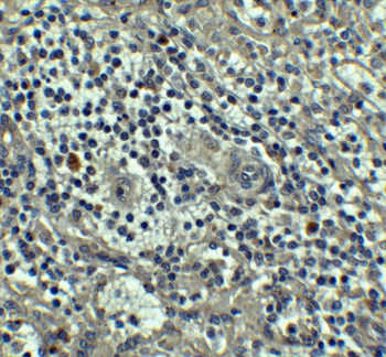
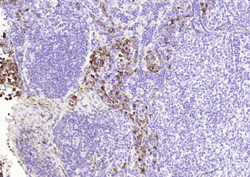
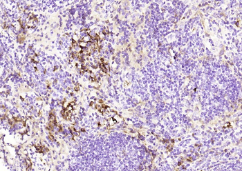
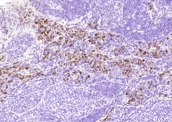
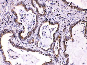
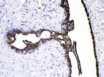
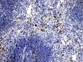
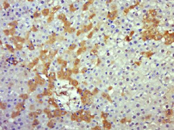
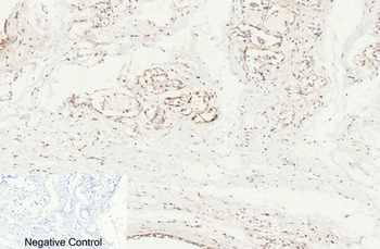
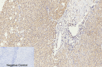
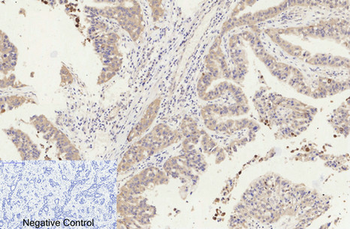
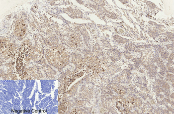
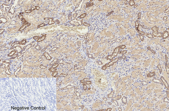
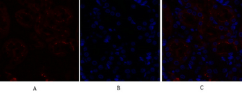
Immunohistochemistry of Caspase-8 in human spleen tissue with Caspase-8 antibody at 5 µg/mL.
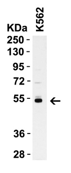
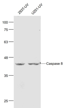
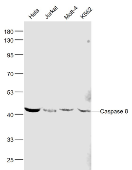
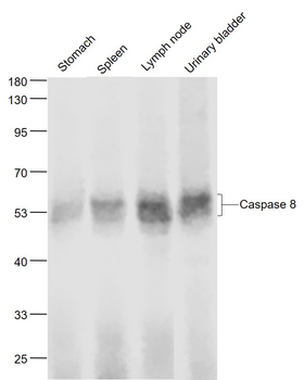
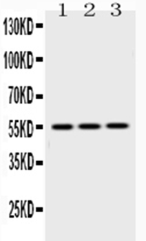
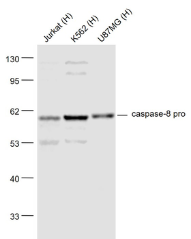
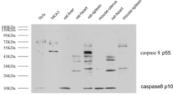
WB Validation in Human K562 Cells. Loading: 15 µg of K562 cell lysate Antibodies: Casp8 orb1239264, 1 µg/mL, 1 h incubation at RT in 5% NFDM/TBST. Secondary: Goat Anti-Rabbit IgG HRP conjugate at 1:10000 dilution.
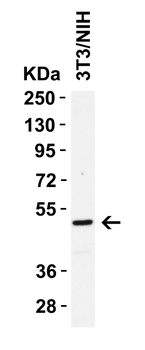
WB Validation in Mouse 3T3/NIH Cells. Loading: 15 µg of 3T3/NIH cell lysate Antibodies: Casp8 orb1239264, 1 µg/mL, 1 h incubation at RT in 5% NFDM/TBST. Secondary: Goat Anti-Rabbit IgG HRP conjugate at 1:10000 dilution.
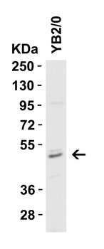
WB Validation in Rat YB2/0 Cells. Loading: 15 µg of YB2/0 cell lysate Antibodies: Casp8 orb1239264, 1 µg/mL, 1 h incubation at RT in 5% NFDM/TBST. Secondary: Goat Anti-Rabbit IgG HRP conjugate at 1:10000 dilution.
Documents Download
Datasheet
Product Information
Request a Document
Protocol Information
WB
Western Blot (IB, immunoblot)
IHC-P
Immunohistochemistry Paraffin
IF
Immunofluorescence
ICC
Immunocytochemistry
ELISA
Enzyme-linked Immunosorbent Assay (EIA)
CASP8 Antibody (orb1239264)

- 0.0
Based on 0 reviews
Participating in our Biorbyt product reviews program enables you to support fellow scientists by sharing your firsthand experience with our products.
Login to Submit a ReviewAvailable Sizes
Select a size below
Free Secondary Antibody (20 ul)0/0
Please add an antibody product to your cart first.