
You have no items in your shopping cart.
Description
Research Area
Cell Biology, Disease Biomarkers, Musculoskeletal & Connective Tissue Research
Images & Validation
−Item 1 of 11
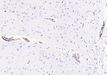
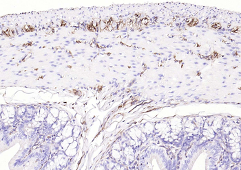
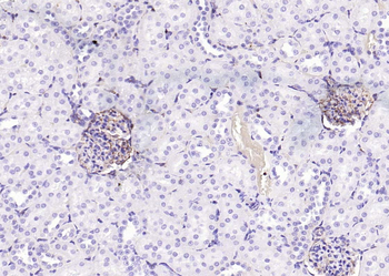
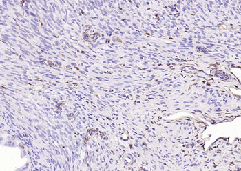
| Tested Applications | ELISA, ICC, IHC, WB |
|---|---|
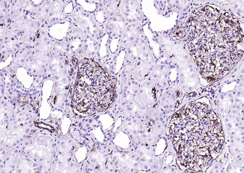
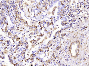
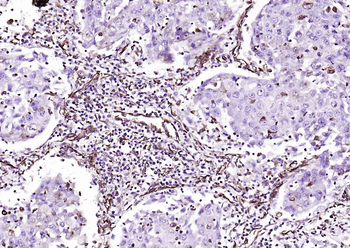
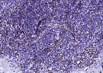
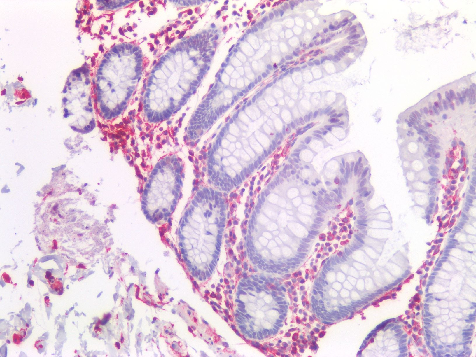
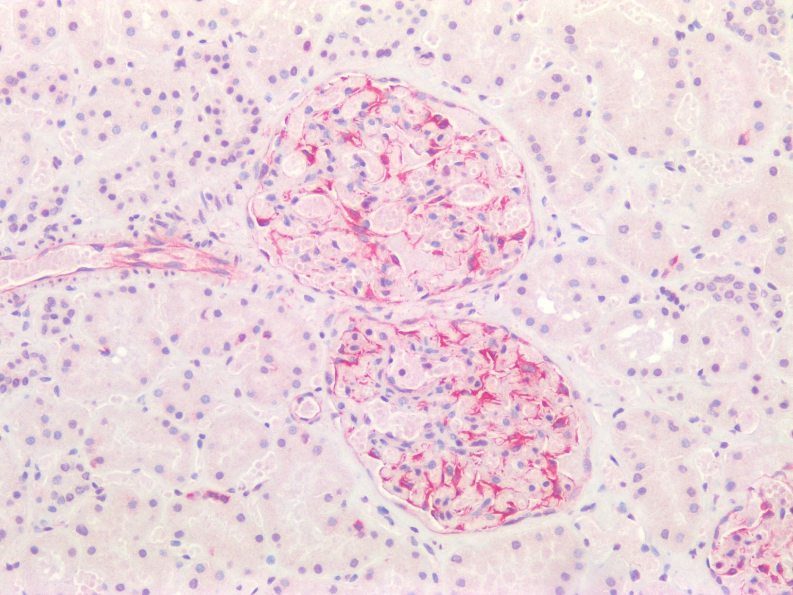
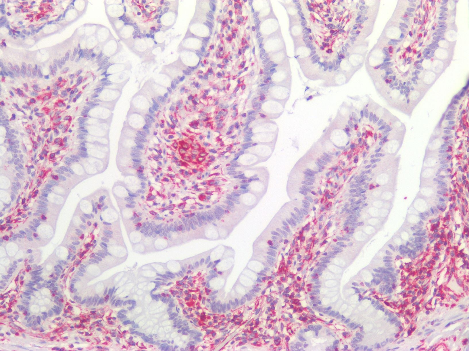
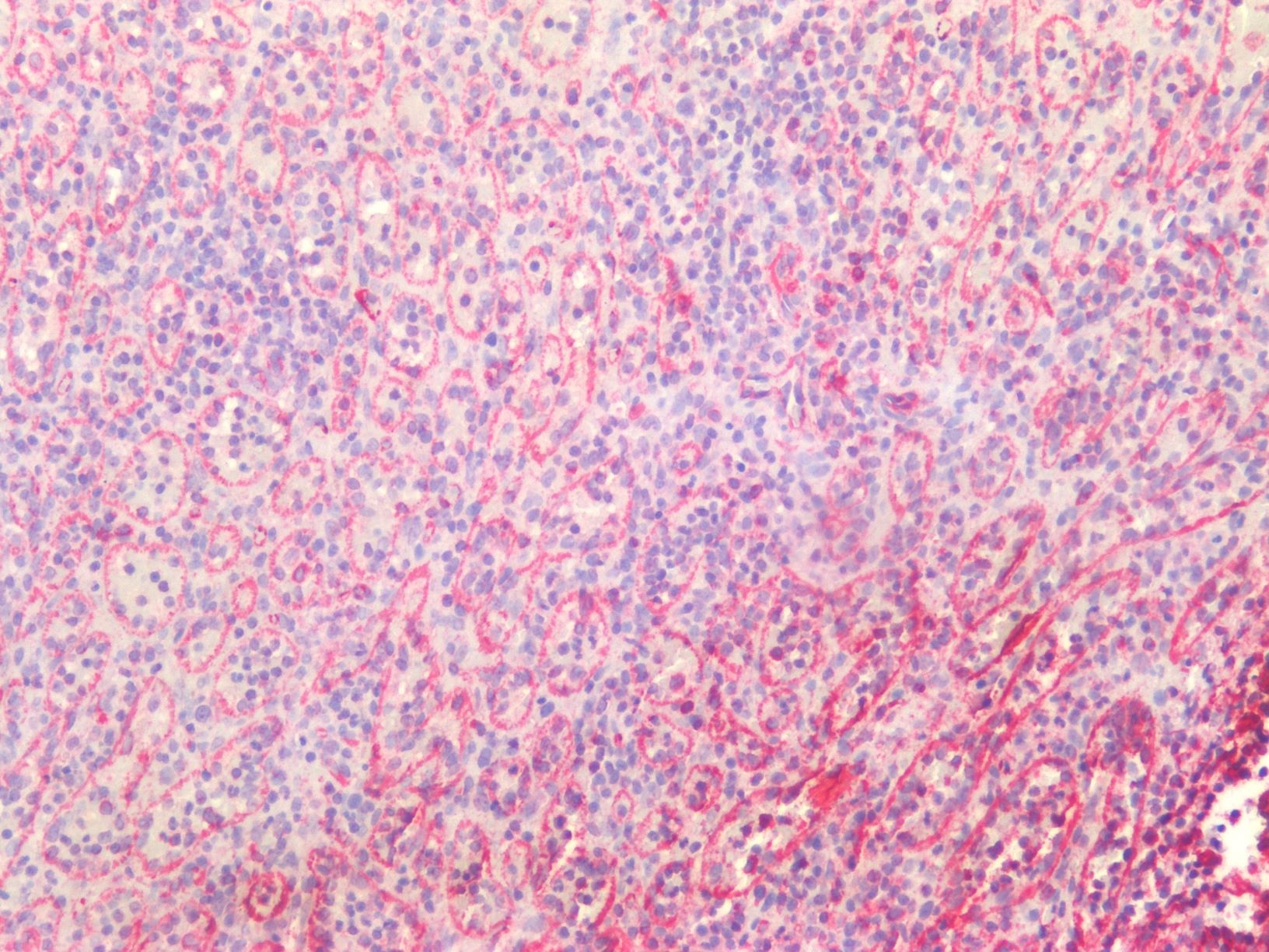
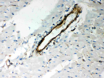
| Dilution Range | WB: 0.736111111, IHC: 1:500-1:5000, ICC: 1:500-1:10,000 |
| Reactivity | Human, Mouse, Rat |
| Predicted Reactivity | Bovine, Canine, Equine, Feline, Goat, Guinea pig, Hamster, Primate, Rabbit, Rodent, Sheep |
Key Properties
−| Antibody Type | Primary Antibody |
|---|---|
| Host | Gallus |
| Clonality | Polyclonal |
| Isotype | IgY |
| Immunogen | Recombinant human vimentin purified from E. coli. |
| Target | Vimentin |
| Molecular Weight | 50 |
| Purification | Total IgY fraction |
| Conjugation | Unconjugated |
Storage & Handling
−| Storage | Maintain refrigerated at 2-8°C for up to 2 weeks. For long term storage store at -20°C in small aliquots to prevent freeze-thaw cycles. |
|---|---|
| Form/Appearance | Liquid |
| Buffer/Preservatives | PBS + 10 mM NaN3. |
| Concentration | 20 mg/ml |
| Expiration Date | 12 months from date of receipt. |
| Disclaimer | For research use only |
Alternative Names
−CTRCT30 antibody, Epididymis luminal protein 113 antibody, FLJ36605 antibody, HEL113 antibody, VIM antibody, VIME_HUMAN antibody, Vimentin antibody
Similar Products
−- Item 1 of 22
Vimentin Rabbit Polyclonal Antibody [orb11559]
FC, ICC, IF, IHC-Fr, IHC-P, KO/KD Validated, WB
Bovine, Gallus, Goat, Porcine
Human, Mouse, Rat
Rabbit
Polyclonal
Unconjugated
50 μl, 100 μl, 200 μl - Item 1 of 13
Vimentin Mouse Monoclonal Antibody [orb499662]
FC, ICC, IF, IHC-Fr, IHC-P, KO/KD Validated, WB
Mouse, Rat
Human
Mouse
Monoclonal
Unconjugated
50 μl, 100 μl, 200 μl, 200 μg - Item 1 of 10
Recombinant mouse vimentin Antibody [orb669776]
ICC, IHC, WB
Human, Mouse
Mouse
Monoclonal
Unconjugated
0.1 mg - Item 1 of 10
Recombinant rabbit vimentin Antibody [orb669777]
ICC, IHC, WB
Human, Mouse
Rabbit
Monoclonal
Unconjugated
0.1 mg - Item 1 of 12
Vimentin Rabbit Polyclonal Antibody [orb251542]
IF, IHC, WB
Human, Mouse, Rat
Rabbit
Polyclonal
Unconjugated
100 μg
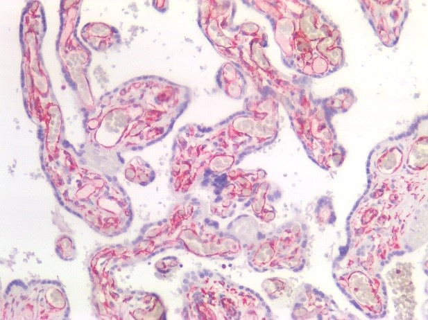
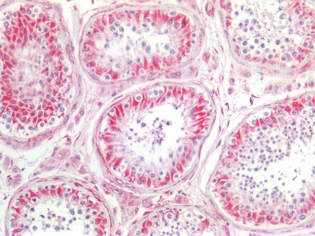
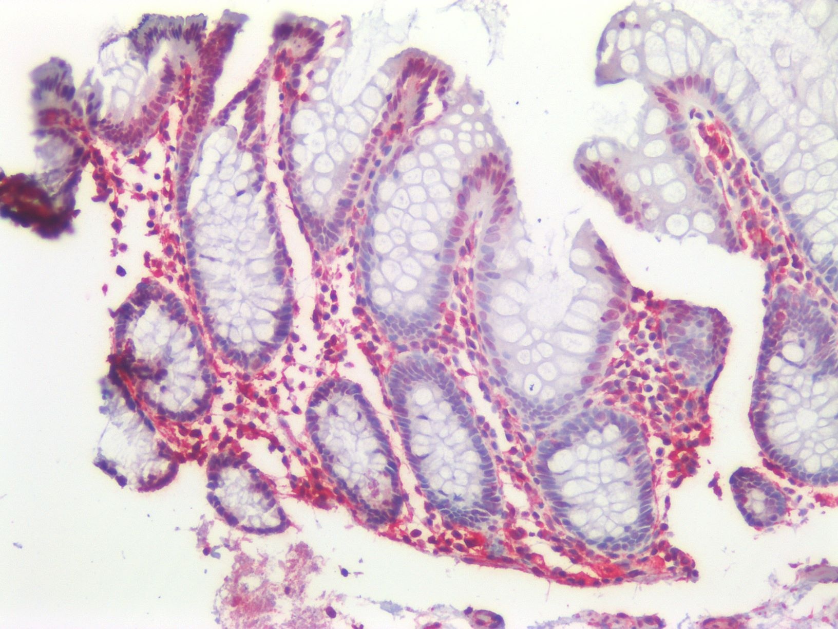
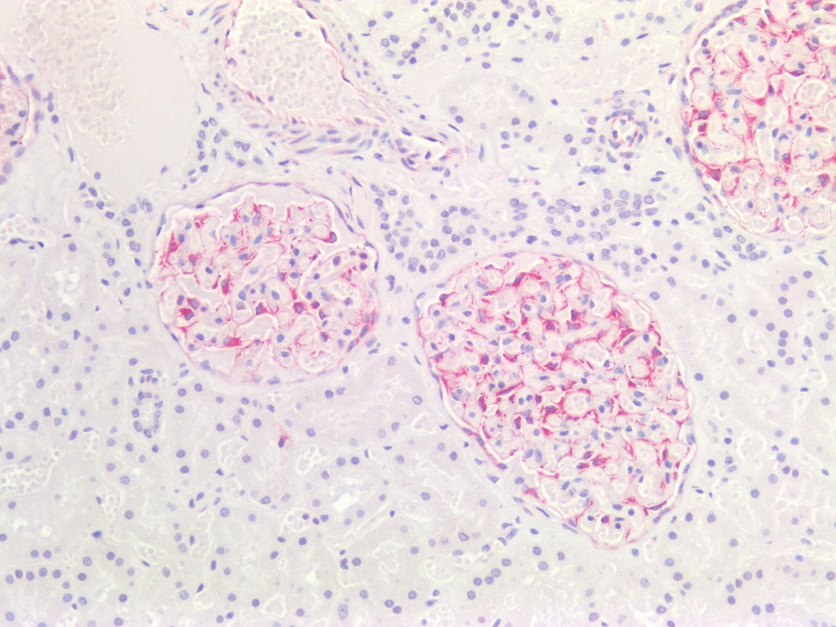


Quality Guarantee
Explore bioreagents carefree to elevate your research. All our products are rigorously tested for performance. If a product does not perform as described on its datasheet, our scientific support team will provide expert troubleshooting, a prompt replacement, or a refund. For full details, please see our Terms & Conditions and Buying Guide. Contact us at [email protected].

Immunoflourescence of HeLa cells labeled with anti-SAP49 (Cat. orb319660, red, 1:1000) and anti-vimentin (Cat. orb319707, green, 1:10,000). The blue is DAPI staining nuclear DNA.

Immunoflourescence of HeLa cells stained with Anti-Park7 (Cat. 1597-PARK, green, 1:1000) showing strong cytoplasmic staining and Anti-Vimentin (Cat. orb319707, red, 1:10,000) labeling cytoplasmic intermediate filaments. The blue is nuclear staining.

Immunoflourescence of mixed fibroblasts and PC12 cells labeled with anti-peripherin (Cat. orb319632, red, 1:500) and anti-vimentin (Cat. orb319707, green, 1:10,000). The blue is Hoechst staining nuclear DNA. Peripherin is labeled in only the PC12 cells cytoplasmic filaments and vimentin is only labeled in the fibroblasts intermediate filaments where peripherin is not present.

Immunofluorescence of HeLa cells showing strong granular nuclear staining of SAP49 (red, 1:1000) and vimentin (Cat. orb319707, green, 1:10,000). The blue is DAPI staining nuclear DNA.

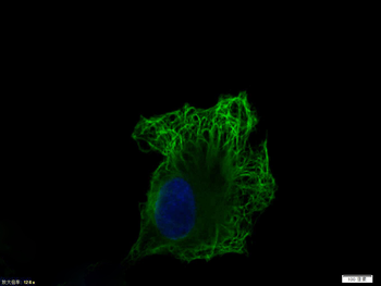
Immunolabeling of HeLa cells stained with anti-nuclear pore complex antibody (Cat. orb319613, red, 1:100) and rabbit anti-vimentin antibody (Cat. orb319707, green, 1:10,000). The blue is DAPI staining nuclear DNA. The anti-nuclear pore complex antibody reveals strong granular labeling of the nuclei corresponding to the protein. The anti-vimentin antibody specifically labels intermediate filaments.

Immunostaining of cultured neonatal rat neurons and glia stained with anti-nestin antibody (Cat. orb319583, red, 1:500) and anti-vimentin antibody (Cat. orb319707, green, 1:500). The blue is Hoechst staining for nuclear DNA. Astrocytes and neuronal stem cells stain strongly and specifically in a clearly filamentous fashion with the anti-Nestin antibody. The presence of Nestin indicates that the cells are developing astrocytes, neuroblasts or undifferentiated neural stem cells.

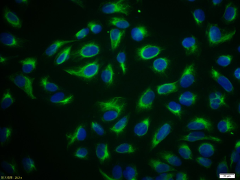
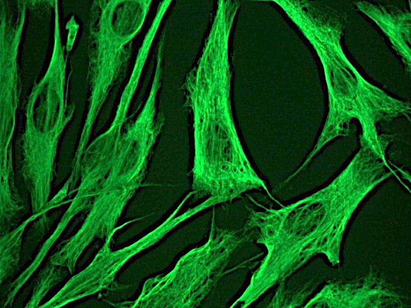
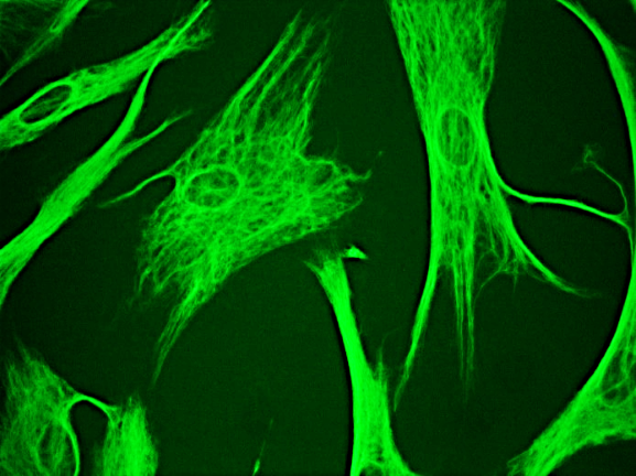
Immunostaining of HeLa cells labelled with anti-vimentin antibody (Cat. orb319707, 1:10,000, green), anti-actin (red), and DAPI (blue) labelling of nuclear DNA. The vimentin antibody labels the intermediate filament network while the actin antibody labels the submembranous cytoskeleton, stress fibers, and bundles of actin associated with cell adhesion sites.

Immunostaining of HeLa cells stained with anti-nuclear pore complex antibody (Cat. orb319613, green, 1:100) and rabbit anti-vimentin antibody (Cat. orb319707, red, 1:500).

Immunostaining of mixed neuron/glial cultures stained with anti-Vimentin antibody (Cat. orb319707, 1:500, green) and anti-GFAP antibody (Cat. orb319549, 1:1000, red). Vimentin is expressed alone in fibroblastic and endothelial cells, which are the flattened cells in the middle of the image which appear green. Astrocytes may express primarily GFAP, or GFAP and vimentin, and so appear red (GFAP only) or golden yellow (GFAP and Vimentin).

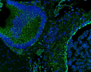
Immunostaining of tumors and precursor PanIN lesions from 4 week old LSL-KrasG12D; Cdkn2alox/lox; p48Cre (KIC) mice that were treated with 2G8 and/or gemcitabine, Gem. (Anti-Vimentin, cat. orb319707, green; Anti-E-cadherin, red).

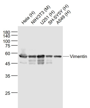
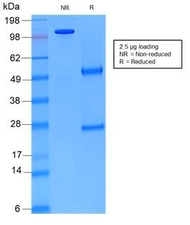
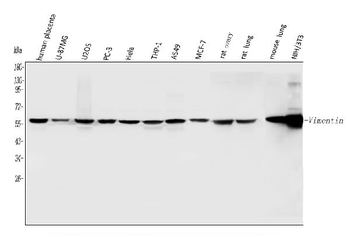
Western blot of NIH 3T3 cell lysate showing specific immunolabeling of the ~50 kDa Vimentin protein.
Quick Database Links
Gene Symbol
Vimentin
UniProt
UniProt Details
− No UniProt data available
Documents Download
Datasheet
Product Information
Request a Document
Protocol Information
WB
Western Blot (IB, immunoblot)
IHC
Immunohistochemistry
ICC
Immunocytochemistry
ELISA
Enzyme-linked Immunosorbent Assay (EIA)
Vimentin Antibody (orb319707)

- 0.0
Based on 0 reviews
Participating in our Biorbyt product reviews program enables you to support fellow scientists by sharing your firsthand experience with our products.
Login to Submit a ReviewAvailable Sizes
Select a size below
Choose Conjugation or Carrier Free Version
Free Secondary Antibody (20 ul)0/0
Please add an antibody product to your cart first.
 Usually dispatched within 5-10 working days