You have no items in your shopping cart.
Cart summary
Item 1 of 4
Item 1 of 4
MCP-1/MCAF Antibody
Catalog Number: orb1272402

| Catalog Number | orb1272402 |
|---|---|
| Category | Antibodies |
| Description | MCP-1/MCAF Antibody |
| Target | Ccl2 |
| Clonality | Polyclonal |
| Conjugation | Unconjugated |
| Reactivity | Rat |
| Form/Appearance | Lyophilized |
| Concentration | batch dependent |
| Purification | Anti-Rat MCP-1/MCAF specific antibody was purified by affinity chromatography employing immobilized Rat MCP-1/MCAF matrix. |
| Immunogen | Produced from sera of rabbits pre-immunized with highly pure (>98%) recombinant Rat MCP-1/MCAF (Macrophage/Monocyte chemotactic protein-1). |
| UniProt ID | P14844 |
| Tested applications | ELISA, NeA, WB |
| Antibody Type | Primary Antibody |
| Storage | Maintain refrigerated at 2-8°C for up to 2 weeks. For long term storage store at -20°C in small aliquots to prevent freeze-thaw cycles. |
| Alternative names | MCP-1, Scya2, Sigje, Je, Mcp1, C-C motif chemokine Read more... |
| Note | For research use only |
| NCBI | P14844 |
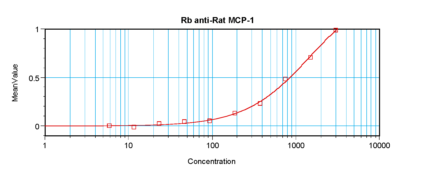
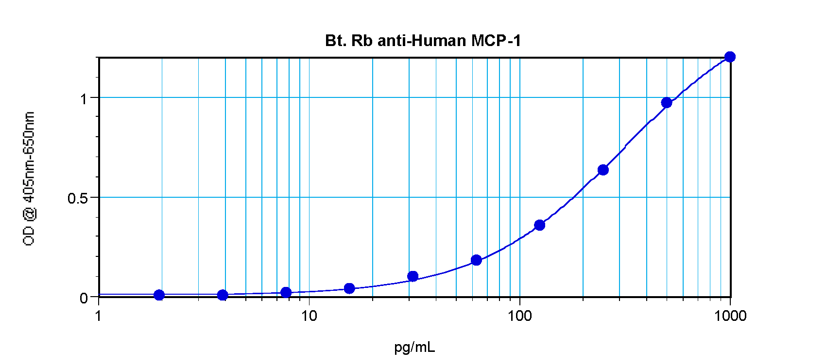
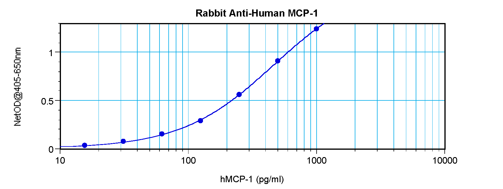
To detect Rat MCP-1 (MCAF) by sandwich ELISA (using 100 ul/well antibody solution) a concentration of 0.5 - 2.0 ug/ml of this antibody is required. This antigen affinity purified antibody, in conjunction with Anti-Rat MCP-1 (MCAF) (orb1272401) as a detection antibody, allows the detection of at least 0.2 - 0.4 ng/well of recombinant Rat MCP-1 (MCAF).

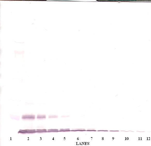
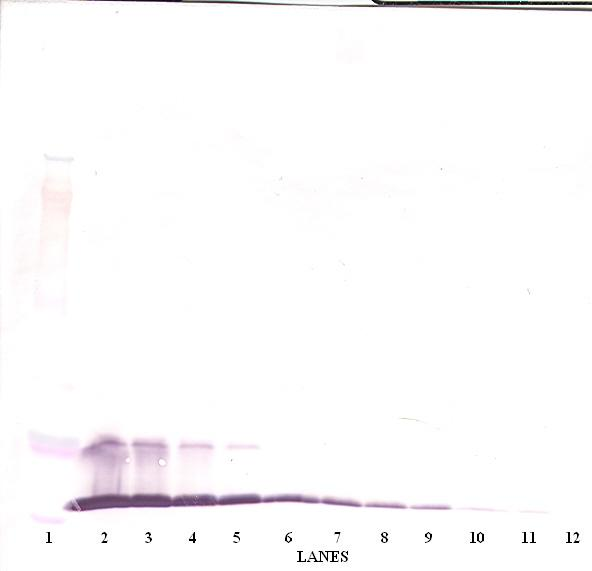
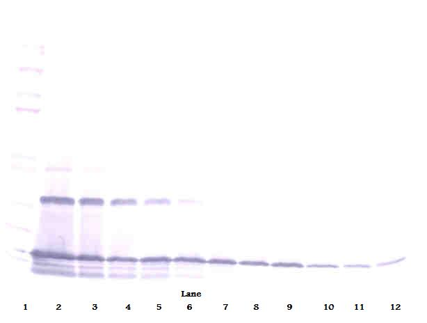
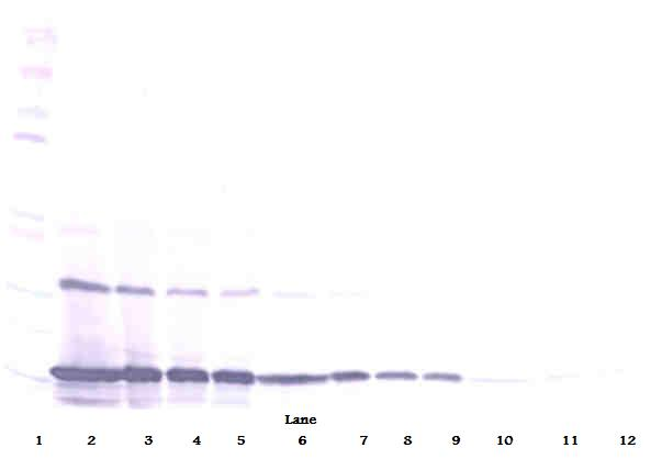
To detect Rat MCP-1 (MCAF) by Western Blot analysis this antibody can be used at a concentration of 0.1 - 0.2 ug/ml. Used in conjunction with compatible secondary reagents the detection limit for recombinant Rat MCP-1 (MCAF) is 1.5-3.0 ng/lane, under either reducing or non-reducing conditions.

To detect Rat MCP-1 (MCAF) by Western Blot analysis this antibody can be used at a concentration of 0.1 - 0.2 ug/ml. Used in conjunction with compatible secondary reagents the detection limit for recombinant Rat MCP-1 (MCAF) is 1.5-3.0 ng/lane, under either reducing or non-reducing conditions.
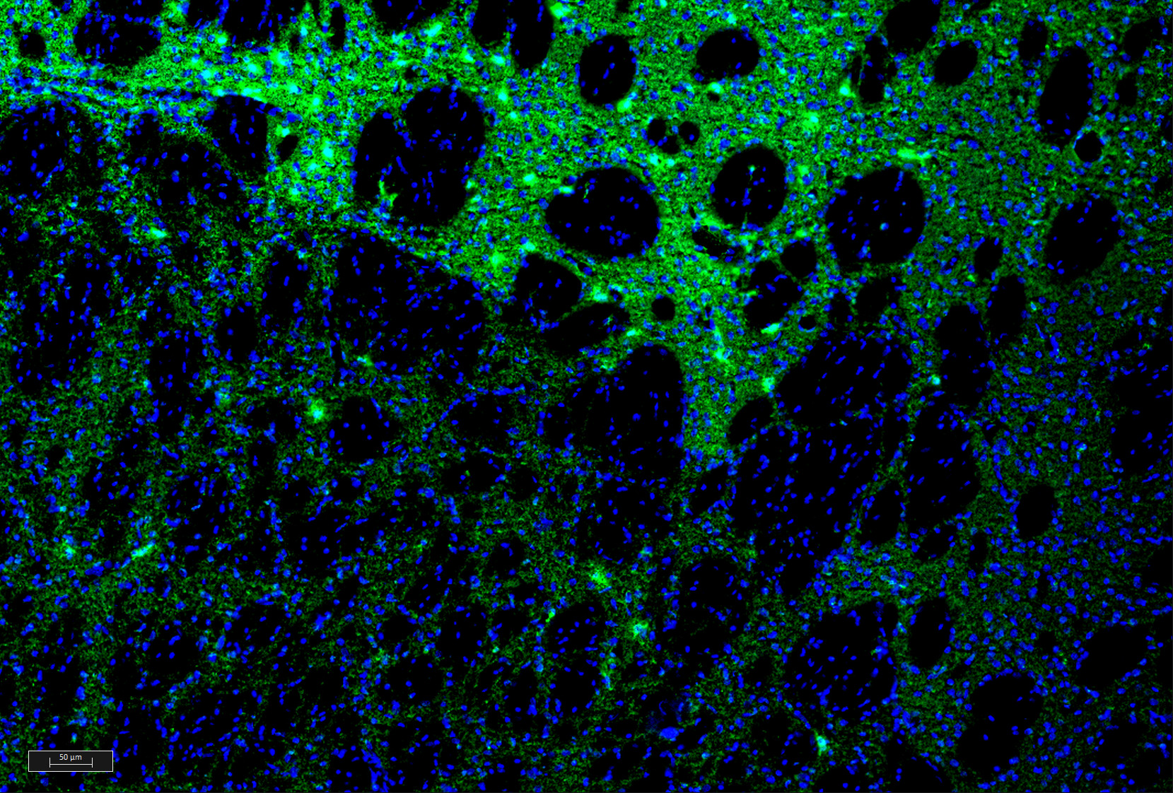
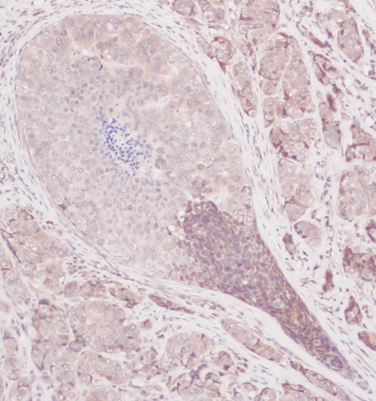
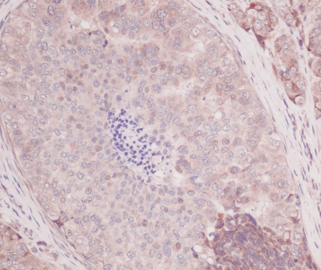
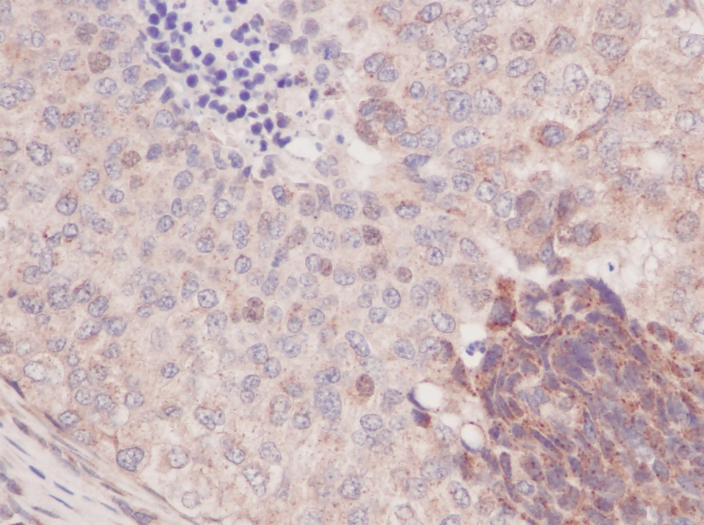
This antibody stained colchicine injected rat brain (including the caudate putamen) tissue. The primary antibody was incubated at 1.0 ug/ml overnight at 4°C. This was followed by a peroxidase conjugated secondary antibody and then a fluorescein Tyramide Signal Amplification (TSA) reagent. Optimal concentrations and conditions may vary.
- Item 1 of 6
- Item 1 of 3
- Item 1 of 2
- Item 1 of 1

![MCP-1 / MCAF antibody [PD-5]](/images//pub/media/catalog/product/NewWebsite/12/orb1973108_1.png)
![MCP-1 / MCAF antibody [PD-5]](/images/pub/media/catalog/product/NewWebsite/12/orb1973108_2.png)