You have no items in your shopping cart.
Cart summary
Item 1 of 4
Item 1 of 4
LCK Antibody
Catalog Number: orb1262980
| Catalog Number | orb1262980 |
|---|---|
| Category | Antibodies |
| Description | LCK Antibody |
| Species/Host | Rabbit |
| Clonality | Polyclonal |
| Tested applications | FC, IF, IHC-P, WB |
| Reactivity | Human, Mouse |
| Isotype | Rabbit Ig |
| Immunogen | This LCK antibody is generated from rabbits immunized with a KLH conjugated synthetic peptide between 23-52 amino acids from the N-terminal region of human LCK. |
| Concentration | batch dependent |
| Dilution range | For WB starting dilution is: 1:1000For IHC-P starting dilution is: 1:50~100For IF starting dilution is: 1:10~50For FACS starting dilution is: 1:10~50 |
| Form/Appearance | Liquid |
| Conjugation | Unconjugated |
| MW | 58 kDa |
| Target | LCK |
| UniProt ID | P06239 |
| NCBI | P06239 |
| Storage | Store at 4°C for three months and -20°C, stable for up to one year. As with all antibodies care should be taken to avoid repeated freeze thaw cycles. Antibodies should not be exposed to prolonged high temperatures. |
| Buffer/Preservatives | Supplied in PBS with 0.09% (W/V) sodium azide. |
| Alternative names | Tyrosine-protein kinase Lck, Leukocyte C-terminal Read more... |
| Note | For research use only |
| Application notes | For WB starting dilution is: 1:1000For IHC-P starting dilution is: 1:50~100For IF starting dilution is: 1:10~50For FACS starting dilution is: 1:10~50 |
| Expiration Date | 12 months from date of receipt. |
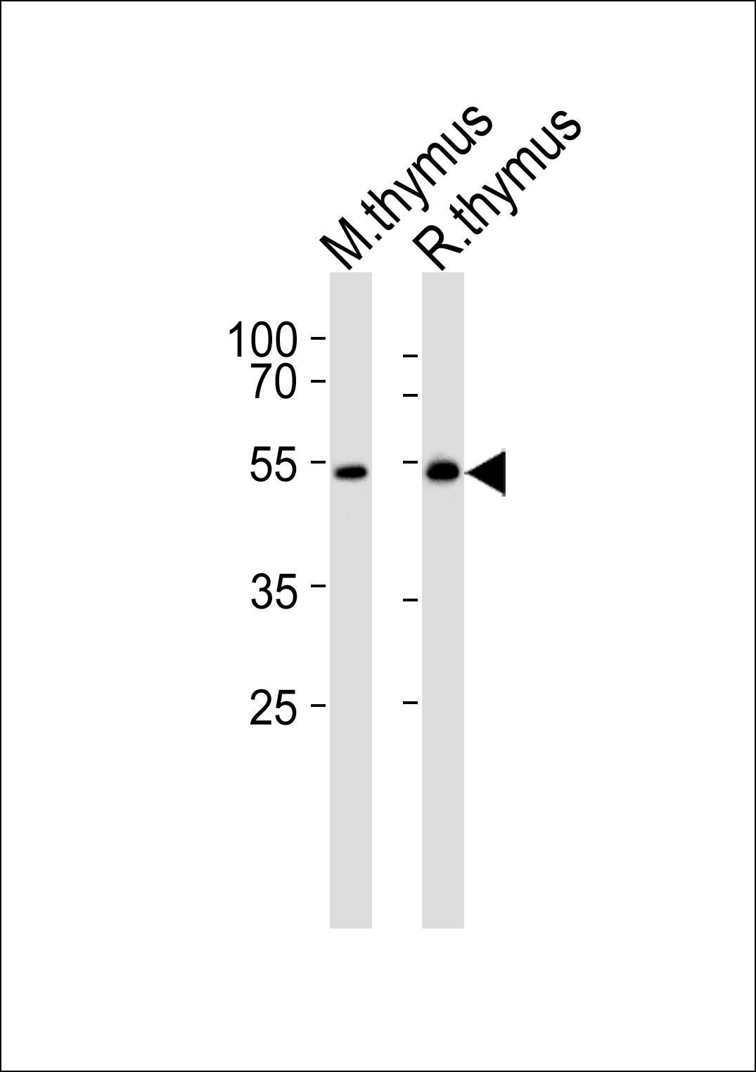
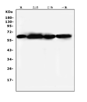
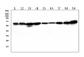
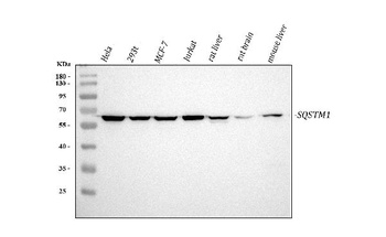
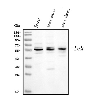
Western blot analysis of lysates from mouse thymus and rat thymus tissue lysate (from left to right), using LSK Antibody (I37) at 1:1000 at each lane.
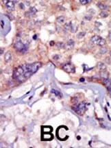
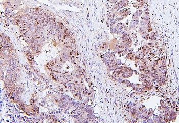
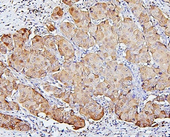
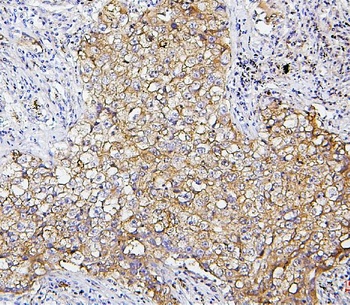
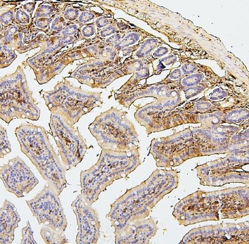
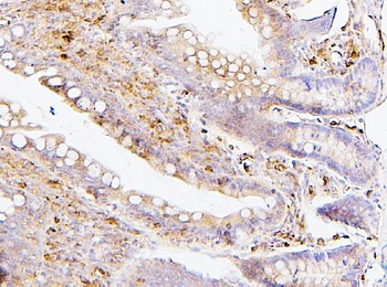
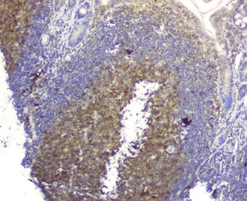
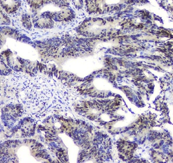
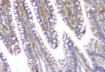
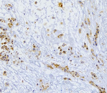
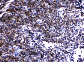
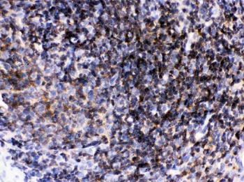
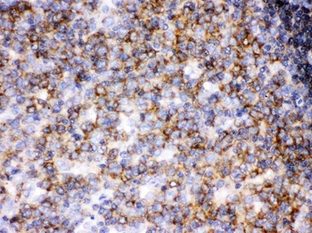
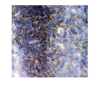
Formalin-fixed and paraffin-embedded human cancer tissue reacted with the primary antibody, which was peroxidase-conjugated to the secondary antibody, followed by AEC staining. BC = breast carcinoma; HC = hepatocarcinoma.
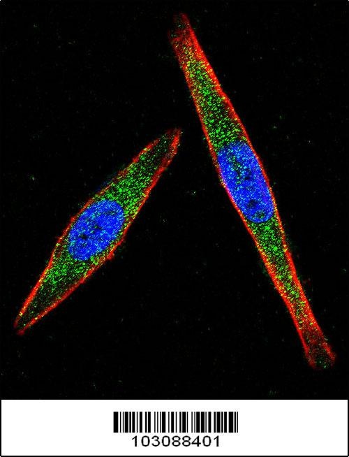
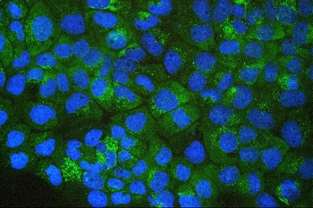
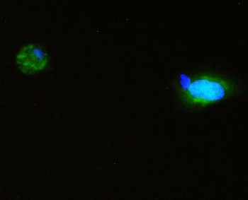
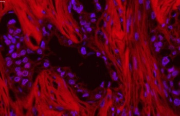
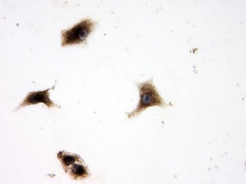
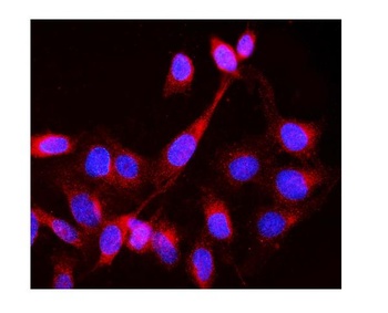
Confocal immunofluorescent analysis of LCK Antibody with A2058 cell followed by Alexa Fluor 488-conjugated goat anti-rabbit lgG (green).Actin filaments have been labeled with Alexa Fluor 555 phalloidin (red).DAPI was used to stain the cell nuclear (blue).
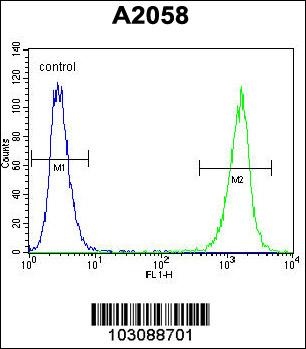
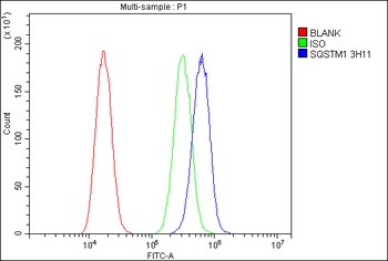
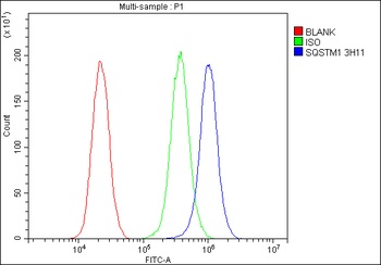
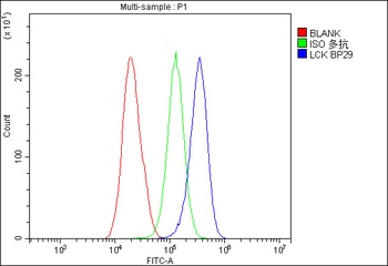
Flow cytometric analysis of A2058 cells (right histogram) compared to a negative control cell (left histogram). FITC-conjugated goat-anti-rabbit secondary antibodies were used for the analysis.
- Item 1 of 11
SQSTM1/p62 Antibody (monoclonal, 3H11) [orb570318]
FC, ICC, IF, IHC, WB
Human, Mouse, Rat
Mouse
Monoclonal
Unconjugated
10 μg, 100 μg - Item 1 of 8
SQSTM1/p62 Antibody [orb507551]
ICC, IF, IHC, WB
Hamster
Human, Mouse, Rat
Rabbit
Polyclonal
Unconjugated
10 μg, 100 μg - Item 1 of 7
Lck Antibody [orb334540]
FC, ICC, IF, IHC, IHC-Fr, WB
Human, Mouse, Rat
Rabbit
Polyclonal
Unconjugated
10 μg, 100 μg - Item 1 of 6
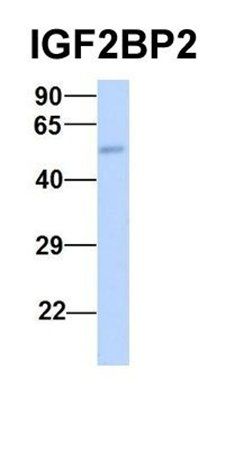
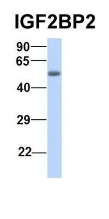
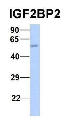
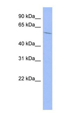
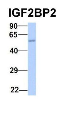
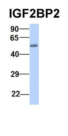
IGF2BP2 antibody [orb330141]
WB
Animal, Bovine, Canine, Guinea pig, Human, Mouse, Rabbit, Rat, Yeast
Canine, Equine, Guinea pig, Human, Mouse, Rat, Yeast
Rabbit
Polyclonal
Unconjugated
100 μl - Item 1 of 6
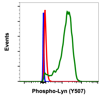
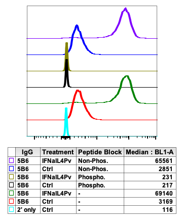
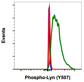
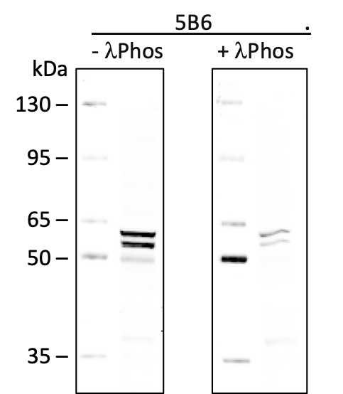
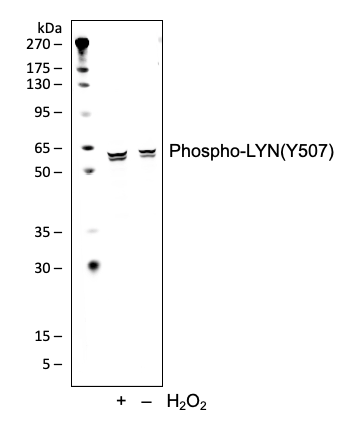
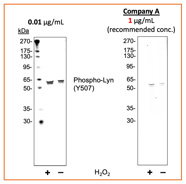
Phospho-Lyn (Tyr507) (5B6) rabbit mAb Antibody [orb1946031]
FC, WB
Human, Mouse
Monoclonal
Unconjugated
200 μl, 20 μl

Submit a review
Filter by Rating

- 5 stars

- 4 stars
- 3 stars
- 2 stars
- 1 stars