You have no items in your shopping cart.
Rabbit p16INK4a Recombinant Monoclonal Antibody
SKU: orb2281287
Description
Research Area
Cancer Biology, Immunology & Inflammation, Neuroscience
Images & Validation
−Item 1 of 4
| Tested Applications | FC, ICC, IHC, IP, WB |
|---|---|
| Dilution Range | WB-1:1,000 FC-Fixed in 4% formaldehyde and permeabilized with 90% methanol. 0.5 µl per 1 x 10^6 cells. IP-12 µl/mg lysate IHC-1:100 to 1:500 ICC-1:100 to 1:500. |
| Reactivity | Human |
Key Properties
−| Antibody Type | Primary Antibody |
|---|---|
| Host | Rabbit |
| Clonality | Recombinant |
| Isotype | IgG |
| Clone No. | BLR318M |
| Immunogen | Between 106 and C-terminus |
| Target | p16INK4a |
| Purification | Purified |
| Conjugation | Unconjugated |
Storage & Handling
−| Storage | Maintain refrigerated at 2-8°C for up to 2 weeks. For long term storage store at -20°C in small aliquots to prevent freeze-thaw cycles. |
|---|---|
| Form/Appearance | Liquid |
| Buffer/Preservatives | Phosphate Buffered Saline (PBS) with 0.1% rAlbumin and 0.09% Sodium Azide |
| Concentration | 500 µg/ml |
| Expiration Date | 12 months from date of receipt. |
| Disclaimer | For research use only |
Alternative Names
−alternative reading frame;ARF;CDK4 inhibitor p16-INK4;CDK4I;CDKN2;cell cycle negative regulator beta;CMM2;cyclin-dependent kinase 4 inhibitor A;cyclin-dependent kinase inhibitor 2A;cyclin-dependent kinase inhibitor 2A (melanoma, p16, inhibits CDK4);INK4;INK4A;MLM;MTS1;MTS-1;multiple tumor suppressor 1;P14;P14ARF;P16;P16INK4;p16-INK4;P16INK4A;P16-INK4A;P19;P19ARF;TP16
Similar Products
−- Item 1 of 3
CDKN2A/p14ARF Recombinant Rabbit Monoclonal Antibody [orb1152020]
ICC, WB
Human
Rabbit
Recombinant
Unconjugated
25 μl, 50 μl, 100 μl 
Rabbit p16INK4a Recombinant Monoclonal Antibody [orb2281286]
FC, ICC, IHC, IP, WB
Human
Rabbit
Recombinant
Unconjugated
100 μg (BSA-free)Rabbit p14ARF Recombinant Monoclonal Antibody [orb2302670]
FC, ICC, IHC, IP, WB
Human
Rabbit
Recombinant
Unconjugated
100 μl, 10 μlRabbit p14ARF Recombinant Monoclonal Antibody [orb2302669]
FC, ICC, IHC, IP, WB
Human
Rabbit
Recombinant
Unconjugated
100 μg (BSA-free)
Quality Guarantee
Explore bioreagents carefree to elevate your research. All our products are rigorously tested for performance. If a product does not perform as described on its datasheet, our scientific support team will provide expert troubleshooting, a prompt replacement, or a refund. For full details, please see our Terms & Conditions and Buying Guide. Contact us at [email protected].
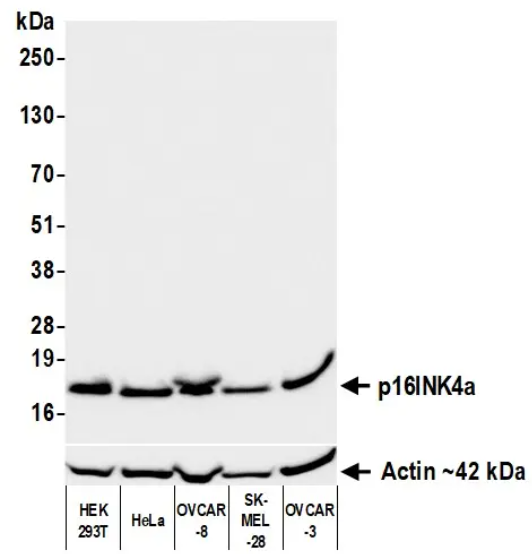
Detection of human p16INK4a by western blot. Samples: Whole cell lysate (50 µg) from HEK293T, HeLa, OVCAR-8, SK-MEL-28, and OVCAR-3 cells prepared using NETN lysis buffer. Antibody: Rabbit anti-p16INK4a recombinant monoclonal antibody used at 1:1000. Secondary: HRP-conjugated goat anti-rabbit IgG. Detection: Chemiluminescence with an exposure time of 3 seconds
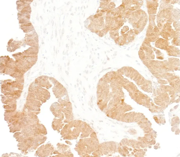
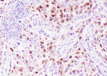
Detection of human p16INK4a by immunohistochemistry. Sample: FFPE section of ovarian carcinoma. Antibody: Rabbit anti-p16INK4a recombinant monoclonal antibody. Secondary: HRP-conjugated goat anti-rabbit IgG

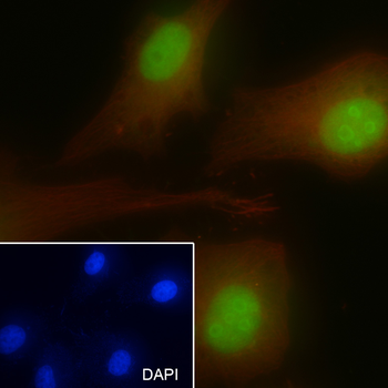
Detection of human p16INK4a by immunocytochemistry. Sample: FFPE section of HEK293T cells. Antibody: Rabbit anti-p16INK4a recombinant monoclonal antibody. Secondary: HRP-conjugated goat anti-rabbit IgG
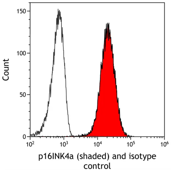
Detection of human p16INK4a (shaded) in HeLa cells by flow cytometry. Antibody: Rabbit anti-p16INK4a recombinant monoclonal antibody or isotype control (unshaded). Secondary: DyLight® 650-conjugated goat anti-rabbit IgG
Quick Database Links
UniProt Details
− No UniProt data available
NCBI Reference Sequences
−Associated Accession Numbers
Curated reference sequences for the gene transcript and protein product| Protein | NP_478104.2 |
|---|
Documents Download
Datasheet
Product Information
Request a Document
Protocol Information
WB
Western Blot (IB, immunoblot)
IHC
Immunohistochemistry
FC
Flow Cytometry
ICC
Immunocytochemistry
IP
Immunoprecipitation
Rabbit p16INK4a Recombinant Monoclonal Antibody (orb2281287)

- 0.0
Based on 0 reviews
Participating in our Biorbyt product reviews program enables you to support fellow scientists by sharing your firsthand experience with our products.
Login to Submit a ReviewAvailable Sizes
Select a size below