You have no items in your shopping cart.
Featured
Description
Research Area
Immunology & Inflammation
Images & Validation
−Item 1 of 3
| Tested Applications | FC, IF, IHC, IP, WB |
|---|---|
| Dilution Range | Flow-Cyt Live or cells fixed in 4% formaldehyde and permeabilized with 90% methanol. 1 µl per 1 x 10^6 cells. Immunohistochemistry (IHC) 1:100 - 1:500. Epitope retrieval with Tris-EDTA pH 9.0 is recommended for FFPE tissue sections. Immunoprecipitation (IP) 6 µl/mg lysate. Multiplex Immunofluorescence (mIF) 1:250. Western Blot (WB) 1:1000 |
| Reactivity | Human |
| Application Notes |
Key Properties
−| Antibody Type | Primary Antibody |
|---|---|
| Host | Rabbit |
| Clonality | Recombinant |
| Isotype | IgG |
| Clone No. | BL-155-1C11 |
| Immunogen | Residues 26-397 (ECD) |
| Target | CD4 |
| Purity | Recombinant antibody was purified from cell culture supernatant |
| Purification | Purified |
| Conjugation | Unconjugated |
Storage & Handling
−| Storage | 2 - 8°C |
|---|---|
| Form/Appearance | Liquid |
| Buffer/Preservatives | Borate Buffered Saline (BBS) pH 8.2 with 0.09% Sodium Azide, rAlbumin-Free |
| Concentration | 1000 µg/ml |
| Expiration Date | 12 months from date of receipt. |
| Disclaimer | For research use only |
Alternative Names
−CD antigen CD4; CD4 antigen (p55); CD4 receptor; CD4mut; T-cell surface antigen T4/Leu-3; T-cell surface glycoprotein CD4
Similar Products
−- Item 1 of 14
CD4 Recombinant Rabbit Monoclonal Antibody [orb612230]
FC, IF, IHC-Fr, IHC-P, WB
Human
Rabbit
Recombinant
Unconjugated
50 μl, 100 μl, 25 μl - Item 1 of 4
Rabbit CD4 Recombinant Monoclonal Antibody [orb1519574]
IHC, IP, WB
Mouse
Rabbit
Recombinant
Unconjugated
100 μl, 10 μl - Item 1 of 2
Recombinant CD8 Antibody (alpha chain) / Rabbit Monoclonal [orb386200]
IHC-P
Human
Rabbit
Recombinant
Unconjugated
100 μg, 20 μg - Item 1 of 2
Recombinant CD8 Antibody (alpha chain) / Rabbit Monoclonal [orb2640339]
IHC-P
Human
Rabbit
Recombinant
Unconjugated
7 ml - Item 1 of 2
Recombinant CD8 Antibody (alpha chain) / Rabbit Monoclonal [orb2640340]
IHC-P
Human
Rabbit
Recombinant
Unconjugated
100 μg


Quality Guarantee
Explore bioreagents carefree to elevate your research. All our products are rigorously tested for performance. If a product does not perform as described on its datasheet, our scientific support team will provide expert troubleshooting, a prompt replacement, or a refund. For full details, please see our Terms & Conditions and Buying Guide. Contact us at [email protected].
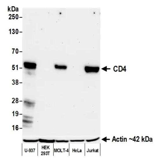
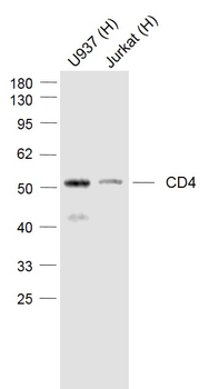
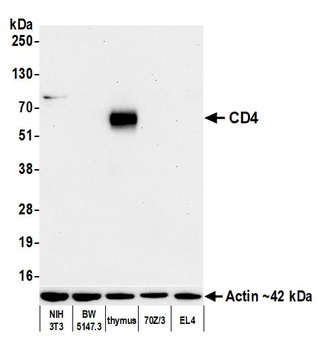
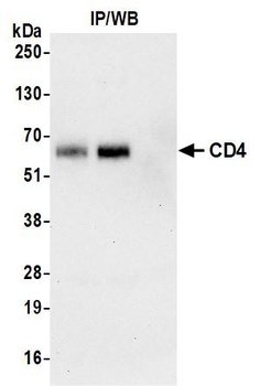
Detection of human CD4 by western blot. Samples: Whole cell lysate (50 µg) from U-937, HEK293T, MOLT-4, HeLa, and Jurkat cells prepared using NETN lysis buffer. Antibody: Rabbit anti-CD4 recombinant monoclonal antibody [BL-155-1C11] used at 1:1000. Secondary: HRP-conjugated goat anti-rabbit IgG. Detection: Chemiluminescence with an exposure time of 75 seconds. Lower Panel: Rabbit anti-Actin recombinant monoclonal antibody
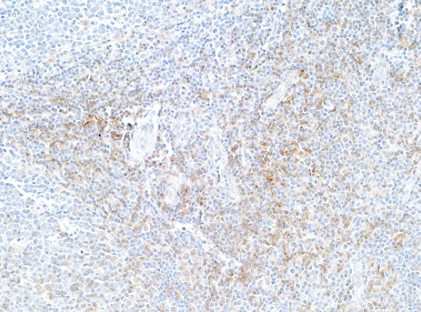
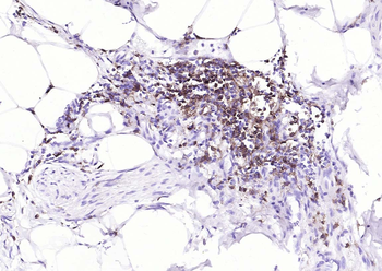
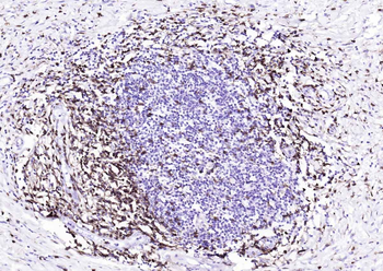
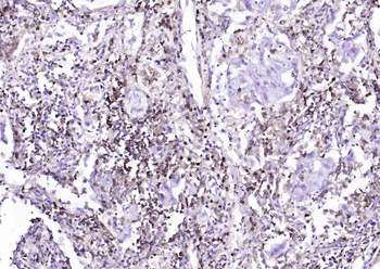
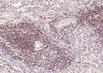
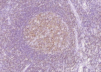
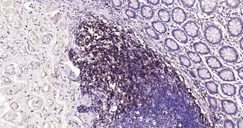
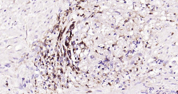
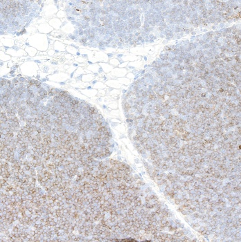
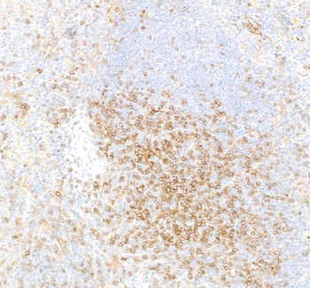
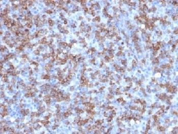
Detection of human CD4 by immunohistochemistry. Sample: FFPE section of tonsil. Antibody: Rabbit anti-CD4 recombinant monoclonal antibody. Secondary: HRP-conjugated goat anti-rabbit IgG
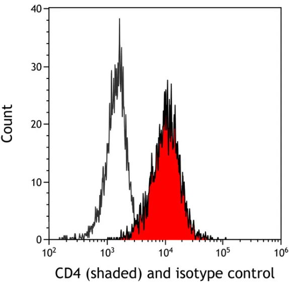
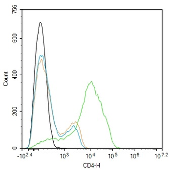
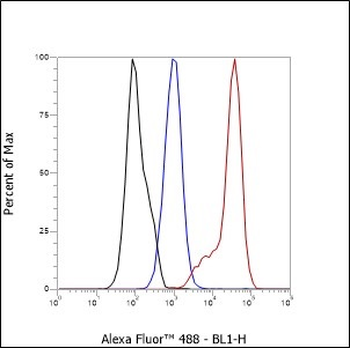
Detection of human CD4 (shaded) in THP-1 cells by flow cytometry. Antibody: Rabbit anti-CD4 recombinant monoclonal [BL-155-1C11] or isotype control (unshaded). Secondary: DyLight® 488-conjugated goat anti-rabbit IgG
Quick Database Links
UniProt Details
− No UniProt data available
NCBI Reference Sequences
−Associated Accession Numbers
Curated reference sequences for the gene transcript and protein product| Protein | NP_000607.1 |
|---|
Documents Download
Datasheet
Product Information
Request a Document
Protocol Information
WB
Western Blot (IB, immunoblot)
IHC
Immunohistochemistry
FC
Flow Cytometry
IF
Immunofluorescence
IP
Immunoprecipitation
CD4 Rabbit Recombinant Monoclonal Antibody (orb1519966)

- 0.0
Based on 0 reviews
Participating in our Biorbyt product reviews program enables you to support fellow scientists by sharing your firsthand experience with our products.
Login to Submit a ReviewAvailable Sizes
Select a size below
Choose Conjugation or Carrier Free Version
Free Secondary Antibody (20 ul)0/0
Please add an antibody product to your cart first.