You have no items in your shopping cart.
Description
Research Area
Cancer Biology, Cell Biology, Protein Biochemistry, Signal Transduction
Images & Validation
−Item 1 of 6
| Tested Applications | FACS, FC, ICC, IF, WB |
|---|---|
| Dilution Range | WB (1:1000), ICC/IF (1:100), FACS (1:250) |
| Reactivity | Bovine, Human, Mouse, Porcine, Rat |
| Application Notes |
Key Properties
−| Host | Mouse |
|---|---|
| Clonality | Monoclonal |
| Isotype | IgG1 |
| Clone No. | 1H11 |
| Immunogen | Human native HSP70 protein |
| Target | HSP70 |
| Purification | Protein G Purified |
| Conjugation | Biotin |
Storage & Handling
−| Storage | Conjugated antibodies should be stored according to the product label |
|---|---|
| Buffer/Preservatives | 136.36mM Ethanolamine, 133.23 mM Chlorides, 9.55mM Phosphates, 9.55mM Sodium Bicarbonate |
| Concentration | 1 mg/ml |
| Expiration Date | 12 months from date of receipt. |
| Disclaimer | For research use only |
Alternative Names
−HSPA1A, HSPA1B, HSPA1, HSP70, HSP70-1, HSP70.1, HSP70-2, HSP72, HSP73, HSX70, Heat shock 70 kDa protein 1A, Heat shock 70 kDa protein 1B
Similar Products
−- Item 1 of 7
HSP70 Antibody (Biotin) [orb151241]
ELISA, FC, ICC, IF, IHC, IP, WB
Bovine, Canine, Fish, Guinea pig, Hamster, Human, Mammal, Monkey, Mouse, Other, Plant, Porcine, Rat, Sheep
Rabbit
Polyclonal
Biotin
100 μg - Item 1 of 6
HSP70/HSC70 Antibody (Biotin) [orb146786]
ICC, IF, IHC, IP, WB
Bovine, Canine, Drosophila, Fish, Frog, Gallus, Guinea pig, Hamster, Human, Mouse, Other, Porcine, Rabbit, Rat, Sheep, Yeast
Mouse
Monoclonal
Biotin
200 μg - Item 1 of 1
Zebrafish HeatShock Protein 70 (HSP-70/HSPA9) ELISA Kit [orb1088148]
Zebrafish
3.13-200 ng/mL
1.33 ng/mL
48 T, 96 T - Item 1 of 1
Goat HeatShock Protein 70 (HSP-70/HSPA9) ELISA Kit [orb1146821]
Goat
3.13-200 ng/mL
1.35 ng/mL
48 T, 96 T - Item 1 of 1
Horse HeatShock Protein 70 (HSP-70/HSPA9) ELISA Kit [orb1146943]
Equine
1.57-100 ng/mL
0.53 ng/mL
48 T, 96 T
Quality Guarantee
Explore bioreagents carefree to elevate your research. All our products are rigorously tested for performance. If a product does not perform as described on its datasheet, our scientific support team will provide expert troubleshooting, a prompt replacement, or a refund. For full details, please see our Terms & Conditions and Buying Guide. Contact us at [email protected].
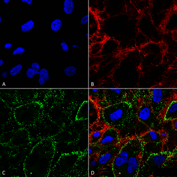
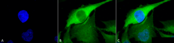
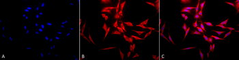
Immunocytochemistry/Immunofluorescence analysis using Mouse Anti-HSP70 Monoclonal Antibody, Clone 1H11. Tissue: HCT116 cells. Species: Human. Fixation: 4% Formaldehyde. Primary Antibody: Mouse Anti-HSP70 Monoclonal Antibody at 1:100. Counterstain: Wheat germ agglutinin Texas red membrane marker; DAPI (blue) nuclear stain. Localization: Cell surface, cell membrane. (A) DAPI nuclear stain. (B) Wheat germ agglutinin Texas red. (C) HSP70 Antibody. (D) Composite.
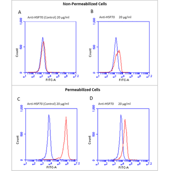
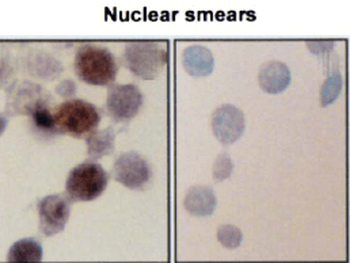
Fluorescence-activated cell sorting analysis using Mouse Anti-HSP70 Monoclonal Antibody, Clone 1H11. Tissue: Jurkat E6.1 cells. Species: Human. Fixation: No fixation. Primary Antibody: Mouse Anti-HSP70 Monoclonal Antibody at 20 μg/ml for 40 min at 4°C. Counterstain: Propidium Iodide nuclear stain at 2.5 μg/ml for 5 min at RT. Isotype Control: Anti-mouse FITC at 1:32 for 15 min at RT (blue line).
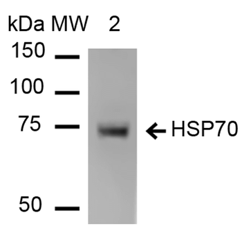
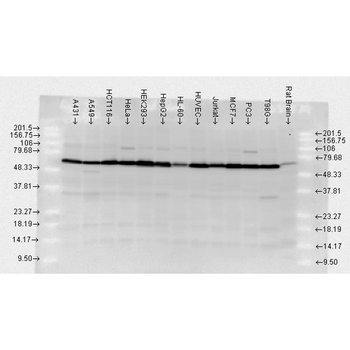
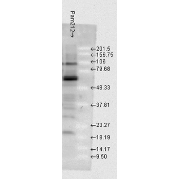
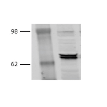
Western Blot analysis of Human Heat Shocked cervical cancer cell line (HeLa) lysate showing detection of HSP70 protein using Mouse Anti-HSP70 Monoclonal Antibody, Clone 1H11. Lane 1: Molecular Weight ladder (MW). Lane 2: HeLa cell lysates. Load: 20 μg. Primary Antibody: Mouse Anti-HSP70 Monoclonal Antibody at 1:1000.
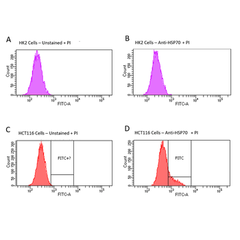
Fluorescence-activated cell sorting analysis using Mouse Anti-HSP70 Monoclonal Antibody, Clone 1H11. Tissue: HCT116 and HK2 cells. Species: Human. Primary Antibody: Mouse Anti-HSP70 Monoclonal Antibody at 1:100 for 90 min at 4°C. Counterstain: Propidium Iodide nuclear stain. (A) HK2 cells unstained. (B) HK2 cells and HSP70 Antibody. (C) HCT116 cells unstained. (D) HCT116 cells and HSP70 Antibody. Shows that HSP70 Antibody binds to the cell surface of tumor cells but not non-tumor cells.
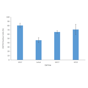
Fluorescence-activated cell sorting analysis using Mouse Anti-HSP70 Monoclonal Antibody, Clone 1H11. Tissue: Jurkat E6.1, U937, MCF7 and HT29 cells. Species: Human. Fixation: No fixation. Primary Antibody: Mouse Anti-HSP70 Monoclonal Antibody at 20 μg/ml for 40 min at 4°C. Counterstain: Propidium Iodide nuclear stain at 2.5 μg/ml for 5 min at 4°C. Shows that binding of HSP70 Antibody is not cell-line specific.

Immunocytochemistry/Immunofluorescence analysis using Mouse Anti-HSP70 Monoclonal Antibody, Clone 1H11. Tissue: HCT116 cells. Species: Human. Fixation: 4% Formaldehyde. Primary Antibody: Mouse Anti-HSP70 Monoclonal Antibody at 1:100. Counterstain: Wheat germ agglutinin Texas red membrane marker; DAPI (blue) nuclear stain. Localization: Cell surface, faint intracellular. (A) DAPI nuclear stain. (B) Wheat germ agglutinin Texas red. (C) HSP70 Antibody. (D) Composite.
Quick Database Links
UniProt Details
− No UniProt data available
NCBI Gene Details
− No NCBI Gene data available
NCBI Reference Sequences
−Associated Accession Numbers
Curated reference sequences for the gene transcript and protein product| Protein | NP_005336.3 |
|---|
Documents Download
Datasheet
Product Information
Request a Document
Protocol Information
WB
Western Blot (IB, immunoblot)
FACS
Fluorescence-Activated Cell Sorting (FC, Flow cytometry)
FC
Flow Cytometry
IF
Immunofluorescence
ICC
Immunocytochemistry
HSP70 Antibody (Biotin) (orb54778)

- 0.0
Based on 0 reviews
Participating in our Biorbyt product reviews program enables you to support fellow scientists by sharing your firsthand experience with our products.
Login to Submit a ReviewAvailable Sizes
Select a size below
Choose Conjugation or Carrier Free Version
Free Secondary Antibody (20 ul)0/0
Please add an antibody product to your cart first.