You have no items in your shopping cart.
Cytokeratin 1 Recombinant Rabbit Monoclonal Antibody
SKU: orb559083
Featured
Description
Research Area
Signal Transduction
Images & Validation
−Item 1 of 4
| Tested Applications | ICC, IF, IHC-Fr, IHC-P, WB |
|---|---|
| Dilution Range | WB=1:500-2000, IHC-P=1:100-500, IHC-F=1:100-500, ICC/IF=1:50-200, IF=1:100-500 |
| Reactivity | Human, Mouse, Rat |
| Predicted Reactivity | Mouse, Rat |
Related Conjugates & Formulations
−Key Properties
−| Antibody Type | Primary Antibody |
|---|---|
| Host | Rabbit |
| Clonality | Recombinant |
| Isotype | IgG |
| Clone No. | 1C2 |
| Immunogen | A synthesized peptide derived from human Cytokeratin 1 (600-644aa) |
| Target | KRT1 |
| Source/Expression System | 293F |
| Molecular Weight | Calculated M.W.: 70 kDa. Observed M.W.: 65 kDa. |
| Purification | Affinity purified by Protein A |
| Conjugation | Unconjugated |
Storage & Handling
−| Storage | Maintain refrigerated at 2-8°C for up to 2 weeks. For long term storage store at -20°C in small aliquots to prevent freeze-thaw cycles. |
|---|---|
| Form/Appearance | Liquid |
| Buffer/Preservatives | 0.01M TBS (pH7.4) with 1% rAlbumin, 0.02% Proclin300 and 50% Glycerol. |
| Concentration | 1mg/ml |
| Expiration Date | 12 months from date of receipt. |
| Disclaimer | For research use only |
Alternative Names
−AEI2; CK1; EHK; EHK1; EPPK; K1; KRT1A; NEPPK; Krt-2.1; Krt2-1; Krt86; Kb1; K2C1_HUMAN; KRT1; 67 kDa cytokeratin; Cytokeratin-1 (CK-1); Hair alpha protein; Keratin-1 (K1); Type-II keratin Kb1; KRTA; K2C1_MOUSE; K2C1_RAT;
Similar Products
−- Item 1 of 6
Cytokeratin 5 Recombinant Rabbit Monoclonal Antibody [orb559074]
ICC, IF, IHC-Fr, IHC-P, WB
Rat
Human, Mouse, Rat
Rabbit
Recombinant
Unconjugated
100 μl, 50 μl - Item 1 of 3
Recombinant Cytokeratin 15 Antibody / Rabbit Monoclonal [orb2640688]
IHC-P
Human
Rabbit
Recombinant
Unconjugated
7 ml - Item 1 of 3
Recombinant Cytokeratin 15 Antibody / Rabbit Monoclonal [orb2640689]
FACS, IHC-P
Human
Rabbit
Recombinant
Unconjugated
100 μg - Item 1 of 3
Recombinant Cytokeratin 15 Antibody / Rabbit Monoclonal [orb606440]
FACS, IHC-P
Human
Rabbit
Recombinant
Unconjugated
100 μg, 20 μg - Item 1 of 2
Recombinant Keratin 16 Antibody / Rabbit Monoclonal [orb2640717]
IHC-P
Human
Rabbit
Recombinant
Unconjugated
7 ml


Quality Guarantee
Explore bioreagents carefree to elevate your research. All our products are rigorously tested for performance. If a product does not perform as described on its datasheet, our scientific support team will provide expert troubleshooting, a prompt replacement, or a refund. For full details, please see our Terms & Conditions and Buying Guide. Contact us at [email protected].
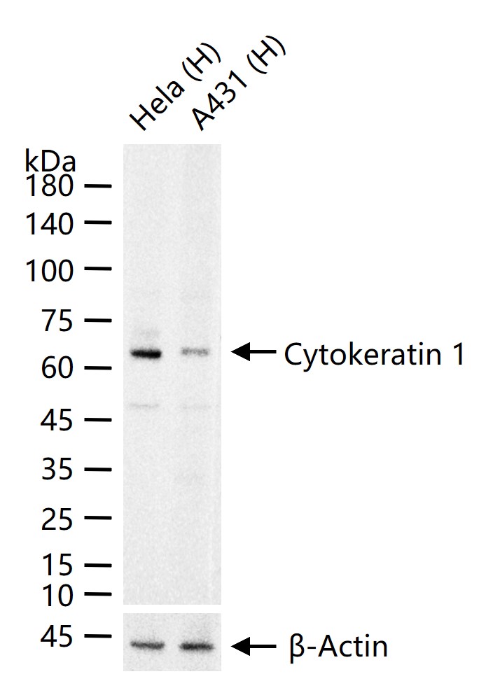
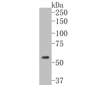
25 ug total protein per lane of various lysates probed with Cytokeratin 1 Recombinant Rabbit Monoclonal Antibody (orb559083) at 1:1000 dilution and 4°C overnight incubation. Followed by conjugated secondary antibody incubation at r.t. for 60 min.
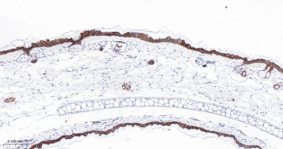
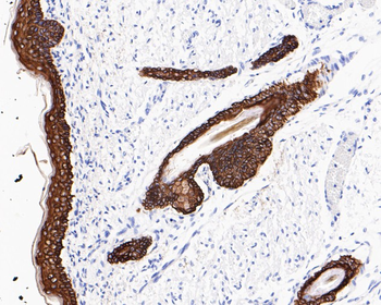
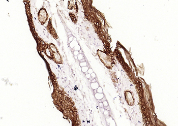
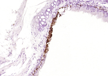
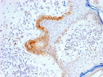
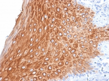
Paraformaldehyde-fixed, paraffin embedded Mouse Skin; Antigen retrieval by boiling in sodium citrate buffer (pH6.0) for 15 min; Antibody incubation with Cytokeratin 1 Recombinant Rabbit Monoclonal Antibody (orb559083) at 1:200 overnight at 4°C, followed by conjugation to the Goat Anti-Rabbit IgG H&L, HRP conjugated (orb572747) and DAB staining.
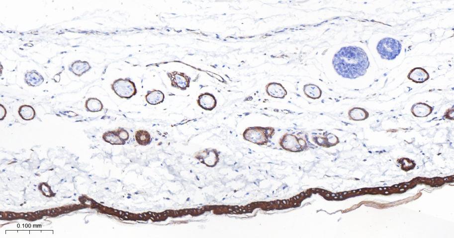
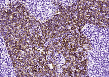
Paraformaldehyde-fixed, paraffin embedded Rat Skin; Antigen retrieval by boiling in sodium citrate buffer (pH6.0) for 15 min; Cytokeratin 1 Recombinant Rabbit Monoclonal Antibody (orb559083) at 1:200 overnight at 4°C, followed by conjugation to the Goat Anti-Rabbit IgG H&L, HRP conjugated (orb572747) and DAB staining.
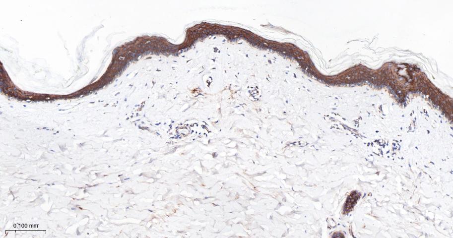
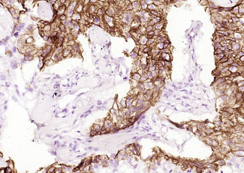
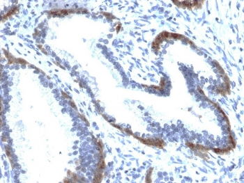
Paraformaldehyde-fixed, paraffin embedded Human Skin; Antigen retrieval by boiling in sodium citrate buffer (pH6.0) for 15 min; Cytokeratin 1 Recombinant Rabbit Monoclonal Antibody (orb559083) at 1:200 overnight at 4°C, followed by conjugation to the Goat Anti-Rabbit IgG H&L, HRP conjugated (orb572747) and DAB staining.
Quick Database Links
Gene Symbol
KRT1
UniProt
UniProt Details
− No UniProt data available
Documents Download
Datasheet
Product Information
Request a Document
Protocol Information
WB
Western Blot (IB, immunoblot)
IHC-P
Immunohistochemistry Paraffin
IHC-Fr
Immunohistochemistry Frozen
IF
Immunofluorescence
ICC
Immunocytochemistry
Cytokeratin 1 Recombinant Rabbit Monoclonal Antibody (orb559083)

- 0.0
Based on 0 reviews
Participating in our Biorbyt product reviews program enables you to support fellow scientists by sharing your firsthand experience with our products.
Login to Submit a ReviewAvailable Sizes
Select a size below