You have no items in your shopping cart.
AURORA KINASE B phospho T232 Antibody
SKU: orb345465
Description
Images & Validation
−Item 1 of 5
| Tested Applications | ELISA, IHC, WB |
|---|---|
| Dilution Range | ELISA: 1:10,000 - 1:30,000, WB: 1:250 - 1:2,000 |
| Reactivity | Human, Monkey |
| Application Notes |
Key Properties
−| Antibody Type | Primary Antibody |
|---|---|
| Host | Rabbit |
| Clonality | Polyclonal |
| Isotype | IgG |
| Immunogen | This affinity purified antibody was prepared from whole rabbit serum produced by repeated immunizations with a synthetic peptide corresponding to an internal region surrounding T232 of Human Aurora Kinase B protein. |
| Purity | Anti-Phospho Aurora B pT232 affinity purified antibody is directed against the phosphorylated form of human Aurora Kinase B at the pT232 residue. The product was affinity purified from monospecific antiserum by immunoaffinity purification. Antiserum was first purified against the phosphorylated form of the immunizing peptide. The resultant affinity purified antibody was then cross-adsorbed against the non-phosphorylated form of the immunizing peptide. Reactivity occurs against human Aurora Kinase B pT232 protein and the antibody is specific for the phosphorylated form of the protein. Reactivity with non-phosphorylated human Aurora Kinase B is minimal by ELISA. No reaction is expected against Aurora Kinase A. However, 100% sequence homology as indicated by BLAST analysis is on record for this protein from human, mouse, rat, cow, pig, dog and chimpanzee. Cross reactivity with Aurora Kinase B from other sources is not known. |
| Conjugation | Unconjugated |
Storage & Handling
−| Storage | Store Phospho Specific Antibody at -20° C prior to opening. Aliquot contents and freeze at -20° C or below for extended storage. Avoid cycles of freezing and thawing. Centrifuge product if not completely clear after standing at room temperature. This product is stable for several weeks at 4° C as an undiluted liquid. Dilute only prior to immediate use. |
|---|---|
| Form/Appearance | Liquid (sterile filtered) |
| Buffer/Preservatives | Preservative: 0.01% (w/v) Sodium Azide. Stabilizer: None; Buffer: 0.02 M Potassium Phosphate, 0.15 M Sodium Chloride, pH 7.2 |
| Concentration | 0.87 mg/mL |
| Expiration Date | 12 months from date of receipt. |
| Dry Ice Shipping | Please note: This product requires shipment on dry ice. A dry ice surcharge will apply. |
| Disclaimer | For research use only |
Alternative Names
−rabbit anti-Aurora B pT232 Antibody, Phospho Aurora B, AIK2 antibody, AIM1 antibody, ARK2 antibody, AurB antibody, AURKB antibody, Aurora 1 antibody, Aurora and Ipl1 like midbody associated protein 1 antibody
Similar Products
−- Item 1 of 5
AURORA KINASE B phospho T232 Antibody [orb345466]
ELISA, IHC, WB
Human, Monkey
Rabbit
Polyclonal
Unconjugated
25 μl - Item 1 of 1
Phospho-Aurora B (Thr232) + Aurora C (Thr198) Rabbit Polyclonal Antibody [orb6923]
WB
Bovine, Canine, Equine, Mouse, Porcine, Rabbit, Rat, Sheep
Human
Rabbit
Polyclonal
Unconjugated
50 μl, 100 μl, 200 μl 
Phospho-Aurora B (Thr232) + Aurora C (Thr198) Rabbit Polyclonal Antibody (Biotin) [orb502115]
WB
Bovine, Canine, Equine, Mouse, Porcine, Rabbit, Rat, Sheep
Human
Rabbit
Polyclonal
Biotin
100 μlPhospho-Aurora B (Thr232) + Aurora C (Thr198) Rabbit Polyclonal Antibody (HRP) [orb504039]
WB
Bovine, Canine, Equine, Mouse, Porcine, Rabbit, Rat, Sheep
Human
Rabbit
Polyclonal
HRP
100 μlPhospho-Aurora B (Thr232) + Aurora C (Thr198) Rabbit Polyclonal Antibody (AP) [orb903416]
WB
Bovine, Canine, Equine, Mouse, Porcine, Rabbit, Rat, Sheep
Human
Rabbit
Polyclonal
AP
100 μl


Quality Guarantee
Explore bioreagents carefree to elevate your research. All our products are rigorously tested for performance. If a product does not perform as described on its datasheet, our scientific support team will provide expert troubleshooting, a prompt replacement, or a refund. For full details, please see our Terms & Conditions and Buying Guide. Contact us at [email protected].

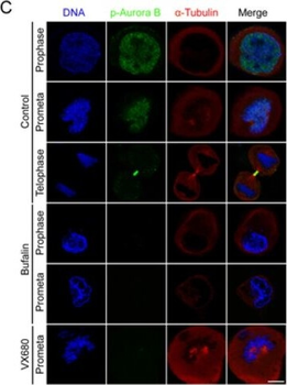
Bufalin prevents Aurora A recruitment to mitotic centrosomes and Aurora B recruitment to unattached kinetochores(A) HeLa cells were synchronized by a single thymidine treatment, released in the presence or absence of bufalin (100 nM) for 9 h, and stained for phospho-Aurora A (Green), α-tubulin (Red) and DNA (Blue). The scale bar represents 10 µm. (B) The phospho-Aurora A (Thr288) staining signals in (A) were normalized to the intensity in a same-size cytoplasmic region for at least five prometaphase cells per condition from three different experiments. ***p < 0.001, versus control prometaphase. Error bar represents SEM. (C) Thymidine-synchronized HeLa cells were treated with or without bufalin (100 nM) for 9 h and then stained for phospho-Aurora B (Green), α-tubulin (Red) and DNA (Blue). The scale bar represents 10 µm. (D) For quantification of the intensity of phospho-Aurora B (Thr232) in (C), more than 88 phospho-Aurora B (Thr232) staining signals from at least five prometaphase cells were analyzed each in control, bufalin (100 nM) and VX680 (0.5 µm) arrest. ***p < 0.001, versus control prometaphase. Error bar represents SEM.

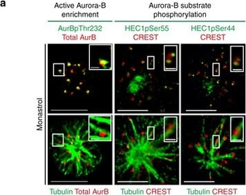
High Aurora-B activity promotes KT attachment to MT-walls. a Representative images show high Aurora-B activity on lateral kinetochores. Monastrol treated cells were immunostained with antibodies against Tubulin, Aurora-BpThr232 and total Aurora-B (AurB) (left panel) or CREST antisera and antibodies against Tubulin and either HEC1pSer55 (middle panel) or HEC1pSer44 (right panel). Cropped images show lateral-kinetochores. Scale: 5 µm in uncropped images; 1 µm in cropped images. b Graphs show higher average signal intensities of HEC1pSer55 (left) and HEC1pSer44 (right) in lateral compared to end-on kinetochores as assessed from at least nine randomly chosen kinetochores from cells in a. CREST signal intensities are used as internal controls. c Experimental regime: Cells transfected with plasmid vectors encoding Mis12-INCENP-GFP were exposed to Monastrol and MG132 with either ZM447439 or DMSO (solvent control), prior to immunostaining. d Images of cells expressing Mis12-INCENP-GFP treated as in c and immunostained with antibodies against Tubulin, SKAP and GFP. White arrowheads in cropped images show ‘Lateral' kinetochore lacking SKAP (upper panel) and ‘End-on' kinetochore enriched with SKAP (lower panel). Scale: 5 µm in uncropped and 2 µm in cropped images. Boxed areas in a and d correspond to cropped images. e Graph shows percentage of lateral, end-on and detached kinetochores in Mis12-INCENP-GFP expressing cells treated as in c. Each circle represents value from one cell. Black horizontal bar marks average values from three independent experimental repeats. ‘*' indicates statistically significant difference on the basis of P-values obtained using unpaired Student's t-test
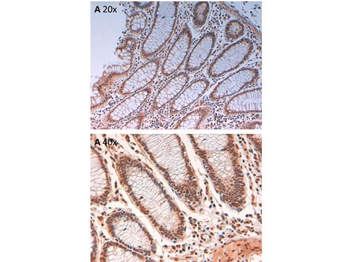
Immunohistochemistry of Rabbit Anti-AuroraB pT232 Antibody. Tissue: human intestine pH9 (A) at 20x and 40x. Fixation: formalin fixed paraffin embedded. Antigen retrieval: not required. Primary antibody: AuroraB pT232 antibody at 10 µg/ml for 1 h at RT. Secondary antibody: Peroxidase rabbit secondary antibody at 1:10000 for 45 min at RT. Localization: AuroraB pT232 is cytoplasmic. Staining: AuroraB pT232 as precipitated brown signal with hematoxylin purple nuclear counterstain.

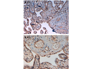
Immunohistochemistry of Rabbit Anti-AuroraB pT232 Antibody. Tissue: human placenta pH9 (A) at 20x and 40x. Fixation: formalin fixed paraffin embedded. Antigen retrieval: not required. Primary antibody: AuroraB pT232 antibody at 10 µg/ml for 1 h at RT. Secondary antibody: Peroxidase rabbit secondary antibody at 1:10000 for 45 min at RT. Localization: AuroraB pT232 is cytoplasmic. Staining: AuroraB pT232 as precipitated brown signal with hematoxylin purple nuclear counterstain.
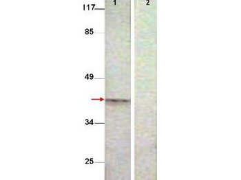
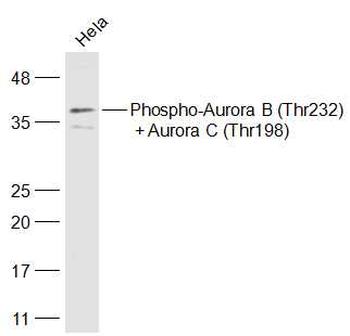
Western Blot shows detection of Aurora B protein at 39 kDa (predicted band size). All lanes : Aurora B (phospho T232) antibody diluted 1:500. Lane 1 : Extract from COS7 cells treated with Nocodazole (1 ug/ml, 16 hrs). Lane 2 : Extract from COS7 cells treated with Nocodazole (1 ug/ml, 16 hrs) and with the phosphopeptide immunogen.
Documents Download
Datasheet
Product Information
Request a Document
Protocol Information
WB
Western Blot (IB, immunoblot)
IHC
Immunohistochemistry
ELISA
Enzyme-linked Immunosorbent Assay (EIA)
AURORA KINASE B phospho T232 Antibody (orb345465)

- 0.0
Based on 0 reviews
Participating in our Biorbyt product reviews program enables you to support fellow scientists by sharing your firsthand experience with our products.
Login to Submit a Review