You have no items in your shopping cart.
Description
Research Area
Cell Biology
Images & Validation
−Item 1 of 6
| Tested Applications | ELISA, ICC, IHC-P, IP, WB |
|---|---|
| Reactivity | Canine, Human, Mouse, Plant, Porcine, Rat |
| Application Notes |
Key Properties
−| Antibody Type | Primary Antibody |
|---|---|
| Clonality | Monoclonal |
| Isotype | Mouse IgM |
| Clone No. | TU-16 |
| Immunogen | Porcine brain microtubule protein MTP-1. |
| Target | alpha-Tubulin |
| Purification | Purified by sequential steps of physicochemical fractionation (differential precipitation and solid-phase chromatography methods). |
| Conjugation | Unconjugated |
Storage & Handling
−| Storage | Maintain refrigerated at 2-8°C for up to 2 weeks. For long term storage store at -20°C in small aliquots to prevent freeze-thaw cycles. |
|---|---|
| Buffer/Preservatives | Tris buffered saline (TBS), pH 8.0, 15 mM sodium azide |
| Concentration | 1 mg/ml |
| Expiration Date | 12 months from date of receipt. |
| Disclaimer | For research use only |
Alternative Names
−TUBA
Similar Products
−- Item 1 of 23
TUBA1B Antibody [orb344425]
ELISA, IF, IHC, Multiplex Assay, WB
Human
Mouse
Monoclonal
Unconjugated
100 μg - Item 1 of 23
TUBA1B Antibody [orb344426]
ELISA, IF, IHC, Multiplex Assay, WB
Human
Mouse
Monoclonal
Unconjugated
25 μl - Item 1 of 11
Tubulin alpha Mouse Monoclonal Antibody [orb738419]
FC, ICC, IF, IHC, WB
Human, Mouse, Rat
Mouse
Monoclonal
Unconjugated
100 μg - Item 1 of 9
alpha-Tubulin Antibody [orb44529]
FC, ICC, IHC-P, IP, WB
Aves, Human, Invertebrate, Mouse, Paramecium, Plant, Porcine, Yeast
Monoclonal
Unconjugated
0.1 mg - Item 1 of 8
alpha Tubulin 1A Rabbit Polyclonal Antibody [orb556641]
ICC, IHC-P, WB
Drosophila, Human, Mouse, Rat
Rabbit
Polyclonal
Unconjugated
100 μl


Quality Guarantee
Explore bioreagents carefree to elevate your research. All our products are rigorously tested for performance. If a product does not perform as described on its datasheet, our scientific support team will provide expert troubleshooting, a prompt replacement, or a refund. For full details, please see our Terms & Conditions and Buying Guide. Contact us at [email protected].
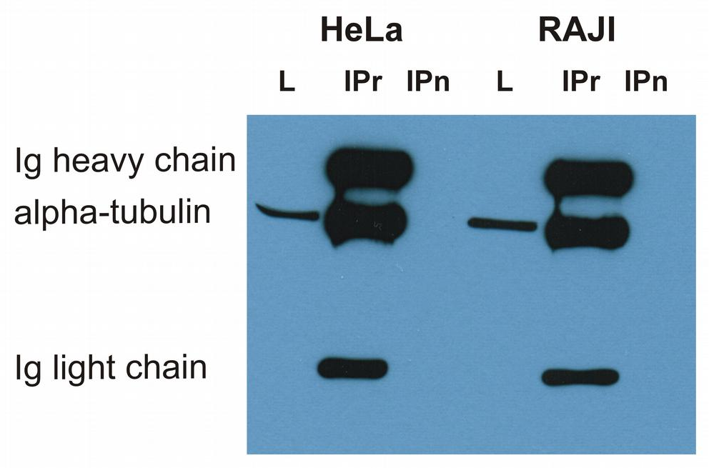
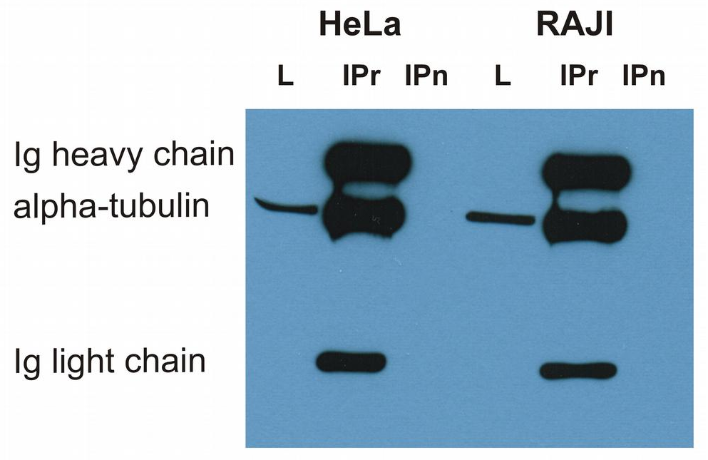
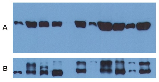
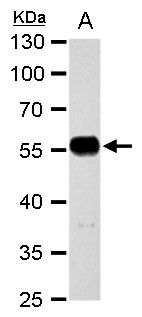
Immunoprecipitation of alpha-tubulin from HeLa and RAJI cell lysate by antibody TU-16 and its detection by antibody TU-01. IgM heavy chain (76-92 kDa) and IgM light chain (25-30 kDa) indicated. Mr of alpha tubulin is around 50 kDa. L = lysate; IPr = immunoprecipitate (reducing conditions).
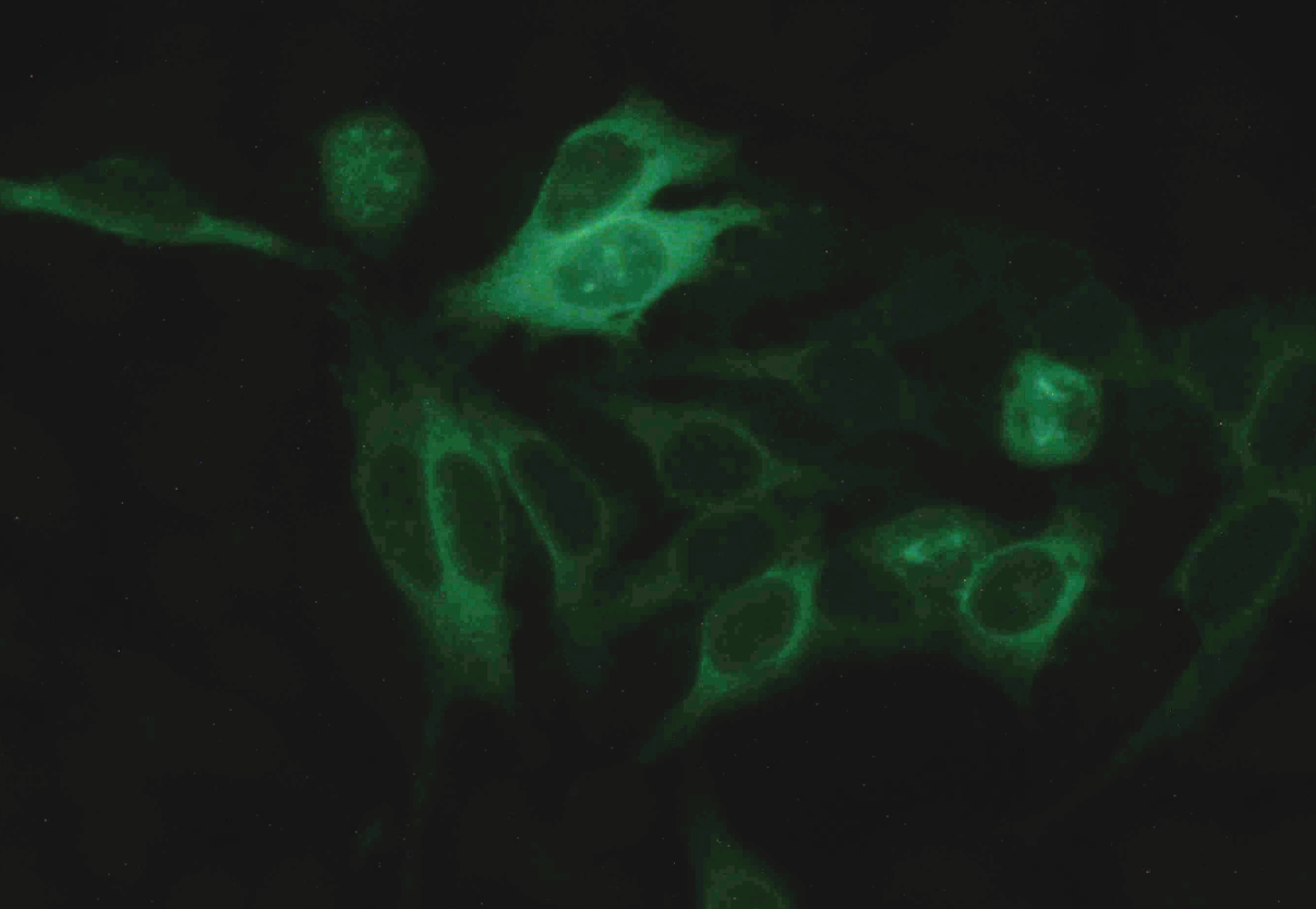
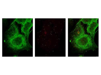
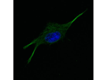
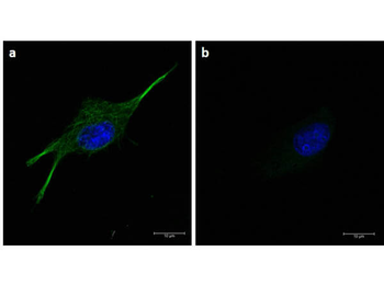
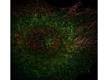
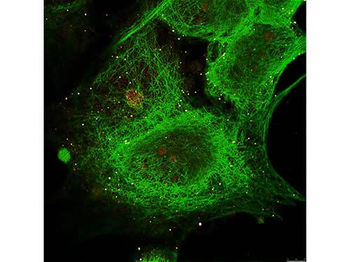
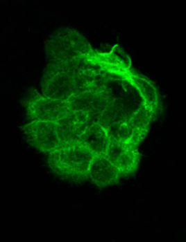
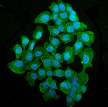
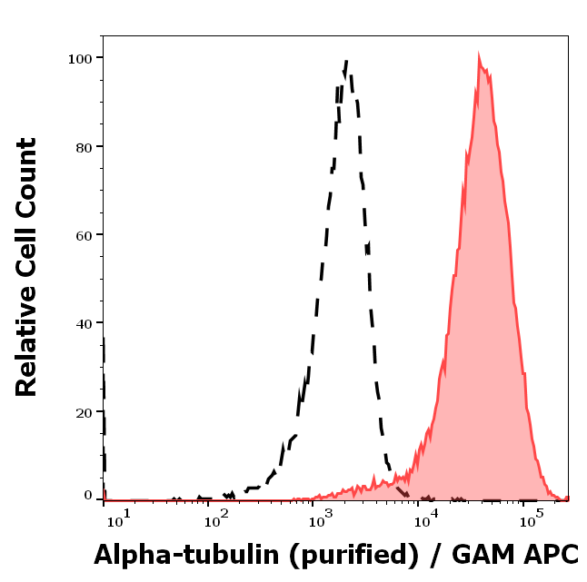
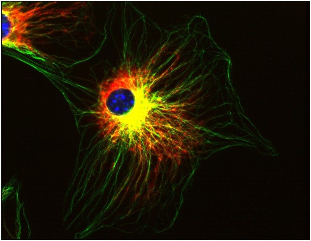
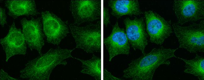
Immunocytochemistry staining of alpha-tubulin in Hep-2 cells using mouse monoclonal antibody TU-16 (diluted 1:400), detected with GAM IgG-Alexa Fluor®488 (diluted 1:200; green).
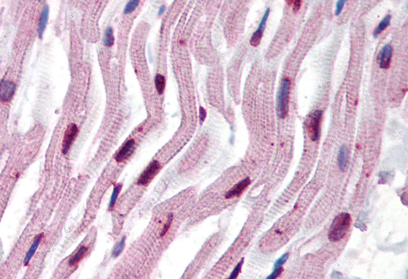
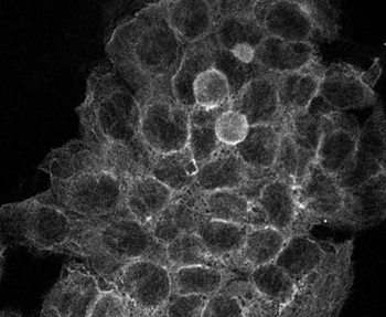
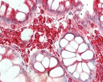
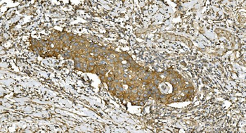
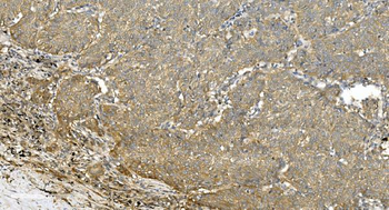
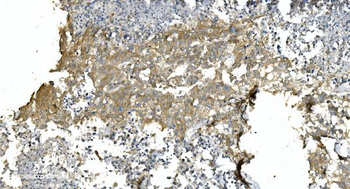
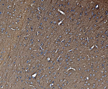
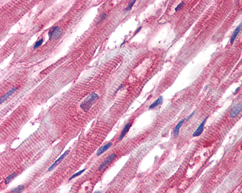
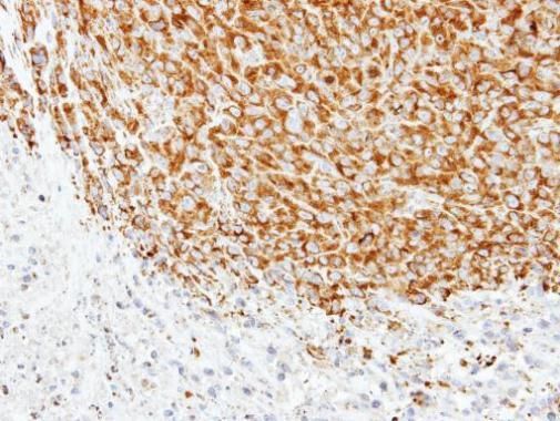
Immunohistochemistry staining of human heart (paraffin sections) using anti-alpha tubulin (TU-16).
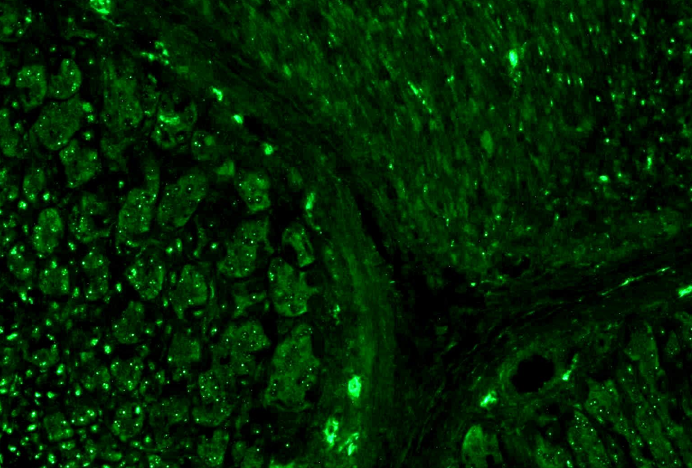
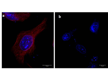
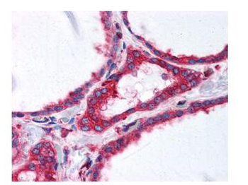
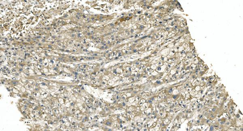
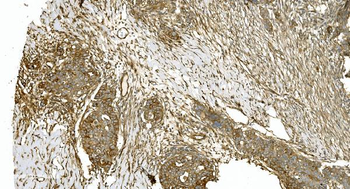
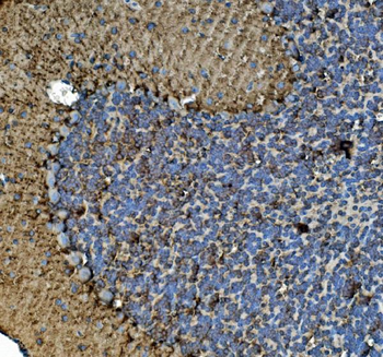
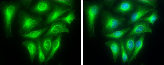
Immunohistochemistry staining (paraffin sections) of alpha-tubulin in human stomach using mouse monoclonal antibody TU-16 (diluted 1:400), detected with GAM IgG-Alexa Fluor®488 (diluted 1:200; green).
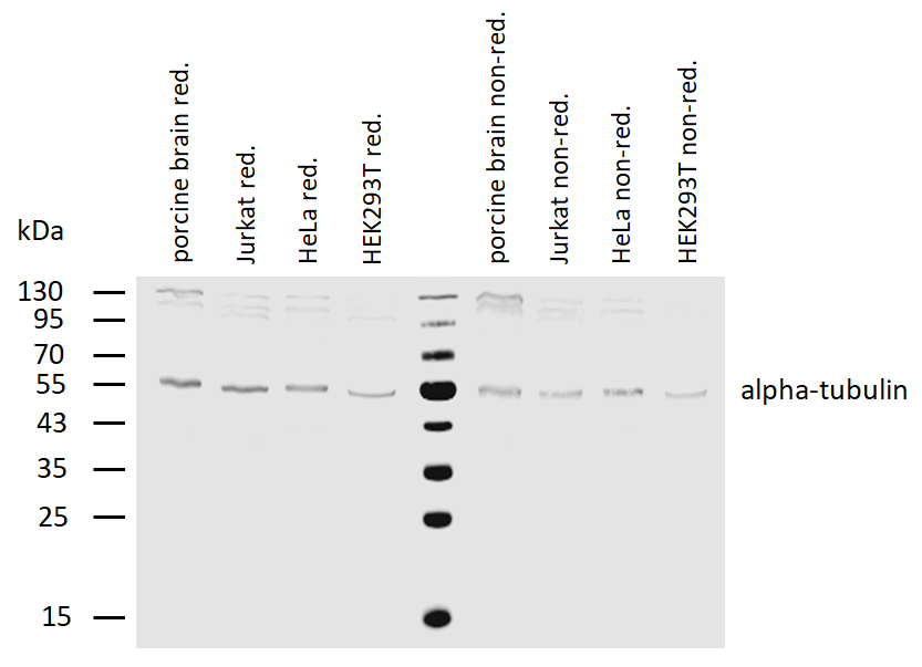
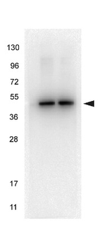
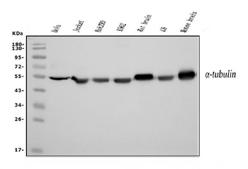
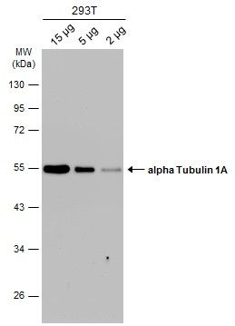
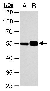
Western blotting analysis of human alpha-tubulin using mouse monoclonal antibody TU-16 on lysates of various cell lines and porcine brain under reducing and non-reducing conditions. Nitrocellulose membrane was probed with 2 µg/ml of mouse anti-alpha-tubulin monoclonal antibody followed by IRDye800-conjugated anti-mouse secondary antibody. A specific band was detected for alpha-tubulin at approximately 54 kDa, nonspecific minor bands above 100 kDa do not interfere with specific signal.
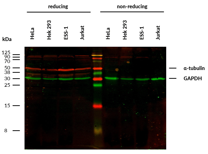
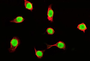
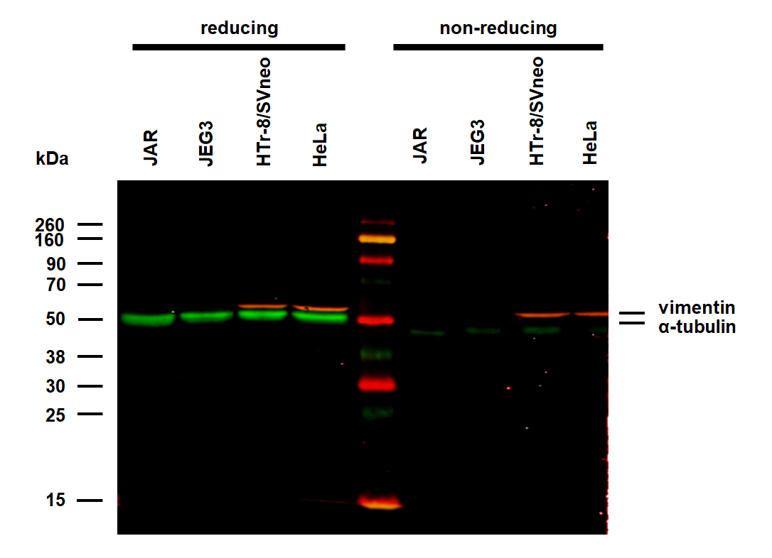
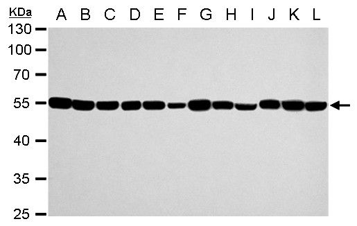
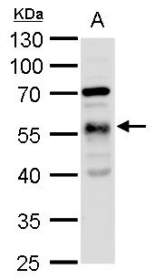
Anti-alpha-Tubulin Purified (TU-16) works in WB application under reducing conditions. Western blotting analysis was performed on whole cell extracts (RIPA lysis buffer) of HeLa, HEK 293, ESS-1 and Jurkat cell lines mixed and heated (100°C, 5 min) with reducing (2-mercaptoethanol) or non-reducing SDS-loading buffer. Samples were resolved using 12% Tris-glycine SDS gel electrophoresis. Nitrocellulose membrane blot was probed simultaneously with mouse IgM monoclonal antibody TU-16 (1 µg/ml) and mouse IgG1 anti-GAPDH monoclonal antibody FF26A (1 µg/ml) used as the loading control. Subclass-specific secondary antibodies IRDye 680RD Goat-anti-Mouse IgM (red) and IRDye 800CW Goat-anti-Mouse IgG (green) were used for multiplex fluorescent Western blot detection. Alpha-tubulin was detected at ~50 kDa in all tested cell lines.
Documents Download
Datasheet
Product Information
Request a Document
Protocol Information
WB
Western Blot (IB, immunoblot)
IHC-P
Immunohistochemistry Paraffin
ICC
Immunocytochemistry
ELISA
Enzyme-linked Immunosorbent Assay (EIA)
IP
Immunoprecipitation
alpha-Tubulin Antibody (orb44543)

- 0.0
Based on 0 reviews
Participating in our Biorbyt product reviews program enables you to support fellow scientists by sharing your firsthand experience with our products.
Login to Submit a ReviewAvailable Sizes
Select a size below
Free Secondary Antibody (20 ul)0/0
Please add an antibody product to your cart first.