You have no items in your shopping cart.
Cart summary
Item 1 of 1
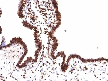
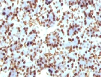
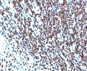
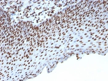
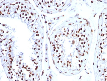
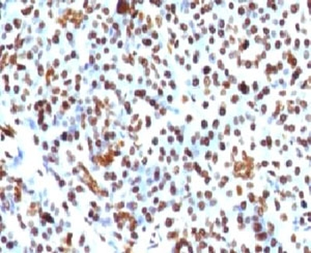
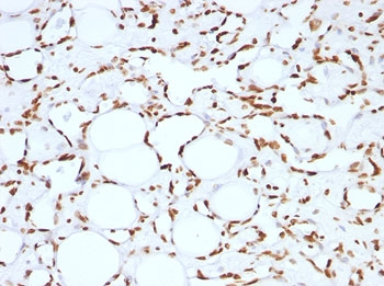
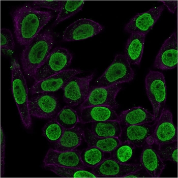
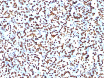
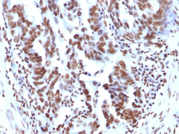
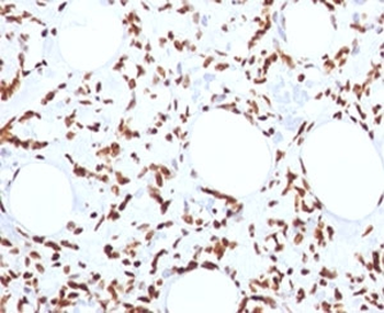
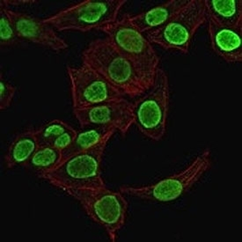
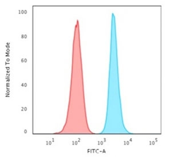
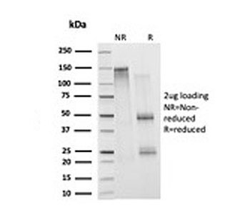
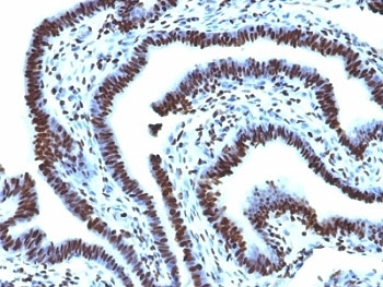
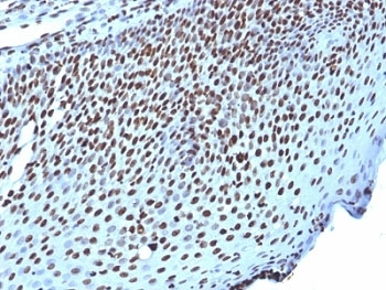
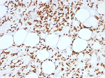
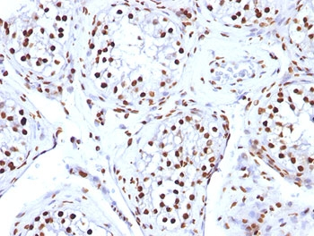
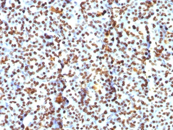
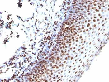
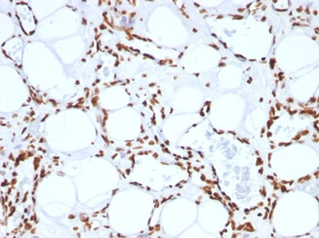
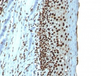
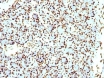
Histone H2B antibody
Catalog Number: orb6151

| Catalog Number | orb6151 |
|---|---|
| Category | Antibodies |
| Description | Histone H2B antibody |
| Species/Host | Rabbit |
| Clonality | Polyclonal |
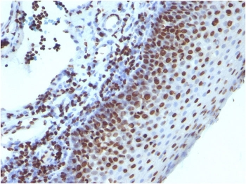
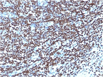
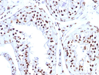
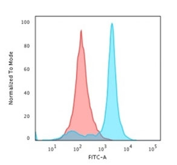
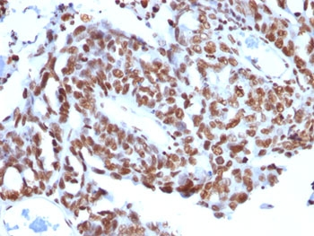
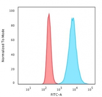
| Tested applications | FC, IHC-P |
| Predicted Reactivity | Bovine, Canine, Equine, Gallus, Mouse, Porcine, Rabbit |
| Reactivity | Human, Rat |
| Isotype | IgG |
| Immunogen | KLH conjugated synthetic peptide derived from human Histone H2B (21-126/126 aa) |
| Concentration | 1mg/ml |
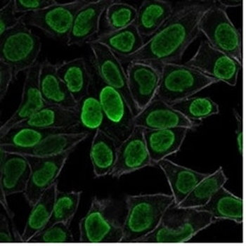
| Dilution range | IHC-P=1:100-500, IHC-F=1:100-500, ICC=1:100-500, IF=1:100-500, ELISA=1:5000-10000 |
| Form/Appearance | Liquid |
| Conjugation | Unconjugated |
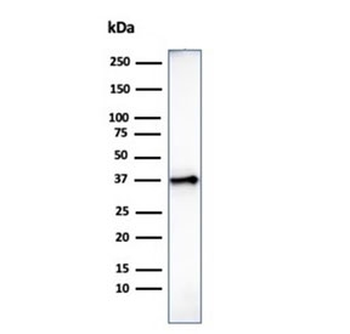
| MW | 14 kDa |
| Target | Histone H2B/HIST1H2BD |
| UniProt ID | P33778 |
| RRID | AB_10926513 |
| Storage | Shipped at 4°C. Store at -20 °C for one year. Avoid repeated freeze/thaw cycles. |
| Buffer/Preservatives | 0.01M TBS(pH7.4) with 1% rAlbumin, 0.03% Proclin300 and 50% Glycerol. |
| Alternative names | H2B.1; H2B.1 B; H2B.b; H2B.c; H2B.d; H2B.e; H2B.f; Read more... |
| Note | For research use only |
| Expiration Date | 12 months from date of receipt. |

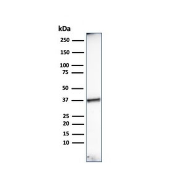
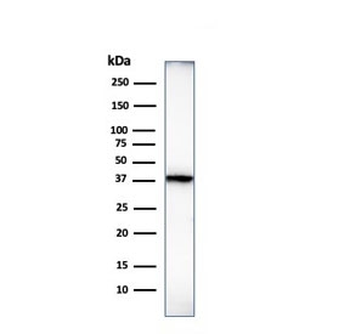
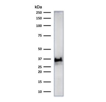
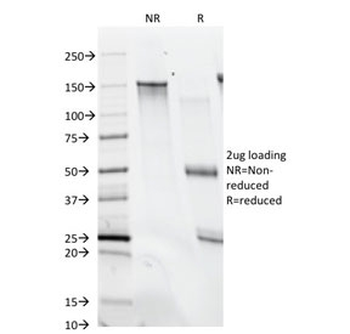
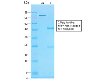
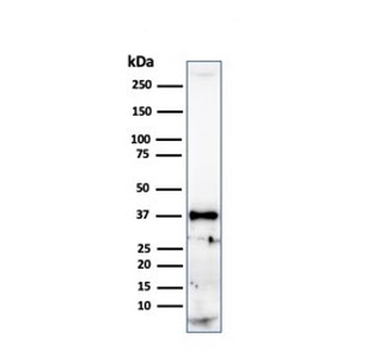
Western blot analysis of mouse liver lysates (Lane1), mouse testicular (Lane2) using Histone H2B antibody
- Item 1 of 10
Histone H1 Antibody [orb749552]
FACS, IF, IHC-P, WB
Human, Rat
Mouse
Monoclonal
Unconjugated
20 μg, 100 μg - Item 1 of 10
Histone H1 Antibody [orb317050]
FACS, IF, IHC-P, WB
Human, Mouse, Rat
Mouse
Monoclonal
Unconjugated
20 μg, 100 μg - Item 1 of 8
Histone H1 Antibody [orb749550]
FACS, IF, IHC-P, WB
Human, Mouse, Rat
Mouse
Monoclonal
Unconjugated
20 μg, 100 μg - Item 1 of 7
Histone H1 Antibody [orb749551]
FACS, IF, IHC-P, WB
Human, Mouse, Rat
Mouse
Monoclonal
Unconjugated
20 μg, 100 μg - Item 1 of 6
Histone H1 Antibody [orb606517]
FACS, IF, IHC-P, WB
Human, Mouse, Rat
Mouse
Recombinant
Unconjugated
20 μg, 100 μg


Submit a review
Filter by Rating

- 5 stars

- 4 stars
- 3 stars
- 2 stars
- 1 stars
