You have no items in your shopping cart.
Cart summary
Item 1 of 7
Item 1 of 7
Histone H1 Antibody
Catalog Number: orb749551

| Catalog Number | orb749551 |
|---|---|
| Category | Antibodies |
| Description | Eukaryotic histones are basic and water-soluble nuclear proteins that form hetero-octameric nucleosome particles by wrapping 146 base pairs of DNA in a left-handed super-helical turn sequentially to form chromosomal fiber. Two molecules of each of the four core histones (H2A, H2B, H3, and H4) form the octamer; formed of two H2A-H2B dimers and two H3-H4 dimers, forming two nearly symmetrical halves by tertiary structure. Over 80% of nucleosomes contain the linker Histone H1, derived from an intronless gene that interacts with linker DNA between nucleosomes and mediates compaction into higher order chromatin. Histones are subject to posttranslational modification by enzymes primarily on their N-terminal tails, but also in their globular domains. Such modifications include methylation, citrullination, acetylation, phosphorylation, sumoylation, ubiquitination and ADP-ribosylation. |
| Clonality | Monoclonal |
| Species/Host | Mouse |
| Isotype | Mouse IgG2a, kappa |
| Conjugation | Unconjugated |
| Reactivity | Human, Mouse, Rat |
| Immunogen | Nuclei of human leukemia biopsy cells were used as the immunogen for the anti-Histone H1 antibody. |
| UniProt ID | P07305 |
| Tested applications | FACS, IF, IHC-P, WB |
| Dilution range | Immunofluorescence: 1-2ug/ml,Immunohistochemistry (FFPE): 1-2ug/ml for 30 min at RT,Western blot: 1-2ug/ml,Flow cytometry: 1-2ug/million cells |
| Application notes | Optimal dilution of the anti-Histone H1 antibody should be determined by the researcher.1. Staining of formalin/paraffin tissues requires boiling tissue sections in pH 9 10mM Tris with 1mM EDTA for 10-20 min followed by cooling at RT for 20 min.2. The prediluted format is supplied in a dropper bottle and is optimized for use in IHC. After epitope retrieval step (if required), drip mAb solution onto the tissue section and incubate at RT for 30 min. |
| Antibody Type | Primary Antibody |
| Clone Number | SPM256 |
| Formula | 0.2 mg/ml in 1X PBS with 0.1 mg/ml BSA (US sourced) and 0.05% sodium azide |
| Storage | Maintain refrigerated at 2-8°C for up to 2 weeks. For long term storage store at -20°C in small aliquots to prevent freeze-thaw cycles. |
| Research Area | Epigenetics |
| Note | For research use only |
| Expiration Date | 12 months from date of receipt. |
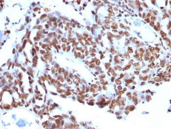
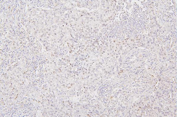
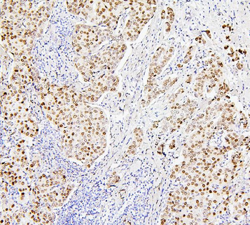
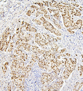
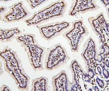
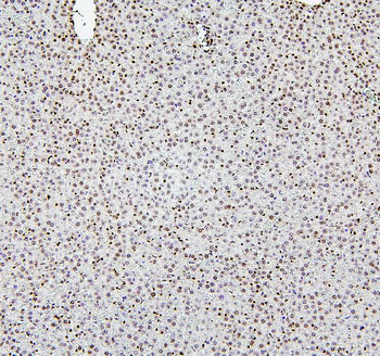
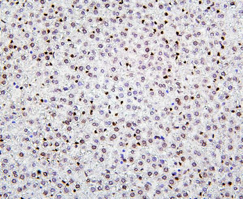
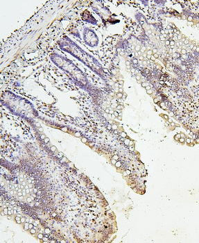
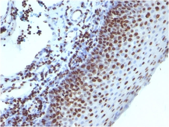
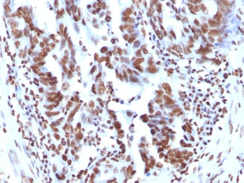
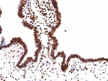
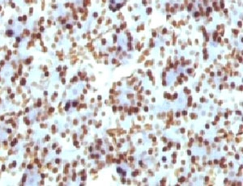
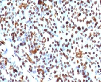
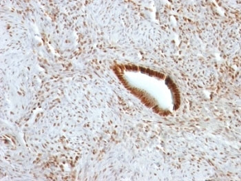
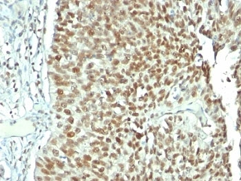
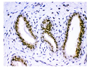
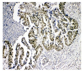
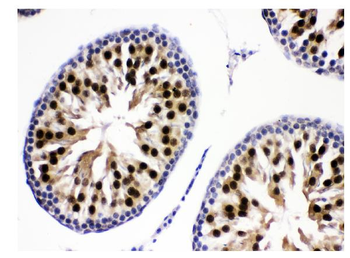
IHC: FFPE human ovarian carcinoma tested with anti-Histone H1 antibody (clone SPM256).
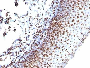
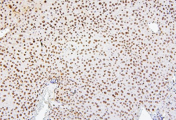
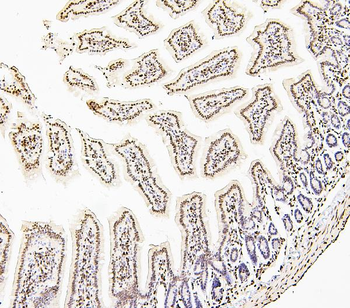
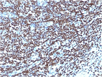
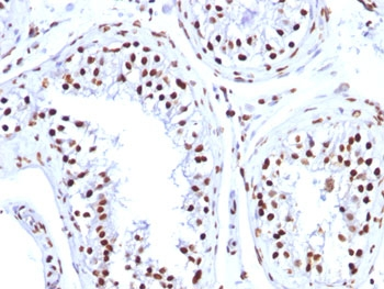
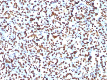
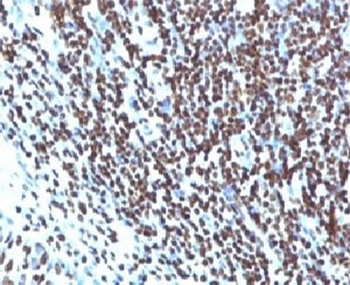
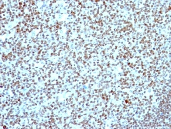
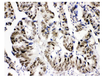
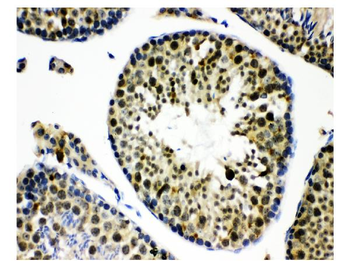
IHC: Formalin-fixed, paraffin-embedded human tonsil stained with anti-Histone H1 antibody (clone SPM256).
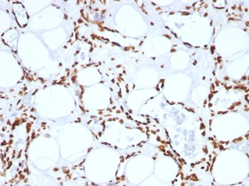
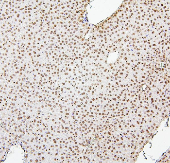
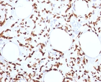
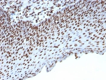
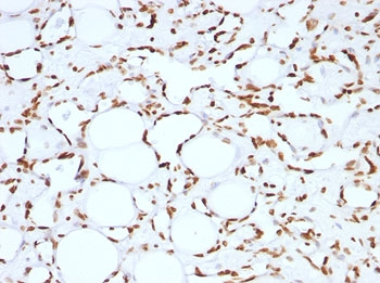
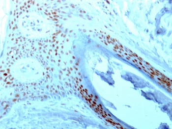
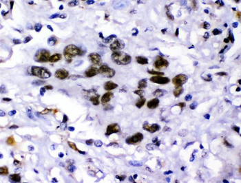
IHC: FFPE human angiosarcoma tested with anti-Histone H1 antibody (clone SPM256).

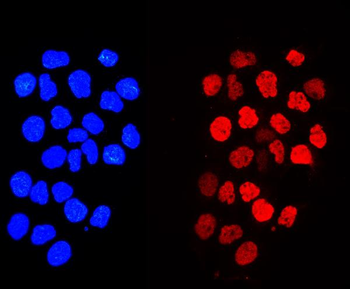
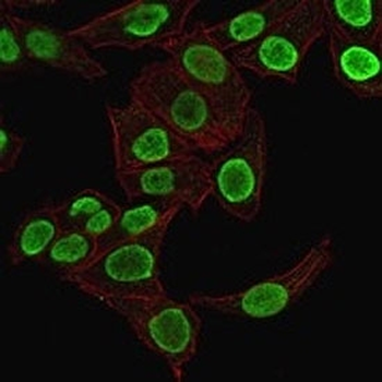
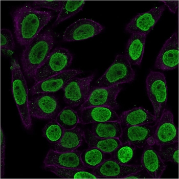
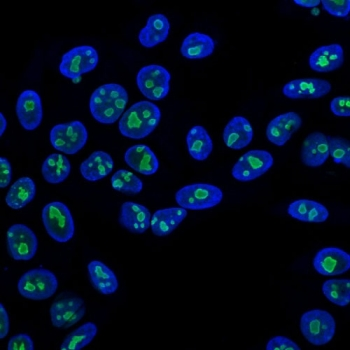
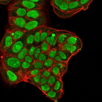
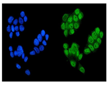
Immunofluorescent staining of permeabilized human HeLa cells with anti-Histone H1 antibody (clone SPM256, green) and Phalloidin (red).
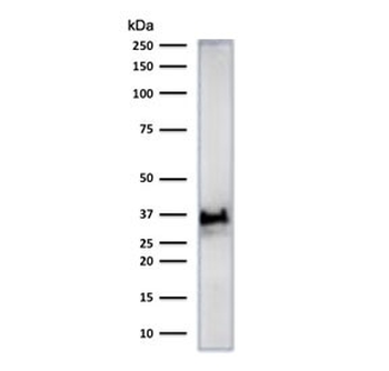
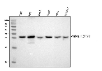
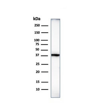
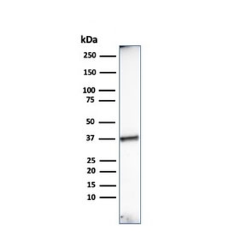
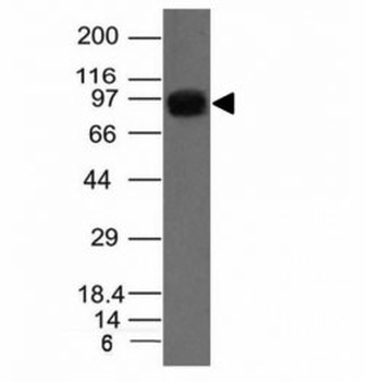
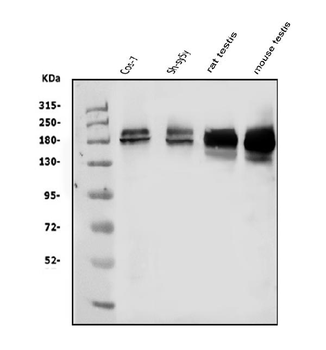
Western blot test of human heart lysate with anti-Histone H1 antibody (clone SPM256).
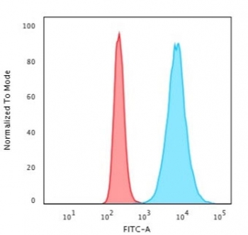
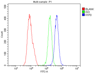
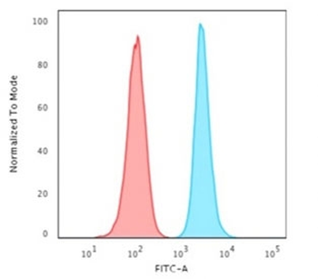
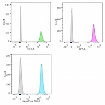
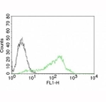
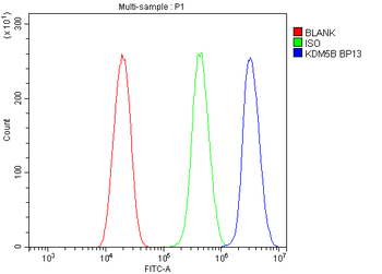
Flow cytometry testing of permeabilized human HeLa cells with anti-Histone H1 antibody (clone SPM256); Red = isotype control, Blue = anti-Histone H1 antibody.
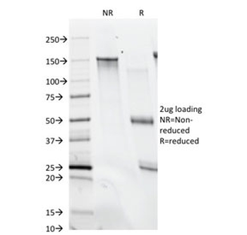
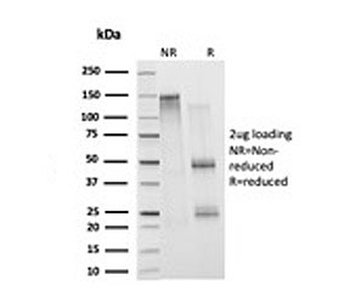
SDS-PAGE analysis of purified, BSA-free anti-Histone H1 antibody (clone SPM256) as confirmation of integrity and purity.
- Item 1 of 13
Histone H1.0/H1F0 Antibody [orb570388]
ELISA, FC, ICC, IF, IHC, WB
Human, Mouse, Rat
Rabbit
Polyclonal
Unconjugated
100 μg - Item 1 of 10
Histone H1 Antibody [orb317050]
FACS, IF, IHC-P, WB
Human, Mouse, Rat
Mouse
Monoclonal
Unconjugated
20 μg, 100 μg - Item 1 of 10
Histone H1 Antibody [orb749552]
FACS, IF, IHC-P, WB
Human, Rat
Mouse
Monoclonal
Unconjugated
20 μg, 100 μg - Item 1 of 10
Nucleolin Antibody [orb749711]
FACS, IF, IHC-P, WB
Human, Mouse, Rat
Mouse
Monoclonal
Unconjugated
20 μg, 100 μg - Item 1 of 9
KDM5B/PLU1/Jarid1B Antibody [orb381075]
FC, ICC, IF, IHC, WB
Human, Monkey, Mouse, Rat
Rabbit
Polyclonal
Unconjugated
100 μg