You have no items in your shopping cart.
Featured
KO/KD
Validated
Validated
Description
Research Area
Cell Biology
Images & Validation
−Item 1 of 10
| Tested Applications | ELISA, IF, IHC-P, KO/KD Validated, WB |
|---|---|
| Reactivity | Human, Rat |
| Predicted Reactivity | Mouse |
Key Properties
−| Antibody Type | Primary Antibody |
|---|---|
| Host | Rabbit |
| Clonality | Polyclonal |
| Isotype | IgG |
| Immunogen | Anti-PUMA antibody (orb1239919) was raised against a peptide corresponding to 14 amino acids near the amino terminus of human PUMA isoform 1. The immunogen is located within the first 50 amino acids of PUMA. |
| Target | BBC3 |
| Molecular Weight | Predicted: 21kD Observed: 23kD |
| Purification | PUMA Antibody is affinity chromatography purified via peptide column. |
| Conjugation | Unconjugated |
Storage & Handling
−| Storage | Maintain refrigerated at 2-8°C for up to 2 weeks. For long term storage store at -20°C in small aliquots to prevent freeze-thaw cycles. |
|---|---|
| Form/Appearance | Liquid |
| Buffer/Preservatives | PUMA Antibody is supplied in PBS containing 0.02% sodium azide. |
| Concentration | 1 mg/mL |
| Expiration Date | 12 months from date of receipt. |
| Disclaimer | For research use only |
Alternative Names
−PUMA Antibody: JFY1, PUMA, JFY-1, Bcl-2-binding component 3
Similar Products
−- Item 1 of 9
BBC3 Antibody [orb1239911]
ELISA, ICC, IF, KO/KD Validated, WB
Rat
Human, Mouse
Rabbit
Polyclonal
Unconjugated
0.02 mg, 0.1 mg - Item 1 of 1
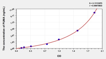
Human p53 Upregulated Modulator of Apoptosis (PUMA) ELISA Kit [orb776538]
Human
0.16-10 ng/mL
0.058 ng/mL
48 T, 96 T - Item 1 of 1
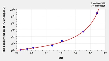
Rat p53 Upregulated Modulator of Apoptosis (PUMA) ELISA Kit [orb779906]
Rat
0.16-10 ng/mL
0.048 ng/mL
48 T, 96 T - Item 1 of 4
Puma BH3 Domain Antibody [orb1936871]
FC, IF, IHC-P, WB
Mouse, Rat
Human
Rabbit
Polyclonal
Unconjugated
50 μl, 100 μl - Item 1 of 2
BBC3/PUMA Rabbit Polyclonal Antibody [orb10176]
IF, IHC-Fr, IHC-P, WB
Bovine, Canine, Mouse, Porcine
Human, Rat
Rabbit
Polyclonal
Unconjugated
50 μl, 100 μl, 200 μl

Quality Guarantee
Explore bioreagents carefree to elevate your research. All our products are rigorously tested for performance. If a product does not perform as described on its datasheet, our scientific support team will provide expert troubleshooting, a prompt replacement, or a refund. For full details, please see our Terms & Conditions and Buying Guide. Contact us at [email protected].
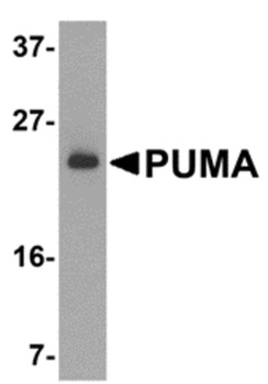
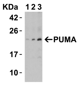
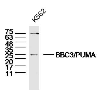
Western Blot Validation of PUMA in K562 Cells. Loading: 15 µg of lysates per lane. Antibodies: orb1239919 (2 µg/mL), 1 h incubation at RT in 5% NFDM/TBST. Secondary: Goat anti-rabbit IgG HRP conjugate at 1:10000 dilution.
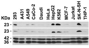
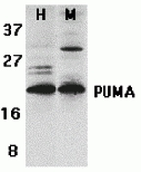
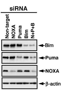
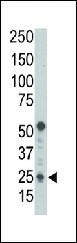
Independent Antibody Validation (IAV) via Protein Expression Profile in Human Cells. Loading: 20 µg of lysates per lane. Antibodies: orb1239911 (3 µg/mL), orb1239919 (2 µg/mL), beta-actin (1 µg/mL) and GAPDH (0.02 µg/mL), 1 h incubation at RT in 5% NFDM/TBST. Secondary: Goat anti-rabbit IgG HRP conjugate at 1:10000 dilution.
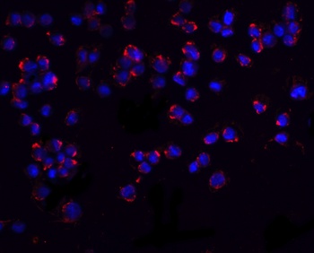
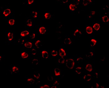
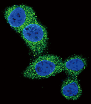
Immunofluorescence Validation of PUMA in K562 Cells. Immunofluorescent analysis of 4% paraformaldehyde-fixed K562 cells labeling PUMA with orb1239919 at 20 µg/mL, followed by goat anti-rabbit IgG secondary antibody at 1/500 dilution (red) and DAPI staining (blue).
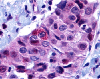
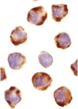
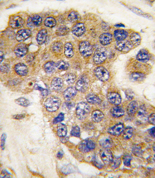
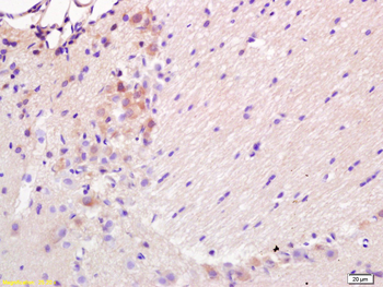
Immunohistochemistry Validation of PUMA in Human Breast Carcinoma. Immunohistochemical analysis of paraffin-embedded human breast carcinoma tissue using anti-PUMA antibody (orb1239919) at 10 µg/ml. Tissue was fixed with formaldehyde and blocked with 10% serum for 1 h at RT; antigen retrieval was by heat mediation with a citrate buffer (pH6). Samples were incubated with primary antibody overnight at 4°C. A goat anti-rabbit IgG H&L (HRP) at 1/250 was used as secondary. Counter stained with Hematoxylin.
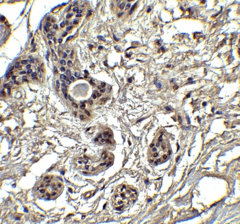
Immunohistochemistry Validation of PUMA in Human Breast Tissue. Immunohistochemical analysis of paraffin-embedded human breast tissue using anti-PUMA antibody (orb1239919) at 2.5 µg/ml. Tissue was fixed with formaldehyde and blocked with 10% serum for 1 h at RT; antigen retrieval was by heat mediation with a citrate buffer (pH6). Samples were incubated with primary antibody overnight at 4°C. A goat anti-rabbit IgG H&L (HRP) at 1/250 was used as secondary. Counter stained with Hematoxylin.
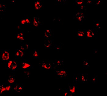
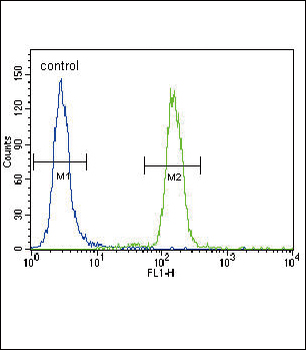
Immunofluorescence Validation of PUMA in K562. Immunofluorescent analysis of 4% paraformaldehyde-fixed K562 cells labeling PUMA with orb1239919 at 10 µg/mL, followed by goat anti-rabbit IgG secondary antibody at 1/500 dilution (red). Image showing cytosol staining on K562 cells.
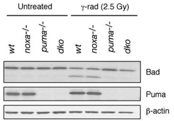
KO Validation of PUMA in Mouse Thymocytes (Michalak et al., 2008). Western blot analysis ofthymocytes from wt, noxa knockout, puma knockout and noxa/puma double knockout mice cultured for 7 h in the presence or absence of 2.5 Gy g-irradiation.PUMA expression was not detected in puma KO and double KO mice with anti-PUMA antibodies (orb1239919).
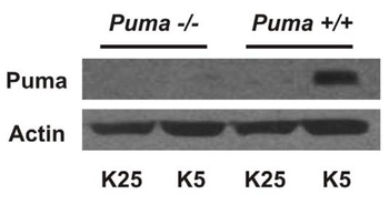
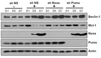
KO Validation of PUMA in Mouse Cerebellar Neurons (Ambacher et al., 2012). Puma expression is induced by potassium withdrawal in cerebellar granule neurons. After 7 days in culture CGNs were either maintained in media containing 25 mM potassium (K25) or switched to low potassium medium containing 5 mM potassium (K5). PUMA protein levels were analyzed by western blot with anti-PUMA antibodies (orb1239919). PUMA expression was not detected in PUMA KO mice and was increased after treatment in WT.
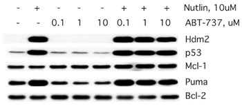
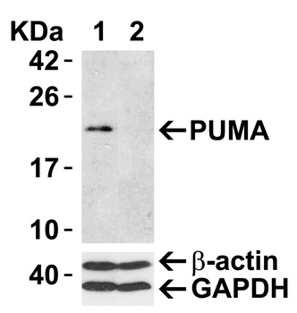
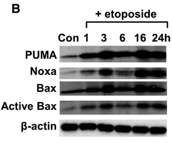
Induced Expression of PUMA in MCF7 cells (Wade et al., 2008). Western analysis of MCF7 treated with the indicated dose of Nutlin-3a orABT-737 for 24h. Note that Puma is induced following Nutlin-3a treatment in these cells and PUMA expression was detected by anti-PUMA antibodies (orb1239919).
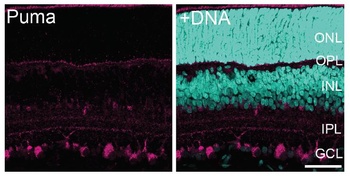
Immunofluorescence Validation of PUMA in Rat Retina (Wakabayashi et al., 2012). PUMA expression in the rat retina detected by anti-PUMA antibodies (orb1239919). The specimens were counterstained with Hoechst 33258 to visualize nuclei (+DNA). GCL, ganglion cell layer; INL, inner nuclear layer; IPL, inner plexiform layer; ONL, outer nuclear layer; OPL, outer plexiform layer; P, postnatal day.
Documents Download
Datasheet
Product Information
Request a Document
Protocol Information
WB
Western Blot (IB, immunoblot)
IHC-P
Immunohistochemistry Paraffin
IF
Immunofluorescence
ELISA
Enzyme-linked Immunosorbent Assay (EIA)
BBC3 Antibody (orb1239919)

- 0.0
Based on 0 reviews
Participating in our Biorbyt product reviews program enables you to support fellow scientists by sharing your firsthand experience with our products.
Login to Submit a ReviewAvailable Sizes
Select a size below
Free Secondary Antibody (20 ul)0/0
Please add an antibody product to your cart first.