You have no items in your shopping cart.
Cart summary
Item 1 of 2
Item 1 of 2
Phosphotyrosine Antibody / P-Tyr
Catalog Number: orb2639674

| Catalog Number | orb2639674 |
|---|---|
| Category | Antibodies |
| Description | Protein phosphorylation is a fundamental event in the regulation of a large number of intracellular processes. Phosphorylation of specific tyrosine residues is the result of activation or stimulation of their respective protein tyrosine kinases. The phosphorylated proteins can be auto-phosphorylated kinases or certain cellular protein substrates. Tyrosine-phosphorylated proteins are involved in signal transduction and in the regulation of cell proliferation. Antibody to phosphotyrosine provides an excellent tool for the detection, characterization, and purification of phosphotyrosine containing proteins. This mAb shows no cross-reaction with other phosphoamino acids and is superb for multiple applications including staining of formalin/paraffin tissues. |
| Species/Host | Mouse |
| Clonality | Monoclonal |
| Clone Number | PY793 |
| Tested applications | FACS, IF, IHC-P |
| Isotype | Mouse IgG2b, kappa |
| Immunogen | P-Tyr conjugated to BSA was used as the immunogen for the Phosphotyrosine antibody. |
| Antibody Type | Primary Antibody |
| Dilution range | Flow cytometry: 0.5-1ug/million cells,Immunofluorescence: 1-2ug/ml,Immunohistochemistry (FFPE): 1-2ug/ml for 30 min at RT |
| Purity | Protein G affinity chromatography |
| Conjugation | Unconjugated |
| Formula | 0.2 mg/ml in 1X PBS with 0.1 mg/ml BSA (US sourced) and 0.05% sodium azide |
| Hazard Information | This Phosphotyrosine antibody is available for research use only. |
| Storage | Maintain refrigerated at 2-8°C for up to 2 weeks. For long term storage store at -20°C in small aliquots to prevent freeze-thaw cycles. |
| Buffer/Preservatives | 0.2 mg/ml in 1X PBS with 0.1 mg/ml rAlbumin (US sourced) and 0.05% sodium azide |
| Note | For research use only |
| Application notes | Optimal dilution of the Macrophage / Histiocytoma Marker antibody should be determined by the researcher.1. Staining of formalin-fixed tissues requires boiling tissue sections in 10mM Citrate buffer, pH 6.0, for 10-20 min followed by cooling at RT for 20 min.2. The prediluted format is supplied in a dropper bottle and is optimized for use in IHC. After epitope retrieval step (if required), drip mAb solution onto the tissue section and incubate at RT for 30 min. |
| Expiration Date | 12 months from date of receipt. |
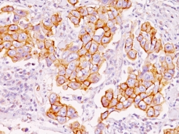
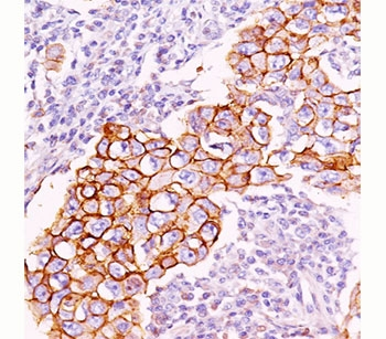
IHC: Formalin-fixed, paraffin-embedded human breast carcinoma stained with Phosphotyrosine antibody (clone PY793).
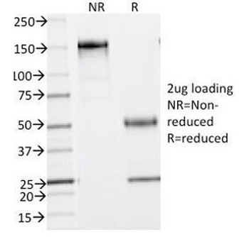
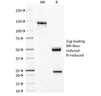
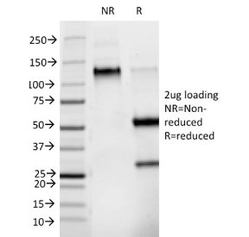
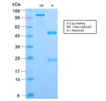
SDS-PAGE Analysis of Purified, BSA-Free Phosphotyrosine Antibody (clone PY793). Confirmation of Integrity and Purity of the Antibody.
- Item 1 of 2
Phosphotyrosine Antibody / P-Tyr [orb749422]
FACS, IF, IHC-P, IP, WB
Mouse
Monoclonal
Unconjugated
20 μg - Item 1 of 2
- Item 1 of 2
Phosphotyrosine Antibody / P-Tyr [orb2638092]
FACS, IF, IHC-P, IP, WB
Mouse
Monoclonal
Unconjugated
100 μg - Item 1 of 1
- Item 1 of 1