You have no items in your shopping cart.
Cart summary
Item 1 of 4
Item 1 of 4
PGP9.5 antibody
Catalog Number: orb33715
| Catalog Number | orb33715 | ||||
|---|---|---|---|---|---|
| Category | Antibodies | ||||
| Description | Rabbit polyclonal antibody to PGP9.5 | ||||
| Species/Host | Rabbit | ||||
| Clonality | Polyclonal | ||||
| Clone Number | RB7379 | ||||
| Tested applications | FC, IF, IHC-P, WB | ||||
| Predicted Reactivity | Equine, Porcine | ||||
| Reactivity | Human, Mouse, Rat | ||||
| Isotype | Rabbit IgG | ||||
| Immunogen | Synthetic Peptide | ||||
| Dilution range | WB: 1:1000, IHC-P: 1:50-100, FACS: 1:10-50, IF/ICC: 1:10-50 | ||||
| Form/Appearance | Purified polyclonal antibody supplied in PBS with 0.09% (W/V) sodium azide. This antibody is purified through a protein A column, followed by peptide affinity purification. | ||||
| Conjugation | Unconjugated | ||||
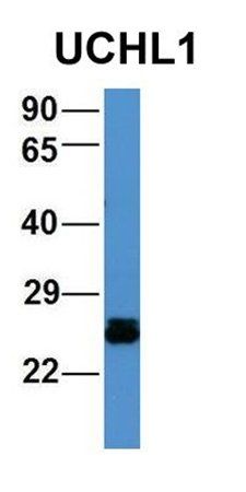
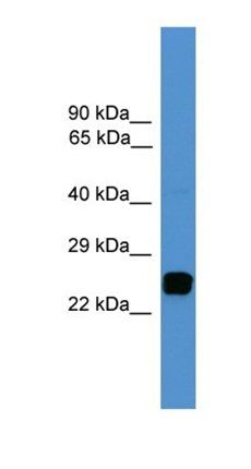
| MW | 24824 | ||||
| Target | UCHL1 | ||||
| UniProt ID | P09936 | ||||
| NCBI | NP_004172.2 | ||||
| Alternative names | anti-Epididymis luminal protein 117 antibody, anti Read more... | ||||
| Note | For research use only | ||||
| Application notes | Protein InformationName | UCHL1Function | Ubiquitin-protein hydrolase involved both in the processing of ubiquitin precursors and of ubiquitinated proteins. This enzyme is a thiol protease that recognizes and hydrolyzes a peptide bond at the C-terminal glycine of ubiquitin. Also binds to free monoubiquitin and may prevent its degradation in lysosomes. The homodimer may have ATP-independent ubiquitin ligase activity. Cellular Location | Cytoplasm. Endoplasmic reticulum membrane; Lipid-anchor. Note=About 30% of total UCHL1 is associated with membranes in brainTissue Location | Found in neuronal cell bodies and processes throughout the neocortex (at protein level). Expressed in neurons and cells of the diffuse neuroendocrine system and their tumors Weakly expressed in ovary. Down-regulated in brains from Parkinson disease and Alzheimer disease patients |
| Expiration Date | 12 months from date of receipt. |
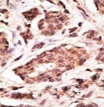
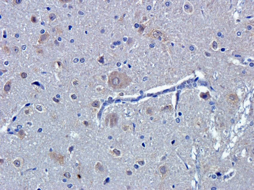
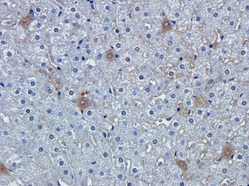
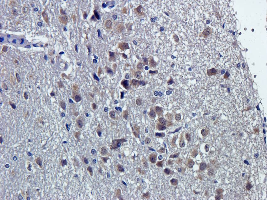
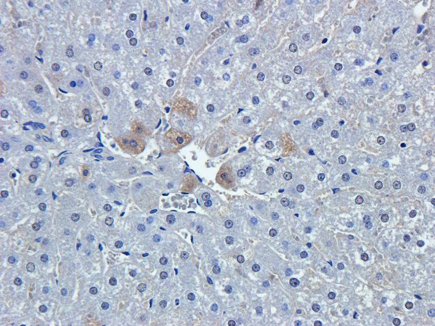
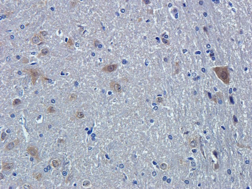
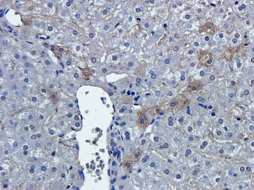
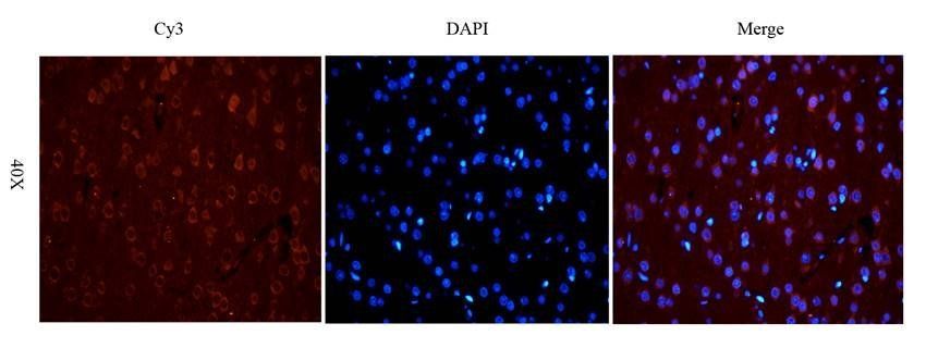
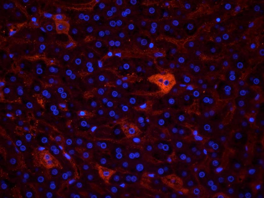
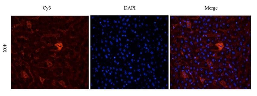
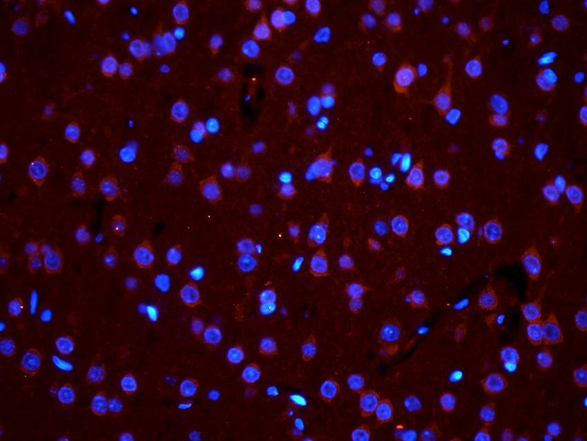
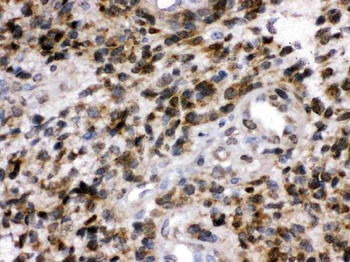
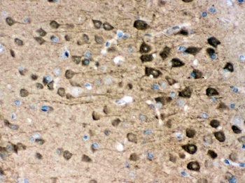
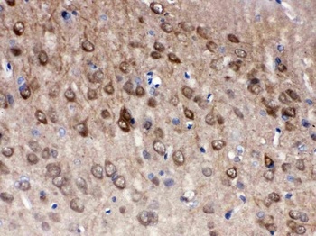
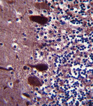
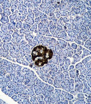
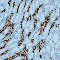
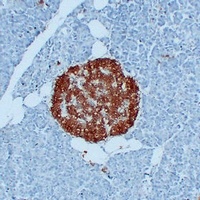
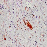
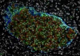
Immunohistochemical analysis of formalin-fixed paraffin embedded human cancer tissue using PGP9.5 antibody (Dilution at :1:200)
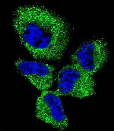
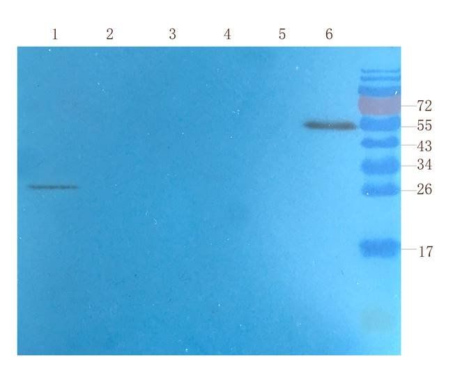
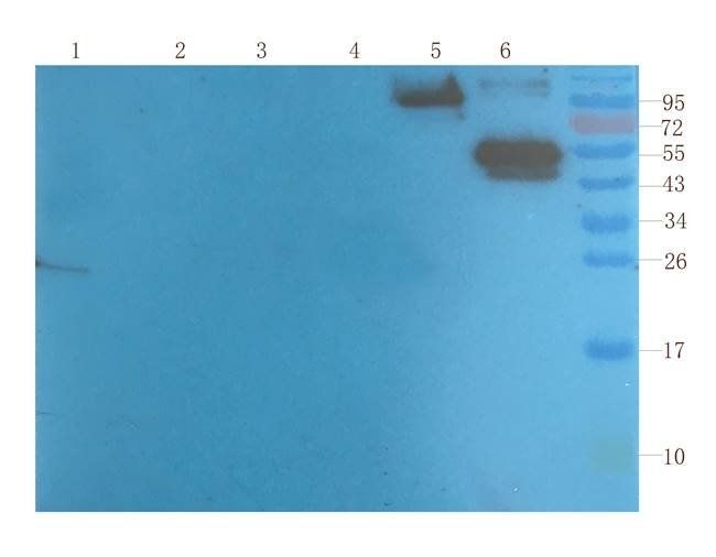
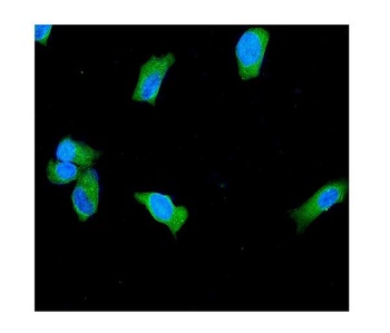
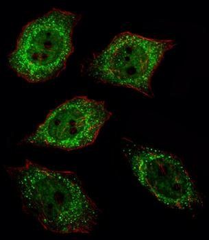
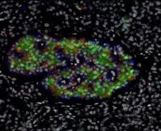
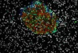
Immunofluorescense analysis of NCI-H460 cell lysate using PGP9.5 antibody
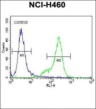
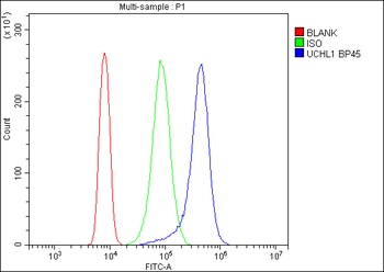
Flow cytometric analysis of NCI-H460 cells (right histogram) compared to a negative control cell (left histogram) using PGP9.5 antibody
- Item 1 of 12
PGP9.5 antibody [orb6713]
ELISA, ICC, IF, IHC-P, WB
Human, Mouse, Rat
Rabbit
Polyclonal
Unconjugated
200 μg, 100 μg - Item 1 of 6
PGP9.5/UCHL1 Antibody [orb334572]
FC, ICC, IF, IHC, WB
Hamster
Human, Mouse, Rat
Rabbit
Polyclonal
Unconjugated
10 μg, 100 μg - Item 1 of 5
PGP9.5 antibody [orb33716]
IF, IHC-P, WB
Equine, Mouse, Porcine
Human, Rat
Rabbit
Polyclonal
Unconjugated
80 μl - Item 1 of 4
- Item 1 of 5
PGP9.5 antibody [orb331010]
IHC, WB
Animal, Bovine, Canine, Guinea pig, Human, Mouse, Rabbit, Rat, Zebrafish
Canine, Equine, Guinea pig, Human, Mouse, Rat, Zebrafish
Rabbit
Polyclonal
Unconjugated
100 μl
Submit a review
Filter by Rating

- 5 stars

- 4 stars
- 3 stars
- 2 stars
- 1 stars